
Cutaneous malignancies accounts for most cancers across the world and they are primarily divided into melanoma (MSC) and nonmelanoma (NMSC) skin cancers [1]. According to the Globocan (Global Cancer Observatory) 2022 data, non-melanoma skin cancer ranks as the fifth most commonly diagnosed cancer worldwide, with over 1.2 million new cases reported [2]. However, despite its high incidence, NMSC ranks only 22nd in terms of cancer-related mortality, highlighting the generally favorable prognosis of NMSC compared to other malignancies [2].
The incidence of head and neck MSC and NMSC is rising all over the world and can be the source of significant morbidity and mortality [3]. The necessity of a multidisciplinary team that includes dermatology, plastic and reconstructive surgery, medical and radiation oncology remains a financial problem in many countries, due to the comprehensive care that needs to be provided for patients with head and neck cutaneous malignancies [3,4].
The vast majority of non-melanoma skin cancers include basal cell carcinoma (BCC), followed by squamous cell carcinoma (SCC), both showing a rapidly increasing trend in their incidence in the last years and a significant health burden in many countries [5,6]. Although the exact global incidence of these two is unknown, an overview of the last years carried out by World Health Organization concluded that BCC comprises about 80% of all NMSC, while SCC comprise about 20%, with a BCC to SCC ratio ranging from 2 to 4:1 [5,6]. While this is a classic agreed ratio, the recent studies suggest a possible changing in the pattern due to aging of the population, with equal numbers of SCC and BCC being treated in the future [6,7].
Multiple etiological risk factors related to the individual or the environment were studied for understanding the cause of NMSC, but the main driving carcinogen remains ultraviolet radiation (UVR) [5,6]. This explains why over 80% of these malignancies occur on the sun exposed skin of the head and neck (followed by the hands and forearms), with higher prevalence of BCCs in the nasal area, while SCCs are preferentially located on the auricle [5,8,9]. UVR are responsible for the induced dinucleotide mutations at pyrimidine bases, exhibiting strong transcription strand bias and also showing a high mutational burden with the mutant genes varying between BCCs and SCCs [6].
Although its mortality is low and it rarely results in metastatic disease, BCCs can cause significant health burden through his great morbidity, generated by the destructive local spread [9]. These tumors are slow growing but can, however, develop large, causing locally tissular destruction and spreading into vital structures such as the central nervous system or important blood vessels [10]. Traditionally, the five most commonly accepted histological subtypes of BCC, described by Lang and Maize, include superficial, nodular, micronodular, infiltrative and morpheaform or sclerosing BCC [10,11]. Of note, a particular form of BCC, called the basosquamous cell carcinoma (BSCC) or the metatypical BCC, may arguably be a SCC subtype, as it shows both basal cell and squamous cell cancer differentiation in a continuous fashion [10,11]. The metatypical BCC, together with morpheiform and infiltrative BCC belong to the aggressive-growth subsets, while the others are considered indolent-growth variants [11]. Regarding their epidemiology, in one large retrospective study on 1039 consecutive cases of BCC, the histological analysis demonstrated 21% were nodular, 17% superficial, 15% micronodular, 7% infiltrative and just 1% morpheic [12].
In contrast with basal cell carcinoma, the squamous cell carcinoma has metastatic potential, and when present, the general prognosis is rather poor due to the mortality associated with regional and nodal metastases [9]. It is the most common skin cancer after BCC and generally has a good prognosis, the overall metastasis rate varying from 1% to 4% [13]. There are several risk factors attributed with the regional metastatic potential, like a poor differentiation, a perineural invasion, the presence of recurrency, a depth of invasion greater than 6 mm or a size bigger than 2 cm [14]. The SCC typically develop in a progressive manner, beginning with premalignant lesion -actinic keratosis (AK)-to in situ SCC (Bowen’s disease) and eventually to invasive squamous cell carcinoma [13]. Usually, only a small proportion of the precursors develop into invasive cutaneous SCC [13,15]. Despite that, the high mortality of the metastatic variant and the high frequency of SCC in general remain of great concern and represent a financial burden for the health care services, as it needs a close clinical surveillance for many years [15].
Unlike the BCC variants, where the biological behaviors show little significant difference, there are a wide histopathological subtype of SCCs which demonstrates not just microscopic differences, but also a very diverse clinical behaviors [16]. There are several histologic subtypes of SCC and each of them carry an important prognostic significance [16,17]. The generic type (the simplex or conventional) has the greatest risk of metastasis, followed by lobular (acantholytic), Bowenoid, spindle (pleomorphic)-all three with a similar risk- and the last, verrucous carcinomas, has the most favorable prognosis [16,17]. Another predicting cancer behavior in cutaneous squamous cell carcinoma are the histopathologic parameters, which stage tumors into four categories primary based on their nuclear pleomorphism [17]. According to Broder, the tumors can be well-differentiated (less than 25% undifferentiated cells), moderately- well differentiated (less than 50% undifferentiated), poor differentiated (less than 75% undifferentiated) and anaplastic or pleomorphic (more than 75% undifferentiated) [17].
Nevertheless, the spectrum of head and neck cutaneous malignancies ranges from fairly well-behaved tumors (most of the BCCs) to potentially very aggressive cancers with high metastatic rate (usually melanoma, but also some histologic types of SCC) [3]. Their increasing incidence, the necessity of a multidisciplinary team and the need of a well-developed early screening evenly distributed among countries and regions, make the topic current and highly important in the present context.
The aim of this paper is to present the incidence of non-melanoma skin cancers of the head and neck in a single tertiary center in a retrospective study, from 2020 to 2024. Also, we’ll describe the correlation between malignancies and gender, age, type of cancer, and its dimensions, together with trends in their frequency. Understanding temporal trends and potential fluctuations in incidence is essential for effective healthcare planning and resource allocation. Seasonal patterns in skin cancer diagnoses suggest that environmental factors, such as ultraviolet exposure, may influence disease occurrence and warrant further investigation. Additionally, variations in lesion characteristics between different demographic groups, including sex and geographic location, highlight the importance of exploring underlying biological, behavioral, and healthcare access factors. Investigating these aspects can contribute to improved prevention strategies, early detection, and tailored treatment approaches for skin cancer patients.
A retrospective observational study was conducted on a database of patients diagnosed and treated for malignant skin tumors at a tertiary care center in Romania between January 2020 and December 2024. Due to the COVID-19 pandemic, there is a notable gap in data collection between April 2020 and June 2021, during which clinical activity was significantly reduced. The goal was to assess demographic distribution, histological types, anatomical localization, and treatment patterns of skin cancer patients in Romania in the last 5 years. Data were extracted from the institution’s electronic medical records and physical registries, curated by the dermatology department.
The patients were selected using the following inclusion criteria: Patients with a clinical diagnosis of a skin lesion suggestive of malignancy; Hospital admission (i.e., full inpatient hospitalization, not outpatient consultation or day-case admissions); Histopathological confirmation of a malignant skin tumor.
Exclusion criteria consists of: Benign skin lesions and suspected malignancies for which no biopsy was performed (e.g., due to patient refusal or other contraindications).
Following the application of the inclusion criteria, a total of 388 malignant non-melanoma skin lesions were identified across 180 patients. While most patients presented with an average of approximately 2–3 lesions, a small subset exhibited over 10 malignant lesions each. Although not all lesions underwent histopathological confirmation, those that were not biopsied shared highly similar macroscopic features with confirmed cases, allowing for a reliable clinical diagnosis based on visual assessment.
The dataset includes both male and female patients residing in Bucharest as well as in 19 other counties across Romania. All patients sought medical evaluation and treatment at the tertiary facility. Of the 180 eligible patients, 125 (69.4%) originated from urban areas, while 55 (30.6%) were from rural settings. Sex distribution was relatively balanced, with 52% of the cohort being female and 48% male. Patient ages ranged from 35 to 93 years, with a median age of 69 years for females and 71 years for males.
The standard clinical pathway involved an initial dermatologic evaluation, during which a provisional diagnosis was made based on the morphological characteristics of the lesion. Patients then underwent a biopsy for histopathological confirmation. Upon receiving the diagnostic result, patients returned for definitive treatment, which typically consisted of complete surgical excision or electrocauterization, depending on the lesion’s type, size, and location.
The following variables were systematically collected for each patient: demographic information (including age, sex, and place of residence); clinical characteristics (lesion location, provisional clinical diagnosis, year and month of diagnosis); histopathological data (tumor type and size); and treatment details, specifically the definitive procedure performed—either surgical excision or electrocauterization—as well as the occurrence of recurrence or the need for re-excision.
All tissue specimens were processed and analyzed by board-certified pathologists within the same department, using standard hematoxylin-eosin (H&E) staining tech-niques.
Data base was created using Microsoft Excel 2019. Statistical analysis was per-formed using JASP 0.19.3 program. Statistical analyses included Chi-square tests to as-sess seasonal distribution differences and Mann-Whitney U tests to compare lesion depth and surface area between demographic subgroups.
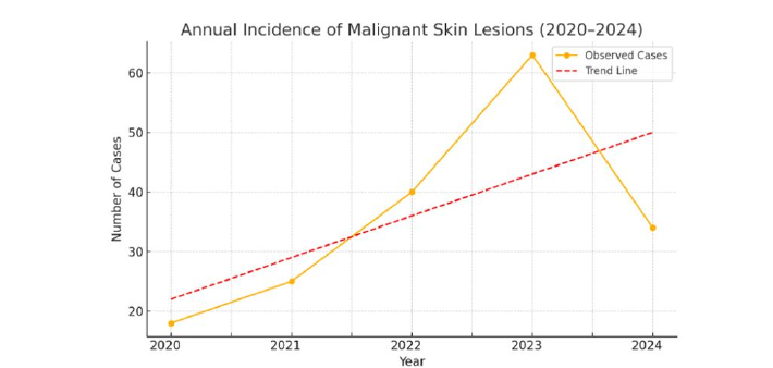
When analyzing the annual incidence of malignant skin lesions, a progressive in-crease was observed over the first four years of the study period: 18 cases in 2020, 25 in 2021, 40 in 2022, and a peak of 63 cases in 2023. This upward trend was followed by a decline in 2024, with 34 cases recorded (Fig. 1). A linear regression analysis revealed an average annual increase of approximately 7 cases (slope = 7), with a coefficient of determination (R²) of 0.64, suggesting a moderate correlation between time and case number. However, the trend did not reach statistical significance (p = 0.244), likely due to the limited number of years analyzed and the variability introduced by external factors such as the COVID-19 pandemic in 2020–2021, the post-pandemic rebound in 2023, and the subsequent decrease observed in 2024.
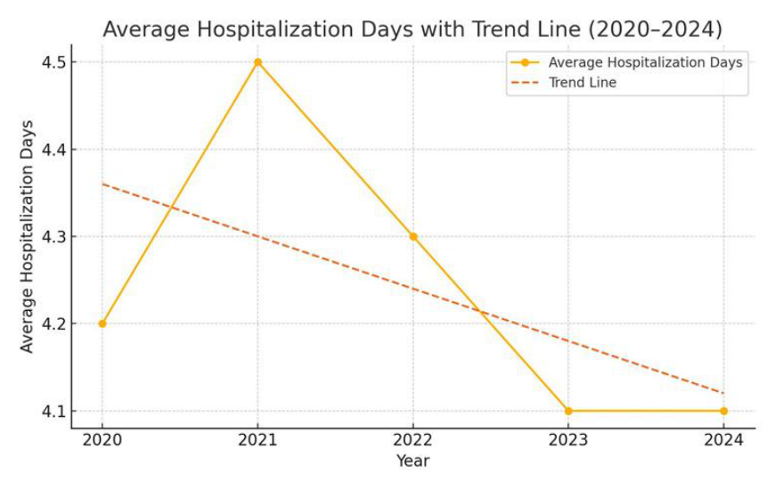
The average hospitalization duration remained relatively stable over the past five years, starting at 4.2 days in 2020, slightly increasing to 4.5 days in 2021, then gradually decreasing to 4.3 days in 2022, and further declining to 4.1 days in both 2023 and 2024 (Fig. 2). The longest hospital stay was recorded in 2020, lasting 19 days, while the shortest stays of 2 days were observed in 2023 and 2024.
Of the 180 patients, 95 were residents of Bucharest, with the highest concentration coming from Sector 3 (n = 35) and Sector 4 (n = 32). Among patients from outside the capital, the neighboring counties of Ilfov (n = 16) and Giurgiu (n = 14) were the most represented.
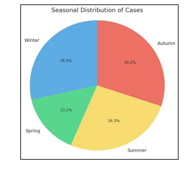
When assessing seasonal distribution over the five-year period, the lowest incidence of diagnosed malignancies was observed in spring, with only 27 cases recorded. The other seasons showed relatively similar frequencies, with 47 cases in summer, 55 in autumn, and 51 in winter (Table 1, Fig. 3). In order to determine whether there is a significand difference between the observed frequencies and expected frequencies, we have applied the Chi-squeare test (χ²). The test revealed a statistically significant difference in the seasonal distribution of skin cancer diagnoses (χ² = 10.31, p = 0.016).
| Presentation period | 2020 | 2021 | 2022 | 2023 | 2024 | TOTAL | No. of cases per season |
|---|---|---|---|---|---|---|---|
| Jan. | 6 | Pandemic | 1 | 9 | 5 | 21 | 51 winter |
| Feb. | 9 | 1 | 5 | 5 | 20 | ||
| Mar. | 3 | 2 | 3 | 6 | 14 | 27 spring | |
| Apr. | Pandemic | 2 | 1 | 3 | 6 | ||
| May | 3 | 1 | 3 | 7 | |||
| Jun. | 5 | 3 | 2 | 10 | 47 summer | ||
| Jul. | 1 | 7 | 7 | 2 | 17 | ||
| Aug. | 10 | 2 | 5 | 3 | 20 | ||
| Sept. | 11 | 4 | 8 | 2 | 25 | 54 autumn | |
| Oct. | 1 | 6 | 6 | 3 | 16 | ||
| Nov. | 1 | 4 | 9 | 0 | 14 | ||
| Dec. | 1 | 3 | 6 | 0 | 10 | 51 winter | |
| TOTAL | 18 | 25 | 40 | 63 | 34 | 180 | 180 |
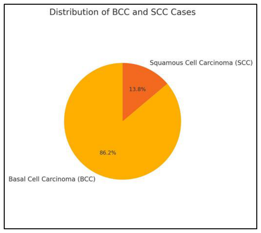
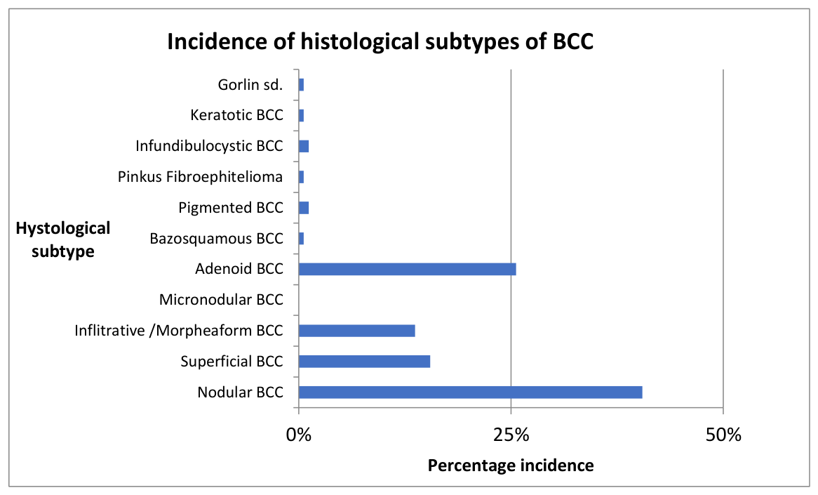
The malignant skin lesions identified in this study were categorized into basal cell carcinoma (BCC), squamous cell carcinoma (SCC), and melanoma. Out of the total 388 lesions, 168 were classified as BCC (Fig. 4). Histopathological evaluation allowed for further subtyping of BCC cases. The nodular subtype was the most prevalent (n = 68), followed by the adenoid type (n = 43), superficial BCC (n = 26), and the infiltrative variant (n = 23). Rare subtypes included basosquamous, pigmented BCC, Pinkus fibroepithelioma, and infundibulocystic BCC, each represented by one or two cases. Additionally, a single case associated with Gorlin syndrome was documented. Notably, no cases of micronodular BCC were observed (Fig. 5). Regarding squamous cell carcinoma, a total of 25 cases were identified. The majority were well-differentiated SCCs (n = 16), followed by moderately differentiated SCCs (n = 8) and poorly differentiated SCCs (n = 3). No anaplastic SCCs were detected during the study period.
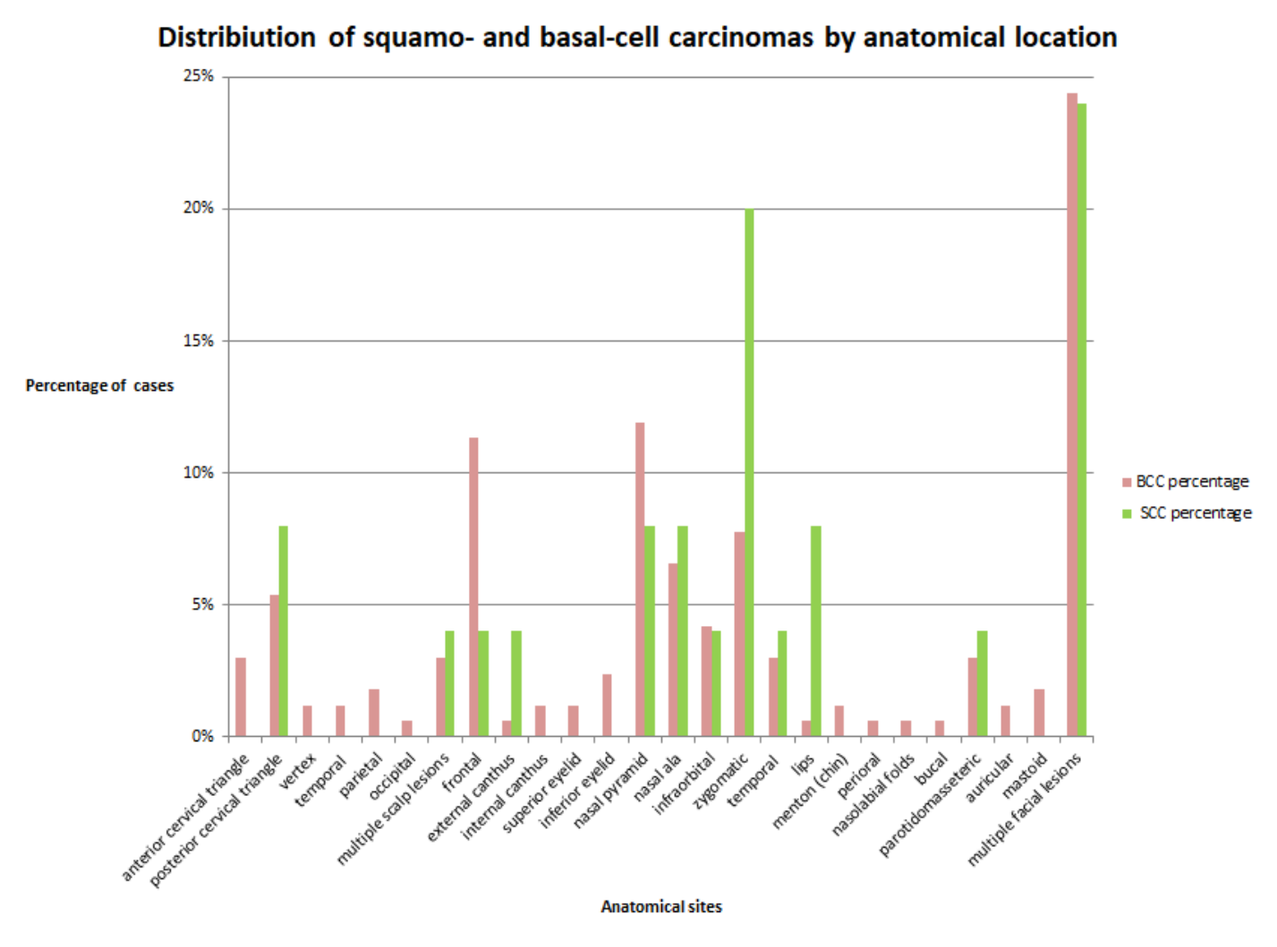
Based on the collected data, the majority of malignant lesions were located on the face (n = 152), a significantly higher number compared to those on the neck (n = 16) and scalp (n = 12). A more detailed anatomical breakdown of facial lesions revealed 20 cases on the forehead (10.3%), 23 on the nasal pyramid (11.8%), and 18 on the zygomatic region (9.2%). Additionally, 23 patients presented with multiple lesions on various facial subregions. Beyond the face, 24 other patients were identified with multiple malignant lesions distributed across the head and neck areas. Regarding the neck, the posterior cervical triangle showed a higher incidence (n = 11) compared to the anterior cervical triangle (n = 5). As for the scalp, lesion distribution was as follows: 2 cases on the vertex, 2 in the temporal region, 4 in the parietal region, 1 occipital lesion, and 3 patients with multiple scalp lesions.
When comparing the anatomical distribution of BCC and SCC, distinct patterns were observed. Several regions—including the anterior cervical triangle, vertex, temporal, parietal, occipital, external canthus, upper and lower eyelids, chin (menton), perioral region, nasal area, auricle, and mastoid region—exhibited only BCC lesions, with no SCC cases recorded in these locations. On the forehead, 11% of all BCCs were located, compared to only 4% of SCCs, indicating a preferential distribution of BCC in this region. In contrast, the zygomatic region accounted for the highest proportion of SCC cases (20%), suggesting a predilection for SCC in this area. Within the posterior cervical triangle, SCC represented 8% of all cases found there, while BCC accounted for 5%, indicating a slightly higher occurrence of SCC in this particular region (Fig. 6).
The vertical dimension of malignant skin lesions showed notable variation across histological subtypes. Among basal cell carcinoma (BCC) types, the infiltrative/morphea form subtype had the highest mean depth (1.46 mm), followed closely by the superficial type (1.35 mm) and adenoid (solid adenoid) variant (1.30 mm). Less invasive subtypes such as nodular BCC had a mean depth of 1.21 mm, while rare variants like pigmented, Pinkus fibroepithelioma, and Gorlin syndrome-associated BCC displayed moderate vertical dimensions (ranging from 1.1 to 1.75 mm). Micronodular BCC was not reported in the dataset. In contrast, squamous cell carcinoma (SCC) subtypes tended to show greater vertical extension. Well-differentiated SCCs had the greatest average depth at 1.92 mm, while moderately differentiated SCCs averaged 0.97 mm, and poorly differentiated SCCs presented an intermediate value of 1.08 mm. These findings suggest a potential correlation between SCC grade and tumor depth.
Temporal analysis revealed fluctuations in lesion depth over the five-year period. For nodular BCC, the vertical dimension ranged from 1.0 mm to 1.49 mm, indicating no clear increasing or decreasing trend. A more pronounced variation was observed in the superficial BCC subtype, which reached a peak of 1.85 mm in 2022 and dropped to 1.06 mm in 2023 before rising again to 1.61 mm in 2024. Infiltrative BCCs peaked at 2.52 mm in 2022—suggesting a possible diagnostic delay or more aggressive presentation in that year. Regarding SCC, well-differentiated lesions consistently displayed greater depth than other SCC subtypes, with a maximum of 2.5 mm recorded in 2022. However, sample sizes for some years and subtypes are limited, warranting cautious interpretation.
A Mann–Whitney U test was performed to compare the vertical depth of BCC and SCC. The median depth in BCC subtypes was 2.20 mm, while SCC subtypes showed a median of 1.32 mm. The test yielded a U statistic of 129.5 and a p-value of 0.797, indicating no statistically significant difference between the two groups.
In order to assess whether there is a correlation between age and the vertical dimension of the lesion, we applied Spearman’s rank correlation test. The results (Spearman’s ρ = 0.023, p = 0.744) suggest that there is no significant association between the two variables, indicating that lesion thickness does not appear to vary meaningfully with patient age in our dataset.
A Mann–Whitney U test (which is non-parametric) was conducted to determine whether there was a difference in vertical dimension between males and females. The results indicated a statistically significant difference between the groups (U = 6473.0, p = 0.009), suggesting that sex may influence vertical dimension. The vertical dimension mean in men was 1.5 mm, while in female was 1.25 mm.
When analyzing the lesion surface area across basal cell carcinoma (BCC) subtypes, we observed considerable variability. The infiltrative/morpheiform subtype demonstrated the largest mean area, with an average of 2.47 mm², followed by the adenoid type (1.58 mm²) and nodular type (1.43 mm²). The superficial BCC subtype had a slightly higher average area (1.85 mm²) than nodular, which may reflect the lateral spread characteristic of this variant. Rare subtypes such as basosquamous/metatypical carcinoma presented a notably high mean area of 74.46 mm², although this finding is based on a single case and should be interpreted cautiously. Other rare variants, including keratotic, fibroepithelioma of Pinkus, and infundibular-cystic subtypes, exhibited relatively small areas ranging from 1.2 to 1.43 mm². The Gorlin syndrome-associated lesion had an average area of 1.55 mm².
For squamous cell carcinoma (SCC), the mean lesion areas were generally larger than most BCC subtypes. Well-differentiated SCCs demonstrated the highest average area, reaching 2.41 mm², while poorly differentiated tumors averaged 1.07 mm². Moderately differentiated SCCs had the smallest mean area among SCC subtypes at 0.77 mm².
A Mann–Whitney U test was also conducted to compare the lesion surface areas between BCC and SCC tumors. The analysis yielded a U statistic of 133.5 with a p-value of 0.60, indicating no statistically significant difference in lesion surface area between the two groups. This suggests that, despite observed differences in average lesion sizes, such as larger surface areas in some well-differentiated SCCs. These differences may be attributed to sample variability rather than a true underlying disparity in tumor size between BCC and SCC in this cohort.
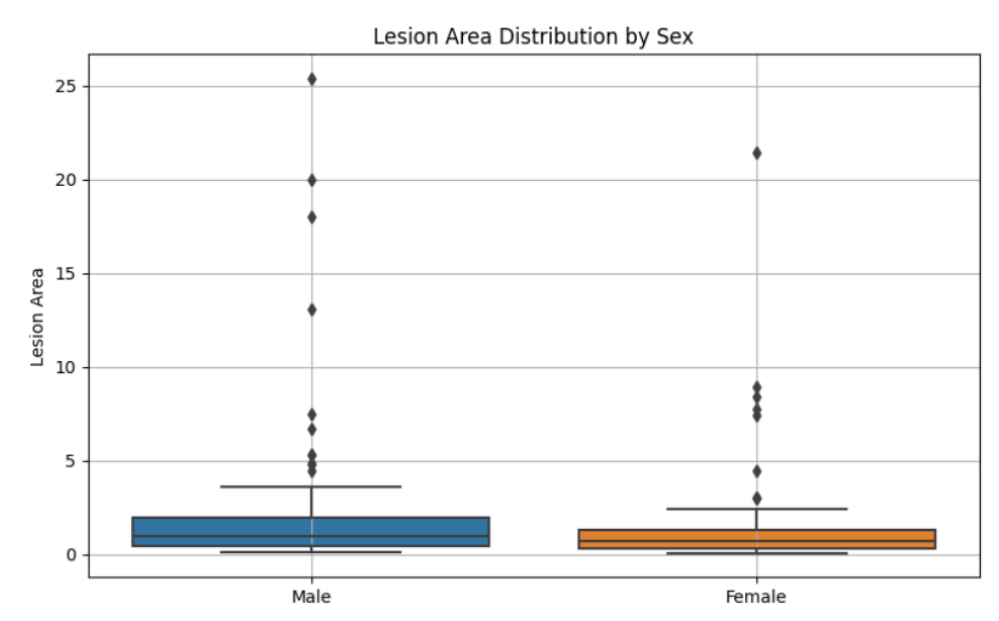
There was a significant difference in lesion area between males and females (U = 6290.0, p = 0.0095) (Fig. 7). While the median lesion area was higher in males (1.0 cm² vs. 0.7 cm²), a rank-based comparison showed a small-to-moderate effect (rank-biserial r = −0.21), likely influenced by high outliers in the female group. A lesion measuring 74.46 mm in a female case was excluded from the analysis to minimize the impact of outliers and ensure that the results more accurately reflect the central tendency and variability within the dataset.
Comparing the data we collected with other clinical studies conducted in recent years in different regions of Romania, a consistent national trend in the incidence of non-melanoma skin cancers can be observed. Both a one-year 2022 study from South-Western part of the country (Craiova city) and a five-years 2014 study from the North-Eastern region (Iasi city) support and validates our findings, confirming their reliability and broader applicability. Thus, both studies indicate a prominence of basal cell carcinomas among the identified NMSC cases, with reported proportions of 55% in the 2014 study and 75,5% in the 2022 study [18,19]. Notably, the 75,5% figure closely aligns with our own findings, where BCC accounted for 86% of cases [18]. This may reflect either an increasing trend in the incidence of BCC over time, or differences in exposure to specific risk factors depending on region. Other similarities to our study include the predominance of the nodular subtype of BCC, which was the most frequently encountered variant in both comparative studies [18,19]. In our study, nodular BCC accounted for 40% of cases, closely matching the 40,4% reported in Iasi [19]. Additionally, all three studies identified the face as the most common anatomical site for tumor occurrence, which may support the correlation between UV exposure and lesion development [18,19]. Moreover, a slight female predominance was observed consistently: women represented 52% of cases in our study and 53% in the Craiova one [18]. These very similar findings highlight the potential role of common environmental and behavioral risk factors and support the need for continued epidemiological monitoring in Romania.
The observed seasonal variation, with higher incidence on non-melanoma skin cancers during autumn and lower numbers in spring, is consistent with the findings of previous studies. A 2017 study from North Carolina concluded that the diagnosis rates of BCC and SCC were highest in October and August, so at the end of the festival season, that confirmed the idea of delayed diagnosis after summer UV exposure [20]. According to Bianconi et al., the Umbria region of Italy had also a higher incidence of NMSC during autumn (mostly October), which was explained as a late cancer progression effect of UV exposure [21]. These similarities support the hypothesis that medical consultation is often delayed due to the clinical appearance of suspicious skin lesions following prolonged summer sun exposure. Consequently, many lesions are diagnosed in the following season- autumn- after cumulative sun damage becomes clinically. Given the seasonal pattern of delayed diagnosis, it is crucial to examine potential preventive measures, such as public health campaigns targeting periods of high UV exposure months. These interventions should aim to raise awareness about the risks of sun damage, promote early skin examinations, and encourage timely medical consultations.
The lack of statistical significance regarding the vertical dimension and area may be due to sample size limitations or uneven subtype representation within groups. For example, rare or extreme outliers like the metatypical BCC area (74 mm²) can skew averages and impact variability. Future studies with larger, more balanced samples may provide clearer insight into surface area differences between malignant skin lesion subtypes.
The recent Covid-19 pandemic impacted in a negative way the worldwide healthcare systems, leading to disruptions in routine medical services. As observed in Table I, there were no admissions of dermatology cases in our department between April 2020 and June 2021, which indicates that dermatological services were not operational through this period. There are several existing factors which contributed to the activity disruption. Firstly, the pandemic’s health care focus likely caused resource redirection. Secondly, institutional policies mandating restriction consults of any case that is not an emergency severely capped support for patients. Another reason may be the fact that considerable sections of the medically unwell population opted not to attend medical facilities due to the high likelihood of contracting the virus, which adds even more weigh. This local experience aligns with global trends observed during the pandemic. A study analyzing data from the American Academy of Dermatology’s DataDerm registry reported a significant decrease in in-person dermatology visits between April 2020 and June 2021, with a corresponding increase in tele-dermatology appointments [22]. However, expanding tele-dermatology services did not ascertain timely diagnosis and prompt treatment for a significant number of patients. Research shows lockdowns led to a diverse range of disruptions that caused cancer diagnoses to be missed on a global scale. The World Health Organization’s International Agency for Research on Cancer estimated that about one million cases of cancer went undetected globally during the lockdown because of Covid-19 [23]. This statement emphasizes the need for strategies to maintain essential healthcare services even during public health crises. The increasing number of diagnoses of cutaneous cancer in the post-pandemic period, could stem from the backlog of cases that went undiagnosed when routine dermatological checkups were either postponed or unavailable.
In the present study, a statistically significant difference was observed in the vertical thickness of non-melanoma skin cancers (NMSC) between male and female patients, with mean measurements of 1.5 mm and 1.25 mm, respectively. This disparity, though numerically modest, holds substantial clinical relevance, as increased tumor thickness is associated with a higher risk of local invasion and recurrence.
Several factors may contribute to this gender-based difference in tumor presenta-tion. One of them could be considered the delayed medical consultation among men. Men are generally less likely to engage in preventive healthcare practices. According to a 2009 study, the white middle-class men were the most affected by melanoma because of the delay in seeking medical attention for skin lesions [24]. This tendency may result in the progression of tumors to greater thickness before diagnosis. Another contributing factor to the gender difference seems to be the sun protection behaviors. The American Association for Cancer Research reported in a 6 years study, that women are more likely to adopt behavioral changes as using sun protection measures like including sunscreen and
protective clothing, to prevent future cutaneous cancers [25]. They would also limit more the outdoor activity and when outside, they would seek shade more than men [25]. This may contribute to earlier detection and thinner tumors at diagnosis.
Although our study shows a higher incidence of NMSC in urban areas, this finding is most likely explained by the location of the study center- a tertiary hospital in Bucharest. The majority of patients included in the study were residents of Bucharest, which leads to an overrepresentation of urban cases. This does not necessarily reflect a higher exposure to risk factors in urban settings, but rather the geographic bias introduced by easier access to specialized dermatological care in the capital. As shown in a retrospective U.S. study with patients recruited from two primary care practices in one rural Maine regional health system, rural patients often face significant barriers to dermatological evaluation and treatment [26]. That may include longer travel distances, limited availability of specialists and socioeconomic constraints.
Another point to discuss is the predominance of basal cell carcinoma in our studied population, with a higher incidence of the nodular subtype (40% of the BCC cases). These findings are aligning with the worldwide trend, as the BCC is well-known the most frequent skin cancer in adult patients with fair skin, the nodular lesions accounting for more than 60% of all BCCs [27]. Nodular subtype are usually slow growing, non-aggressive tumors, belonging to the indolent-growth subset [10]. Thus, its predominance in the studied population may indicate that most cutaneous neoplasms in fair-skin adults are having a favorable outlook. This may be explained by the commonly less aggressive nature of the nodular variant, or by the fact that they are being detected in an early stage, before becoming invasive. The last one is not a well-supported hypothesis, as we have to emphasize the frequent delay in making a definitive diagnostic due to several contributing factors like the still existing need for access to a good dermatological care for all socioeconomic groups.
Regarding squamous cell carcinoma, our study revealed a greater average vertical dimension of well-differentiated lesions (1,92mm), compared to all the mean depths of the different subtypes of BCCs. The bigger vertical growth of SCC can have potential implications in the tumor outcome, as may be an indicator for the cancer aggressiveness. According to a 2024 literature review, the cutaneous squamous cell carcinoma of the head and neck can present metastasis in approximative 2,5% of patients, most often localized in the level II lymph nodes and the parotid glands [28,29]. Unlike BCC, cSCC can spread both nearby and distant locations, one of the highrisk criteria for the metastasis being represented by a depth invasion greater than 6 mm [28,29]. Although none of the studied lesions were deeper than 6 mm, it is necessary to highlight the importance of correlating depth patterns of cSCCs with their metastatic rates and with the right choice of treatment. The primary method of treatment remains the surgical resection, but the tumor depth and size can dictate the type of therapy need to be used. The vertical dimension of cSCCs influences also the follow-up protocols, as patients with greater tumor depths (so high-risk groups) requires a tighter follow-up [29].
Lastly, although most NMSC cases are treated successfully with local excision, ad-vanced or recurrent head and neck lesions (particularly large or deep infiltrating squamous cell carcinomas) may require complex surgical resections. In such cases, reconstruction becomes critical not only for functional and aesthetic restoration but also for maintaining the patient’s quality of life. Free flaps and other advanced reconstructive techniques, such as the radial forearm or anterolateral thigh flap, offer valuable options. The success of these procedures underscores the importance of surgical expertise and a multidisciplinary approach, as emphasized in recent scoping reviews. [30]
Our study’s possible limitations should be taken into account when interpreting the results as they may have introduced bias. Since only 180 patients were chosen from a single tertiary medical center, the study had a relatively small sample size and was carried out over a five-year period. Since it ignores possible variations in clinical, histo-pathological, or demographic characteristics among various populations or geographical areas, this may lower its statistical power and have an impact on the findings’ generalizability. Furthermore, the COVID-19 pandemic coincided with a portion of the study, which disrupted data collection and resulted in fewer patients having access to medical services. Clinical activity was interrupted by these contextual factors, resulting in missing or insufficient data. Notwithstanding these drawbacks, our research still provides insightful information.
Nevertheless, we emphasize the very actual need of further prospective studies which should assess the targeted screening programs influence in early cancer detection and diagnosis, and the effectiveness of different treatment strategies in patients with head and neck non-melanoma skin cancers.
The study identified a progressive increase in the incidence of malignant skin lesions from 2020 to 2023, followed by a decrease in 2024. This trend could be attributed to factors such as the impact of the COVID-19 pandemic on healthcare access and a possible postpandemic surge in cases. Continued monitoring is essential to validate whether this trend persists in subsequent years.
The analysis revealed a statistically significant variation in skin cancer diagnoses across seasons, with the lowest incidence observed in spring and relatively similar fre-quencies in summer, autumn, and winter.
The face was the most common location for malignant lesions, emphasizing the significant role of sun exposure in the etiology of NMSC. BCC was the most frequent histological subtype, with nodular BCC being particularly prevalent. In contrast, SCC, although less common, demonstrated greater metastatic potential and depth, especially the well-differentiated subtype.
Our analysis revealed statistically significant differences in lesion size between sexes, with both area and vertical dimensions being greater in males compared to fe-males.
The majority of NMSC cases were recorded in urban areas, particularly Bucharest, suggesting a potential link between urban living and higher skin cancer incidence or, conversely, a lack of adequate healthcare access in rural regions. Additionally, while the sex distribution was relatively balanced, males exhibited significantly greater lesion depths compared to females.
The authors declare no conflict of interest. This research received no external funding.
Authors’ contribution
Conceptualization, M.M., M.D. and A.C.; methodology, I.A.T. and G.M.; software, A.Z.C.A. and D.V.; validation, M.M., S.R.G. and A.C.; formal analysis, M.D. and I.A.T.; investigation, M.M. and S.R.G.; resources, G.M. and A.Z.C.A.; data curation, M.D. and D.V.; writing—original draft preparation, M.M. and M.D.; writing—review and editing, M.M. and I.A.T.; visualization, G.M. and D.C.; supervision, S.R.G. and D.C.; project administration, A.C. and D.V.; funding acquisition, A.Z.C.A. and D.C. All authors have read and agreed to the published version of the manuscript.
The study was conducted in accordance with the Declaration of Helsinki, and approved by the Ethics Committee of Carol Davila University of Medicine and Pharmacy (protocol code 16341 from 1st July 2025).
Patient consent for publication
Not applicable, because it is a retrospective study.