
Crimean-Congo hemorrhagic fever virus (CCHFV) is a tick-borne virus that can cause severe disease in humans and livestock [1]. CCHFV possesses a tri-segmented negative-sense RNA genome and is classified within the Orthonairovirus genus of the Nairoviridae family. Clinical symptoms include fever, diarrhea, fatigue, and drowsiness, with severe cases presenting with kidney lesions, liver failure, and lung damage, and a mortality rate of approximately 30% [1,2]. CCHFV poses a widespread threat to global public health due to its potential prevalence, high mortality, nosocomial infections, and difficulties in treatment and prevention [3]. Currently, there is no licensed vaccine against Crimean-Congo hemorrhagic fever. In 2019, the World Health Organization estimated that 3 billion people worldwide are at risk of CCHFV infection [4]. CCHFV causes acute illness in humans, primarily transmitted through tick bites. In animals, it is asymptomatic but poses a major threat to humans in close contact, especially in farms, clinics, and abattoirs. Previous studies have highlighted the immunogenic potential of the CCHFV nucleoprotein (NP) [3,5,6]. NP is abundantly expressed during infection and plays a critical role in the viral lifecycle by packaging the viral genome and antigenome [7]. The significant immune responses elicited by NP, including neutralizing antibodies and CD4+/CD8+ T cells, indicate its suitability as a vaccine antigen [8, 9]. The current study aims to enhance vaccine design against CCHF by incorporating NP alongside glycoproteins [10]. Utilizing bioinformatics approaches, the identification of conserved, immunogenic epitopes within NP could optimize vaccine formulations and diagnostic assays [11]. This integrated strategy may improve protective efficacy and advance prevention/control efforts for this important pathogen. In summary, NP represents a promising antigen for combating CCHFV through vaccination and diagnosis. The
findings from ongoing research provide insights to guide the rational development of effective countermeasures against this global health threat.
CCHF is one of the most important emerging tick-borne viral diseases, with sporadic human cases regularly reported across various regions globally. While CCHFV circulates naturally among several animal hosts, humans represent a dead end for the virus. The virus is maintained through a silent transmission cycle involving ticks, vertebrates, and subsequent tick reinfection. People in close contact with infected animals or their products face a heightened risk of acquiring the disease. Delayed diagnosis of CCHF can also lead to outbreaks through secondary nosocomial infections [2,6]. CCHFV has an extensive geographic presence as an emerging/re-emerging pathogen. Reported regions include Africa, the Balkans, the Middle East, and Asia. Within the Middle East, cases have been documented in Iran, Iraq, Saudi Arabia, Oman, and the United Arab Emirates. Eastern European nations with local transmission include Albania, Bulgaria, Greece, Kosovo, Turkey, Georgia, and Russia. Its potential for aerosol spreads, high fatality rates, and lack of approved countermeasures qualify CCHFV as a priority pathogen of biosecurity concern [2,8,12].
CCHFV was first recognized in 1944 during an outbreak among military personnel in Crimea, Russia, and later isolated in the Congo region in 1956 [13,14]. Subsequent studies confirmed its identity as a highly pathogenic tick-borne virus, leading to the current designation as CCHFV [6,13,15]. Since then, CCHFV has been established as a major public health threat, particularly in Africa, Asia, and Eastern Europe, where its geographic range and incidence are expanding [3]. Mortality is usually 30–50% and can reach 80% in some outbreaks [4]. Still, no approved vaccine or effective antiviral exists [15], and the risk of hospital transmission shows the urgent need for better tools.
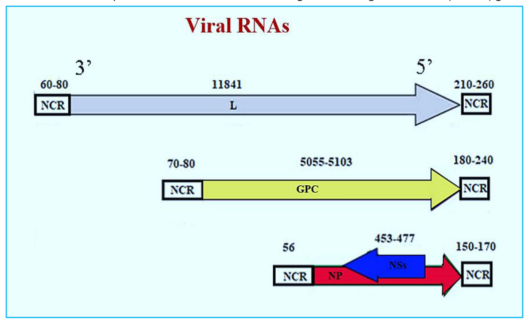
CCHFV possesses a tripartite, linear, single-stranded RNA genome of negative polarity that is encapsidated by viral NP to form distinctive spherical ribonucleoprotein complexes (RNPs) (shown in Figure 1) [3,5]. The small (S) segment is approximately 1,700 nucleotides in length and encodes NP in the viral sense, as well as a non-structural protein (NSs) in the opposite, complementary sense [7]. NP binds to viral RNA and is critical for genome packaging, while NSs act as interferon antagonists to suppress host immune responses [8]. The medium (M) segment measures approximately 5,400 nucleotides [10]. It directs the production of a 160 kDa GPC (glycoprotein precursor) that is cleaved by cellular proteases into the mature glycoproteins glycoprotein N (Gn) and glycoprotein C (Gc), which are found on the virion envelope. The M segment additionally encodes a non-structural protein (NSm) involved in glycoprotein maturation [15]. The largest (L) segment is approximately 12,100 nucleotides in length [1]. It produces an approximately 200 kDa RNA-dependent RNA polymerase with cap-snatching endonuclease and methyltransferase activities essential for transcription and replication of the viral genome [16-18]. Also, the M segment of the virus contains a large GPC, which is processed into seven proteins, including the structural glycoproteins Gn and Gc. These surface glycoproteins are potential vaccine candidates as they have been shown to be targets of neutralizing antibodies. [11,16,19]. GPC also yields a 38 kDa glycoprotein (GP38) and a heavily glycosylated mucin-like domain through additional processing steps [20,21].
CCHFV is an enveloped virus containing a tri-segmented, negative-sense RNA genome [3]. The virion possesses surface glycoproteins Gn and Gc, which facilitate receptor binding and host cell entry. Some evidence suggests an additional protein, GP38, may associate with the viral envelope, though its localization and function remain unclear [7]. The three genomic segments – small (S), medium (M), and large (L) – encode distinct viral proteins. The S segment expresses the NP in one reading frame and an NSs in the opposite frame [8]. Segment M is more complex, directing the production of GPC that host proteases process into glycoproteins Gn and Gc, a mucinlike domain (MLD), GP38, and the NSm involved in glycoprotein maturation [10]. Notably, the L segment of CCHFV is unusually large for bunyaviruses and produces an RNA-dependent RNA polymerase (RdRp) containing an ovarian tumor (OTU) protease domain [1]. The RdRp facilitates transcription and replication of the viral genome [11]. Following receptor-mediated endocytosis, CCHFV undergoes pH-dependent membrane fusion and releases its ribonucleoprotein complexes into the host cytosol [19]. Viral mRNAs initiate translation while the RdRp synthesizes antigens and NP and L proteins package new genomes [3]. Mature virions bud from the Golgi apparatus before exiting via exocytosis [22]. CCHFV proteins also subvert host cell apoptosis and immunity, representing important targets for therapeutic intervention [19].
| 2′-Deoxy-2′- fluorocytidine | Nucleoside analogue antiviral | RNA- dependent RNA polymerase (RdRP) | Preclinical assessment has not yet been performed | No clinical evidence is currently available | Additional preclinical investigations are necessary |
|---|---|---|---|---|---|
| Molnupiravir | Nucleoside analogue antiviral | RNA- dependent RNA polymerase (RdRP) | No significant antiviral activity observed in rodent models | Clinical efficacy data are lacking | Further clinical development appears unlikely |
| Convalescent plasma or survivor- derived antibodies | Neutralizing and non-neutralizing antibody-based therapy | Viral structural proteins | Preclinical efficacy has not been systematically evaluated | Limited and inconclusive clinical findings were reported | Further preclinical and clinical studies are needed to clarify the therapeutic potential |
| Monoclonal antibodies | Neutralizing and non-neutralizing antibody-based therapy | Viral structural proteins | Limited supportive evidence from rodent studies | No validated clinical data available | Comprehensive preclinical and clinical validation is required |
| Corticosteroids | Anti- inflammatory agents | Host immune response modulation | Preclinical investigations have not been reported | Clinical benefit remains uncertain | Current evidence does not support routine use; controlled studies are warranted |
CCHF is a severe, often fatal disease endemic in parts of Africa, Asia, the Middle East, and Southern Europe. Its increasing incidence has led the World Health Organization (WHO) to designate it as a priority for the development of countermeasures [19,24]. While viral envelope glycoproteins are logical vaccine targets due to their role in host cell entry, their antigenic variability facilitates immune evasion. In contrast, the NP is highly conserved and abundantly expressed, making it an attractive vaccine antigen [19]. Multiple vaccine platforms, including subunit, viral vector, DNA, and mRNA vaccines, have been evaluated in preclinical studies, showing variable protective efficacy [1,16,17]. RNA-based vaccines expressing NP alone have achieved complete protection against clinical disease, while combining NP with the glycoprotein precursor further enhanced protection against both disease and viral replication, with even a single 100 ng RNA dose proving protection [25]. Similarly, a DNA vaccine encoding NP and glycoproteins from the CCHFV Hoti strain, administered intramuscularly with electroporation, was well tolerated in cynomolgus macaques and induced robust antibody and T-cell responses. These immune responses correlated with the absence of viremia, reduced tissue viral loads, and improved hematological parameters post challenge [25,26].
The only experimental inactivated CCHFV vaccine to date is the Bulgarian vaccine, developed in the 1970s by propagating a virulent strain in suckling mouse brains, followed by chloroform inactivation and heat treatment at 58 °C to denature viral proteins while preserving conformational epitopes [18,23]. This vaccine has been administered on a limited basis in Bulgaria but remains unapproved by major regulatory authorities. Wider development has been constrained by sporadic case occurrence outside endemic areas and the lack of small animal models that fully mimic human disease [22,27,28]. In a key study, healthy individuals immunized with a fourdose schedule displayed markedly higher frequencies of interferon-gamma (IFN-γ) secreting CD4⁺ and CD8⁺ T cells, as well as increased IgG titers, compared with those receiving a single dose [23]. Neutralizing antibody titers in the four-dose group were two- to four-fold higher than in the single-dose group, suggesting that booster doses are important for optimal protection. While the first dose induced detectable antibodies, subsequent doses significantly enhanced both humoral and cellular immunity. These findings indicate that the inactivated vaccine platform can elicit protective immune responses when optimally administered, and further refinement of production methods and immunization regimens could improve safety, availability, and immunogenicity for the Bulgarian vaccine or novel formulations [23,29].
Adenoviruses are well-characterized viral vectors with high carrying capacity and broad cell tropism, enabling efficient antigen delivery for vaccine development [23]. For CCHFV, adenoviral vectors expressing the N gene induced strong IgG and T-cell responses in mice, providing partial protection against lethal challenge [30]. Despite challenges such as pre-existing immunity and limited transgene expression, continued optimization could enhance their potential for CCHFV vaccine design [23]. In this platform, NP served as the
primary antigen, demonstrating its capacity to induce both humoral and cellular immune responses when delivered via adenoviral vectors.
DNA vaccines are attractive for CCHFV due to their ability to induce protective immunity via in vivo antigen expression, straightforward design, and lack of interference from pre-existing vector immunity [23]. Although no recombinant DNA vaccines have been approved for bunyaviruses, two leading CCHFV candidates have shown strong immunogenicity in animal models [31]. One encodes ubiquitinfused Gn, Gc, and NP, while another uses transcriptionally competent virus-like particles (tc-VLPs). In mice, DNA vaccination incorporating NP elicited a balanced Th1/Th2 response and conferred protection against lethal challenge despite modest neutralizing antibody titers. Boosting with tc-VLPs shifted immunity toward a Th2 profile post-challenge. These results highlight NP’s role in driving cellular immunity, particularly a Th1 response, which may be critical for durable protection. Continued optimization of antigen selection, delivery systems, and immunization regimens could further improve the efficacy of DNA vaccines for CCHFV. This evidence supports including NP as a core antigen in DNA-based CCHFV vaccine strategies to achieve balanced and long-lasting immunity.
The CCHFV envelope polyprotein (EPP) is cleaved into five components: mucin-like variable region, Gp38, Gn, NSm, and Gc, all of which can serve as targets for subunit vaccine design. In one in silico study, protein sequences of EPP and RdRP from the Nigeria/IbAr10200/1970 strain were analyzed for T- and B-cell epitope prediction, MHC binding affinity, and multi-epitope vaccine construct design [32,33]. Selected epitopes were linked with appropriate linkers, fused to a β-defensin adjuvant, and optimized for antigenicity, allergenicity, solubility, and physicochemical stability. Table 2 lists eight predicted cytotoxic T-lymphocyte (CTL) epitopes, and Table 3 summarizes four helper T-lymphocyte (HTL) epitopes with their cytokine-inducing profiles (IL-4, IL-10).
Molecular docking Molecular docking and dynamics simulations confirmed strong and stable binding with human immune receptors (TLR2, TLR3, TLR4), and codon optimization suggested high expression potential in Escherichia coli K12. The final 427–amino acid construct incorporated CTL and HTL epitopes along with ten B-cell epitopes, designed to elicit balanced immune responses, including IL-4–mediated B-cell activation and IL-10–driven regulation of inflammation. Although this construct targeted glycoprotein-derived epitopes rather than NP, incorporating conserved NP epitopes into future subunit designs could broaden immune coverage and durability, since NP has demonstrated strong T-cell immunogenicity in other vaccine platforms [23,32,33].
| Source protein | CTL epitope sequence | Residue range | HLA sup erty pe | Composi te predicti on score | Predicted antigenicit y score | Predicted immunoge nicity score | Toxicity prediction | Allergenicity assessment | Epitope conserva tion (%) | Associated MHC class I alleles | Cross- reactivity evaluation |
|---|---|---|---|---|---|---|---|---|---|---|---|
| EPP | KKIEISGLK | 1438– 1446 | B27 | 0.9605 | 0.9491 | 0.13994 | Predicted to be non- toxic | Not applicable | 100.00 | HLA-A30:01 HLA-A03:01 HLA-A*11:01 | No cross- reactivity detected |
| EPP | HREVEINVL | 411–419 | B39 | 2.5097 | 0.7096 | 0.31548 | Predicted to be non- toxic | Not applicable | 100.00 | HLA-B08:01 HLA-B40:01 | No cross- reactivity detected |
| EPP | TEAIVCVEL | 1231– 1239 | B44 | 1.4217 | 1.1768 | 0.22941 | Predicted to be non- toxic | Not applicable | 100.00 | HLA-B40:01 HLA-B44:03 HLA-B*44:02 | No cross- reactivity detected |
| RDRP | YLYIVITLY | 1411– 1419 | A1 | 1.7622 | 0.9588 | 0.32448 | Predicted to be non- toxic | Not applicable | 100.00 | HLA-B15:01 HLA-A30:02 HLA-A03:01 HLA-A26:01 HLA-A01:01 HLA-B35:01 HLA-A32:01 HLA-B57:01 HLA-A68:01 HLA-B58:01 HLA-A11:01 HLA-B53:01 HLA-A33:01 HLA-A02:01 HLA-A23:01 HLA-B44:02 HLA-A24:02 HLA-B44:03 | No cross- reactivity detected |
| RDRP | VTDIVVGAI | 902–910 | A1 | 1.4576 | 1.0628 | 0.27164 | Predicted to be non- toxic | Not applicable | 100.00 | HLA-A01:01 HLA-B51:01 HLA-A68:02 HLA-A02:06 HLA-A*32:01 | No cross- reactivity detected |
| RDRP | LMSFLNWR V | 3263– 3271 | A2 | 1.3555 | 1.1247 | 0.26439 | Predicted to be non- toxic | Not applicable | 100.00 | HLA-A02:01 HLA-A02:03 HLA-A02:06 HLA-A68:02 HLA-A*32:01 | No cross- reactivity detected |
| RDRP | YHSIAELTM | 42–50 | B39 | 2.0951 | 0.6095 | 0.22589 | Predicted to be non- toxic | Not applicable | 100.00 | HLA-B35:01 HLA-B08:01 HLA-B51:01 HLA-A24:02 HLA-B*53:01 | No cross- reactivity detected |
| HLA-A23:01 HLA-B40:01 | |||||||||
|---|---|---|---|---|---|---|---|---|---|
| RDRP | IQNRITGLY | 2775– 2783 B62 1.4651 | 0.6585 | 0.23824 | Predicted to be non- toxic | Not applicable | 100.00 | HLA-B15:01 HLA-A30:02 HLA-A01:01 HLA-A32:01 HLA-A03:01 HLA-B57:01 HLA-A26:01 HLA-B58:01 HLA-A30:01 HLA-A11:01 HLA-B35:01 HLA-B44:03 HLA-B44:02 HLA-B53:01 | No cross- reactivity detected |
| Protein | Epitopes | Position | Antigen icity | Toxic ity | IC50 (nM) | IL4 pred | IL10 pred | IFN- epitope | Conserv ancy (%) | Interacting MHC class II alleles | Cross- reactivity |
|---|---|---|---|---|---|---|---|---|---|---|---|
| RDRP | EVLINRNSLKARSE | 1240– 1254 | 1.1352 | Non- toxic | 13.3 | Inducer | Indu cer | Positive | 100.00 | HLA-DRB108:02 HLA- DQA102:01/DQB103:01 HLA-DRB501:01 HLA-DRB113:02 HLA-DRB115:01 | Zero |
| RDRP | VHEKRNOPPSVENVO | 700–714 | 1.2275 | Non- toxic | 5 | Inducer | Indu cer | Positive | 80.00 | HLA-DQB102:02 HLA-DRB102:02 HLA-DRB302:02 HLA-DRB109:01 | Zero |
| EPP | KAFSAMPKTSLCFYI | 1508– 1522 | 0.5604 | Non- toxic | 10 | Inducer | Indu cer | Positive | 100.00 | HLA-DRB109:01 HLA-DRB111:01 | Zero |
| EPP | ILFFMFGWRILFCFK | 1610– 1624 | 1.1537 | Non- toxic | 6.3 | Inducer | Indu cer | Positive | 86.67 | HLA- DQA101:01/DQB105:01 HLA-DRB301:01 HLA- DPA101:03/DPB102:01 HLA- DPA103:01/DPB1*04:02 | Zero |
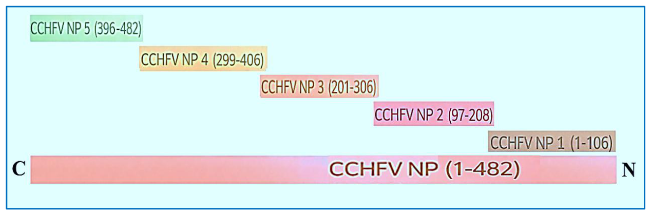
NP can self-assemble into VLPs when overexpressed in insect cells, although the assembly mechanisms remain incompletely understood [4]. VLPs mimic the native virion structure while lacking infectious material, combining high immunogenicity with an excellent safety profile [2,23]. For some viruses, nucleocapsids or glycoproteins autonomously form VLPs during replication, and VLPbased vaccines have demonstrated protective efficacy for other pathogens. The baculovirus expression system supports high-level protein production, facilitating CCHFV VLP formation. The conserved 235–305 amino acid region of NP contains strong antigenic domains, making it a suitable immunogen for VLP design [23]. Preclinical candidates include DNA constructs and modified vaccinia Ankara (MVA) vectors encoding the full GPC or individual NP subunits, which conferred full or partial protection in murine models [19]. MVA, a replication-deficient vaccinia strain, supports high-level transgene expression and has shown promise for CCHFV vaccines, though immune responses require optimization [23–25]. Inactivated virus preparations and combined DNA/VLP regimens achieved partial protection, while subunit or MVA-N vaccines failed to protect [19]. Glycoprotein-focused strategies induced antibodies without protection, underscoring the need to incorporate conserved antigens such as NP [19,27]. Table 4 summarizes all vaccine platforms evaluated for CCHFV to date, and Table 5 provides a comparative overview of these platforms along with the specific role of NP in CCHFV vaccine development. Overall, VLP approaches merit continued development through optimized antigen selection, production processes, and identification of correlates of protection [22,23,29].
NP of CCHFV is highly conserved and abundantly expressed, making it a prime candidate for inclusion in vaccine platforms. It has been evaluated in multiple formats, including recombinant NP subunits, DNA vaccines encoding NP, viral-vectored vaccines (e.g., adenovirus, MVA) expressing NP, and VLPs generated by NP overexpression in baculovirus–insect-cell systems [4,6,7,19,23,25,26]. Preclinical studies demonstrate that NP can induce strong CD4⁺ and CD8⁺ T cell responses and contribute to protection. However, NP alone may not completely block viral replication; combining NP with glycoprotein antigens frequently improves protection against both disease symptoms and viremia [19,23,25,26].
In VLP-based approaches, the conserved NP region (aa 235–305) functions as an immunodominant core that supports particle assembly and enhances antigenicity [4,23]. Thanks to its strong immunogenicity and cross-strain conservation, NP is considered a key component in multi-antigen vaccine designs aimed at eliciting balanced humoral and cellular immune responses. As summarized in Table 5, NP’s specific role across different vaccine platforms underscores its significance in CCHFV vaccine development.
| Type of vaccine | Antigen | Function |
|---|---|---|
| DNA vaccine | M segment | Equally Th1 and Th2 response |
| mRNA vaccine | S segment | Induce both humoral and cellular immune responses |
| Exosome-based vaccine | Whole virus | T-cell activities |
| Inactive vaccine | Whole virus | T-cell activities |
| Plant-expressed vaccine | GN-GC | Antibody response |
| Subunit vaccine | GN-GC | Neutralizing antibody response |
| VSV-based vaccine | GN-GC | Induce both humoral and cellular immune responses |
| MVA vaccine | M, S segment | Induce both humoral and cellular immune responses |
| VLP vaccine | NP | Induce both humoral and cellular immune responses |
| Adenoviral vector vaccine | NP | (a) Upregulated IgG response (b) Protection via CD4+ and CD8+ T cells and non-neutralizing antibody responses |
GN-GC, glycoprotein N-glycoprotein C; MVA, modified vaccinia virus Ankara; NP, nucleoprotein; VLP, virus-like particle; VSV, vesicular stomatitis virus. Information summarized and adapted from the published literature cited in the text.
| Vaccine Platform | Description | Role of NP | Advantages | Limitations/Challenges |
|---|---|---|---|---|
| DNA Vaccine | Plasmid-based delivery of viral antigen genes for in vivo expression. | The NP gene is expressed to induce cellular immunity; it is often combined with glycoproteins for broad immunity. | Induces balanced Th1/Th2 response; easy to produce; no pre- existing vector immunityinterference. | Moderate neutralizing antibody response alone; optimization needed for durability and potency. |
| Subunit Vaccine | Purified recombinant viral proteins (e.g., glycoproteins Gn/Gc) are used as vaccine antigen. | NP may be included as a conserved antigen to enhance T cell responses, though most subunits focus onglycoproteins. | Safe, well- characterized, potentially strong antibody responses. | Subunit of Gn/Gc alone is sometimes insufficient; NP addition is needed to improve protection. |
| Virus-Like Particle (VLP) Vaccine | Non-infectious particles mimicking virion structure, assembled from expressed viral proteins including NP. | NP self-assembles into VLP cores, enhancing immunogenicity by mimicking natural virus morphology and promoting strongcellular immunity. | High immunogenicity without live virus; mimics authentic virion structure. | Complex manufacturing; need to confirm proper assembly and stability. |
| Adenoviral Vector Vaccine | Recombinant adenovirus delivering NP and/or glycoprotein antigens in vivo to stimulate an immune response. | NP expressed to elicit CD4+, CD8+ T cell and antibody responses; partial protection was reported. | Robust induction of cellular immunity; well- studied vector. | Pre-existing anti-vector immunity may reduce efficacy; partial protection from NP alone. |
| Inactivated Vaccine | Chemically or physically inactivated whole virus vaccines. | NP is present in the whole virion; it contributes to cellular immunity with other viral components. | Comprehensive antigen presentation. | Safety concerns, complex production, and lack of widespread approval. |
| mRNA Vaccine | Synthetic mRNA encoding NP and other antigens delivered for in vivo expression. | NP included to enhance cellular immunity alongside glycoproteins (not detailed extensively here but relevant given platform trend). | Rapid design, scalable manufacture, potent immune responses. | Stability and delivery challenges; emerging technology for CCHFV. |
|---|
NP, nucleoprotein; VLP, virus-like particle. Information summarized and adapted from the published literature cited in the text.
The cellular immune response to CCHFV is driven by the activation of both natural killer (NK) cells and cytotoxic T lymphocytes (Tc cells), with NK cells often emphasized due to their prominent role in early infection. Antibodies contribute by recognizing antigens on infected cell surfaces, which may occur through two pathways: (i) intracellular processing of viral proteins, including NP, and presentation via MHC class I; or (ii) antigen presentation during viral budding. NP elicits both humoral and cellular immune responses through multiple mechanisms, including NK cell activation via antibody-dependent cellular cytotoxicity (ADCC), Tc cell stimulation, and balanced Th1/Th2 polarization. When used as a vaccine antigen, NP can also be presented via MHC class II on antigen-presenting cells (APCs), promoting cytokine production (e.g., IL-2, IL-4, IL-6, IL-10, IL-12, TNF-α, IFN-γ, IL-17), memory Tc formation, and crosspresentation for enhanced cytotoxic responses. High NP expression and accessibility on the cell surface reinforce its value as a target for immune monitoring and vaccine development [7,34].
Antibody-mediated mechanisms bridge innate and adaptive immunity in CCHFV infection. ADCC involves NK cell–mediated killing of infected cells via Fc gamma receptor (FcγR) engagement with antigen-specific antibodies, functioning similarly to Tc cells. Antibodydependent cellular phagocytosis (ADCP) is mediated by macrophages and dendritic cells through Fc receptor–dependent uptake of antibody-coated targets, enhancing antigen presentation and activating both cellular and humoral responses. Complementdependent cytotoxicity (CDC) relies on the complement cascade, initially triggered by natural antibodies, to eliminate infected cells prior to full activation of adaptive immunity [7,34].
A study of eleven male survivors of CCHFV infection in South Africa examined peripheral blood mononuclear cells (PBMCs) collected 10 months to 13 years post-infection. Cells were stimulated with 36 peptide pools covering NP, glycoprotein N (GN), and GC. Enzymelinked immunospot (ELISpot) assays detected IFN-γ responses to 16 peptides, mapping to 10 distinct epitopic regions, most within NP.
Two peptides, N262–280 and N298–316, elicited strong responses in multiple survivors, predominantly mediated by CD8⁺ T cells, as confirmed by CD8 depletion assays. No single immunodominant epitope was identified, and response breadth/magnitude did not clearly correlate with time since infection; however, participants tested more than a decade post-infection showed weaker responses. These epitopic regions are illustrated in Figure 3 and summarized in Table 6 [27,20].
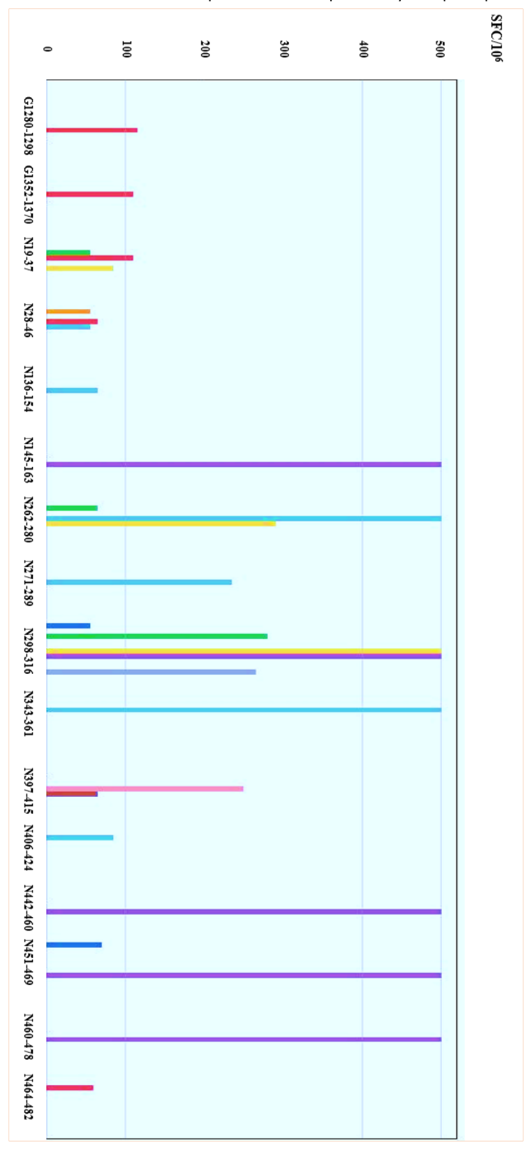
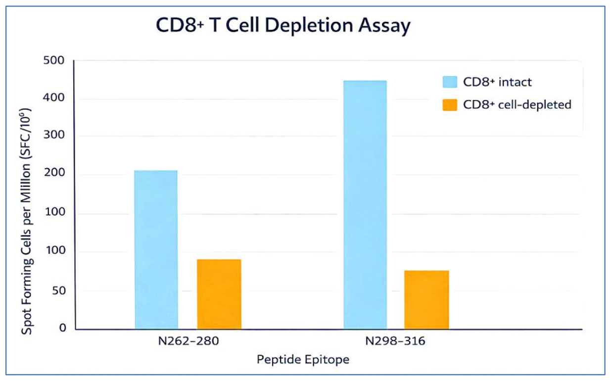
In summary, ten distinct T cell epitopes of CCHFV were identified in survivors, predominantly in the NP, with two in GC. Peptides N262-280 and N298-316 elicited the strongest CD8⁺ T cell responses, as confirmed by depletion assays, which are shown in Figure 4 [27].
Blocking IFN-γ signaling in infected mice significantly increased mortality, underscoring its critical role in viral control [34]. The predominance of NP epitopes, similar to findings in Hantaan virus, may relate to NP abundance during replication and supports its potential as a multi-epitope vaccine target. In contrast, limited GN/GC responses and the failure of previous subunit vaccines suggest glycoproteins alone may be insufficient for T cell–mediated protection. The study identified long-lived T cell responses to CCHFV, focusing on epitope mapping rather than precise quantification. Although 19mer peptides may underestimate responses compared to optimal 9mers, this did not affect the objectives. CD8⁺ T cells were confirmed as the main responders in depletion assays, though broader phenotypic confirmation is needed. Most epitopes were located in NP, with only two in GC and none in GN. Memory T cell responses persisted up to 13 years post-infection, similar to findings in Puumala virus, suggesting potential for durable vaccineinduced protection. The exclusively male cohort reflected past South African epidemiology, with high-risk groups including farmers, veterinarians, and individuals exposed to ticks or infected animal materials. The NP epitopes described here are the first identified after natural CCHFV infection and provide valuable targets for vaccine design and immunogenicity assessment [27].
| Peptide | Number of positive responses | Amino acid sequence | Range of magnitude of responses (SFC/10⁶) |
|---|---|---|---|
| N19–37 | 3 | EFKKGNGLVDTFTNSYSFC | 55–110 |
| N28–46 | 3 | DTFTNSYSFCESVPNLDRF | 55–65 |
| N136–154 | 1 | DIGFRVNANTAALSNKVLA | 65 |
| N145–163 | 1 | TAALSNKVLAEYKVPGEIV | >500 |
| N262–280 | 3 | DKHKDEVDARASADSMITNL | 65–>500 |
| N271–289 | 1 | ASADSMITNLLHKIAKAQE | 235 |
| N298–316 | 5 | RAQGAQIDTAFSSYYWLYK | 55–>500 |
| N343–361 | 1 | KMKKALLSTPMKWGKKLYE | >500 |
| N397–415 | 2 | VANPDDAAQGSGHTKSILN | 65–250 |
| N408–424 | 1 | GSGHTKSILNLRTNTETNN | 85 |
| N442–460 | 1 | NIQDMDIVASEHLLHQSLV | >500 |
| N451–469 | 2 | SEHLLHQSLVGKQSPFQNA | 70–>500 |
| N460–478 | 1 | VGKQSPFQNAYNVKGNATS | >500 |
| N464–482 | 1 | SPFQNAYNVKGNATSANII | 60 |
| G1280–1298 | 1 | TLHPRIIEEGFFDLHVQKV | 115 |
| G1352–1370 | 1 | DGCDLDYYCNMGDWPSCTY | 110 |
<!– Start of picture text –> a<br>a<br>2 s 3 s s 3 2<br>Ss Ss Ss Ss Ss 4<br>gooa“=Soo<br>030QenyOn~woas peras<br>a<br>Ceeoeee<br>4 ——<br>=a<br>4<br>nySP —<br>a-<br>a~Rem“ao<br>3 —<br>CO2oos ee EE<br>6<br>a —————<br>sd2=)<br>aSoeehar — – ee<br>00<br>a<br>oerrry<br>&<br>=<br>aoy———&<br>on<br>aateis~<br>z~sy——————————————as<br>anmIaalo)<br>zaa<br>aoo<br>2<br>—=&osrs)<br><!– End of picture text –>
<!– Start of picture text –> CD8+ T Cell Depletion Assay<br>500<br>CD8+ intact<br>a ~ @® CD8*+ cell-depleted<br>S<br>© 300<br>LL<br>wn<br>& 200<br>=<br>@ 100<br>Qa<br>=<br>S 100<br>oo<br>&<br>=<br>5 50<br>a<br>Ke)<br>“nx 2<br>N262-280 N298-316<br>Peptide Epitope<br><!– End of picture text –>
novel SYBR Green-based one-step real-time RT-PCR assay, have successfully detected CCHFV in clinical samples with high sensitivity, capable of detecting fewer than 20 copies of viral RNA per reaction [36].NP of CCHFV is highly conserved, abundantly expressed, and strongly immunogenic, making it a promising candidate for subunit vaccines. Partial efficacy up to 78% has been reported using a prime-boost vaccination strategy targeting NP [19]. NP can self-assemble into VLPs and stimulate both humoral and cellular immune responses, highlighting its potential as a core vaccine component. Moreover, rNP has proven useful in serosurveillance and diagnostic applications by detecting IgG and IgM antibodies, serving as a safe alternative to whole-virus antigens [3,37]. However, focusing exclusively on NP may be insufficient to induce sterilizing immunity or fully block transmission. Multivalent vaccine strategies incorporating additional conserved antigens, such as the L protein, could provide broader protection. Critical challenges remain in defining which NP epitopes elicit the most protective versus non-neutralizing immune responses, optimizing antigen folding and purity for large-scale production, and establishing correlates of protection in humans and animal models. Ethical and technical limitations hinder direct human challenge studies, further complicating efficacy assessment [5,19].
In conclusion, while NP demonstrates significant promise as a vaccine target and diagnostic tool, further research is needed to refine vaccine design, evaluate combinatorial strategies, and fully understand its protective potential against CCHFV. Continued preclinical and clinical studies are essential to advance safe and effective vaccines and to mitigate the public health impact of this highconsequence pathogen [37].
The authors declare that they have no competing interests. This research received no external funding.
The authors would like to thank the Nanobiotechnology Research Center of Baqiyatallah University of Medical Sciences for their helpful assistance. No generative AI was used for the production of this article. All the figures and tables within the current article have been created or adapted by the authors.
Conceptualization, R.K, M.S.H., M.M.; methodology, R.K.; validation, R.K.; formal analysis, R.K.; investigation, R.K., M.S.H., M.M.; resources, M.M.; data curation, M.S.H.; writing—original draft preparation, R.K.; writing—review and editing, R.K, M.S.H., M.M.; visualization, R.K.; supervision, M.S.H.; All authors have read and agreed to the published version of the manuscript.
Ethics approval and consent to participate
All assessments were conducted in accordance with ethical principles and under the supervision of the University’s Ethics Committee (Ethic NO. IR.BMSU.BLC.1403.001).
| 6. Shrivastava N, Shrivastava A, Ninawe SM, Sharma S, Kumar JS, Alam SI, et al. Development of multispecies recombinant nucleoprotein-based indirect ELISA for high-throughput screening of Crimean-Congo hemorrhagic fever virus-specific antibodies. Front Microbiol. 2019; 10:1822. doi:10.3389/fmicb.2019.01822. |
|---|
| 7. Karaaslan E, Çetin NS, Kalkan-Yazıcı M, Hasanoğlu S, Karakeçili F, Özdarendeli A, et al. Immune responses in multiple hosts to nucleocapsid protein (NP) of Crimean-Congo hemorrhagic fever virus (CCHFV). PLoS Negl Trop Dis. 2021;15(12): e0009973. doi: 10.1371/journal.pntd.0009973. |
| 8. Chinikar S, Persson SM, Johansson M, Bladh L, Goya M, Houshmand B, et al. Genetic analysis of Crimean-Congo hemorrhagic fever virus in Iran. J Med Virol. 2004;73(3):404–11. doi:10.1002/jmv.20106. |
| 9. Raabe VN. Diagnostic testing for Crimean-Congo hemorrhagic fever. J Clin Microbiol. 2020;58(4):e01580-19. doi:10.1128/JCM.01580-19. |
| 10. Wei PF, Luo YJ, Li TX, Wang HL, Hu ZH, Zhang FC, et al. Serial expression of the truncated fragments of the nucleocapsid protein of CCHFV and identification of the epitope region. Virol Sin. 2010;25(1):45–51. doi:10.1007/s12250-010-3067-7. |
| 11. Saijo M, Qing T, Niikura M, Maeda A, Ikegami T, Prehaud C, et al. Recombinant nucleoprotein-based enzyme-linked immunosorbent assay for detection of immunoglobulin G antibodies to Crimean-Congo hemorrhagic fever virus. J Clin Microbiol. 2002;40(5):1587–91. doi:10.1128/JCM.40.5.1587-1591.2002. |
| 12. Samudzi RR, Leman PA, Paweska JT, Swanepoel R, Burt FJ. Bacterial expression of Crimean-Congo hemorrhagic fever virus nucleoprotein and its evaluation as a diagnostic reagent in an indirect ELISA. J Virol Methods. 2012;179(1):70–6. doi: 10.1016/j.jviromet.2011.10.024. |
| 13. Tipih T, Burt FJ. Crimean-Congo hemorrhagic fever virus: advances in vaccine development. Biores Open Access. 2020;9(1):137–50. doi:10.1089/biores.2020.0026. |