
Psoriasis is a chronic, immune-mediated inflammatory disease with an estimated prevalence of approximately 2–3% in the general population (1,2). It is now recognized as a systemic condition, frequently associated with multiple comorbidities, including metabolic, cardiovascular, hepatic, infectious, and neoplastic diseases, which may influence both clinical severity and possible therapeutic options (3,4).
Systemic inflammation plays a central role in the pathogenesis of psoriasis, with complex immunological pathways involved, with the interleukin 23/ T helper 17 axis (IL-23/Th17) playing a central role. These mechanisms have underpinned the development of modern biological therapies, such as tumor necrosis factor alpha (TNF-α) inhibitors, interleukin 17 (IL-17) inhibitors, interleukin 23 (IL-23) inhibitors, and interleukin 12/23 (IL-12/23) inhibitors, which have revolutionized the treatment of moderate and severe forms of the disease (5,6).
In recent years, advances in understanding pathogenic mechanisms, identification of biomarkers, and development of personalized therapeutic strategies have contributed to significant improvements in the prognosis and quality of life of patients with psoriasis (7,8). However, the use of these therapies in certain populations remains limited. Patients with major systemic comorbidities — such as human immunodeficiency virus (HIV) infection, recent oncological history, latent tuberculosis infection, or advanced liver disease — are frequently excluded from randomized clinical trials, which reduces the availability of solid evidence-based recommendations for these categories (6,7). Consequently, therapeutic decisions in these situations are often based on observational data and clinical experience.
The aim of this article is to review immunomodulatory therapeutic strategies in psoriasis associated with severe systemic comorbidities, integrating data from the literature with observations from real-world clinical practice.
This study was designed as a narrative review combined with a retrospective case series, aiming to synthesize current evidence and real-world clinical experience regarding immunomodulatory therapeutic strategies in psoriasis patients with high-risk systemic comorbidities. A literature search was conducted in PubMed/MEDLINE, Scopus, and Web of Science for articles published up to March 2026, using relevant keywords such as “psoriasis,” “biologic therapy,” “HIV,” “malignancy,” “latent tuberculosis,” and “liver disease.” Eligible studies included clinical trials, observational studies, systematic reviews, and case reports addressing treatment approaches and safety in this population, with emphasis on clinically relevant and recent data. Extracted information was qualitatively synthesized according to comorbidity type, therapeutic strategies, and safety outcomes. In parallel, a retrospective case series was realized, including patients with clinically and/or histopathologically confirmed psoriasis and at least one major systemic comorbidity, managed in routine practice. Clinical evaluation included Psoriasis Area and Severity Index score (PASI) and Dermatology Life Quality Index score (DLQI) scores, as well as comorbidity-specific parameters, and therapeutic decisions were individualized based on disease severity and risk–benefit assessment within a multidisciplinary framework. Patients were monitored through regular clinical and laboratory assessments to evaluate treatment response and adverse events. All procedures were conducted in accordance with the Declaration of Helsinki, and written informed consent was obtained from all patients. The clinical components of the study were approved by the Local Ethics Committee of the Dr. Carol Davila University Emergency Central Military Hospital, Bucharest (registration number 750, 22 January 2025).
People living with HIV (PLWH) have an increased incidence of dermatological conditions, many of which are associated with significant morbidity and reduced quality of life. Psoriasis, in particular, tends to have a more severe and prolonged course in this population compared with the general population (9,10). HIV infection is characterized by a progressive decrease in CD4+ T lymphocytes, which makes the increased frequency of psoriasis exacerbations in these patients quite paradoxical. However, psoriasis can occur at any stage of HIV infection and is considered to be the result of immunological dysregulation, particularly through alterations in the Th1/Th17 axis (11). The introduction of combination antiretroviral therapy (cART) has significantly modified the natural history of both HIV infection and psoriasis, contributing to increased life expectancy and improved quality of life for patients. However, moderate and severe forms of psoriasis frequently require systemic or immunomodulatory treatments, raising important safety concerns (4).
Therapeutic options for psoriasis in HIV-positive patients are limited, with first-line treatment usually including topical therapies associated with antiretroviral therapy, followed by phototherapy in more extensive forms (11,12). As a second-line treatment, oral retinoids can be used, either alone or in combination. Also, immunosuppressive therapies and biological therapies represent a useful alternative in difficult cases (9,10,12). On the other hand, in patients with psoriasis and concomitant HIV or other viral infections like hepatitis B or C viruses, there may be a risk of viral reactivation with the use of biological therapies (10). Nonetheless, recent studies suggest that this risk is not significant, especially in patients with well-controlled HIV infection, and that the therapeutic approach only requires interdisciplinary collaboration, particularly with the infectious disease physician (10,11). Moreover, biological agents targeting the IL-23 pathway, such as guselkumab and risankizumab, have shown promising results in small series of cases, with high efficacy and a favorable safety profile, with no observed effects on viral replication or immune parameters (13,14). Furthermore, a recent systematic review, which included multiple biological therapies (TNF-α inhibitors, IL-17 inhibitors and IL-23 inhibitors), highlighted the consistent improvement of cutaneous manifestations of psoriasis in HIV-positive patients, with rare adverse events (11).
However, despite the accumulation of data supporting the safe use of systemic and biological therapies in patients with psoriasis and HIV infection, the management of psoriasis in this context remains a challenge, and therapeutic decisions require interdisciplinary collaboration (15). Next, we present the case of a young HIV positive man with palmoplantar psoriasis encountered in our clinical practice.
A 29-year-old man presented in January 2026 with a painful palmoplantar eruption evolving progressively over approximately six weeks. The initial presentation consisted of dryness and mild palmar erythema, attributed by the patient to occupational exposure and frequent disinfectant use. Over time, lesions evolved into painful pustules with scaling and burning sensations. Plantar involvement led to gait impairment, while palmar involvement affected manual daily activities, contributing to anxiety regarding disease progression. The patient had been diagnosed with HIV infection in 2018 and was receiving combined antiretroviral therapy with dolutegravir/lamivudine (1 tablet/day), with documented excellent adherence since diagnosis. There was no personal or family history of psoriasis and no other known immune-mediated disease. No opportunistic infections were reported in recent years.
At presentation, the immune and virologic status was favorable: CD4 = 1400 cells/mm³ with undetectable viral load. Routine laboratory tests were unremarkable. Dermatologic examination revealed well-demarcated erythematous scaly plaques on both palms, with multiple superficial sterile pustules, some confluent, on an erythematous background (Figure 1). Plantar lesions were more extensive, with hyperkeratosis, painful fissuring, and scattered pustules along the plantar margins and arch. No lesions were observed on the trunk, scalp, or nails.
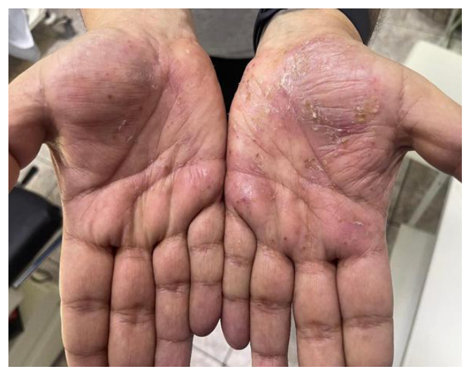
Taking these aspects into consideration, a clinical diagnosis of palmoplantar pustular psoriasis was made. Despite limited body surface area involvement, PASI was 7.8 and DLQI was 15, reflecting the disproportionate functional impact due to the critical localization. The patient reported occupational difficulties and social avoidance due to visible hand lesions. However, given the clinical context — young HIV-positive patient — histopathologic confirmation was necessary to exclude other pustular dermatoses or drug reactions.
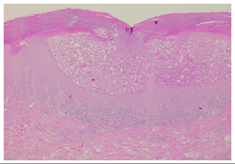
An incisional biopsy was performed under local anesthesia (lidocaine 1%). Histology showed regular acanthosis, elongation of rete ridges, hyperkeratosis with parakeratosis, neutrophilic microabscesses in stratum corneum, and intraepidermal spongiform pustules, consistent with palmoplantar pustular psoriasis (Figure 2).
This diagnosis raised an important therapeutic challenge: HIV-associated psoriasis has been reported to be severe or atypical, possibly related to immune dysregulation and Th1/Th17 axis alterations (7,8,16). Because randomized trials of biologic therapies usually excluded HIV-positive individuals, evidence is limited and decisions must balance cutaneous control against potential immune destabilization. In this case, the presence of significant but localized functional impairment, as well as the absence of systemic involvement, represented factors favoring the choice of a conservative strategy. Despite well-controlled HIV-infection (undetectable viral load and stable immune status), systemic therapies were avoided given the potential risk of immune destabilization. Topical corticosteroids are known to exert potent anti-inflammatory and immunomodulatory effects by binding to intracellular receptors and modulating cellular gene expression (17). Similarly, ultraviolet B phototherapy (UVB) is known to induce apoptosis of pathogenic T lymphocytes, leading to local and systemic immunosuppression (18,19). Therefore, this combination allows effective disease control while providing a safe, non-systemic approach. Based on these considerations, topical treatment with mometasone furoate (1 mg/g) once daily was initiated, combined with narrow band UVB (NB-UVB) phototherapy three times weekly. The choice of NB-UVB was based on its established efficacy in psoriasis and a favorable safety profile in stable HIV patients under appropriate monitoring.
The patient underwent regular dermatologic and infectious diseases follow-up. Clinical evolution was favorable: after approximately four weeks, pustules decreased and erythema improved. By eight weeks, plantar fissuring improved substantially and pain on walking diminished. DLQI decreased to 6. Immunological parameters remained stable, with no decline in CD4+ and no detectable viremia.
This case highlights that severity assessment should include functional and psychosocial impact, not solely extent. In well-controlled HIV infection, a stepwise, non-systemic approach may be effective and safe in localized disease.
The relationship between psoriasis, immunomodulatory therapies, and the risk of malignancy is complex and intensively debated, and the lack of solid evidence leads to reluctance to initiate systemic or biologic therapies in patients with a history of cancer (20). Studies have shown that patients with severe psoriasis are at increased risk for certain types of malignancies, particularly lymphomas and non-melanoma skin cancers (21). In addition, associated risk factors, such as smoking, obesity, and alcohol consumption, may further contribute to this risk. On the other hand, a recent meta-analysis did not reveal a significant increase in the risk of breast cancer in patients with psoriasis, regardless of disease severity (22). These results suggest that although psoriasis is associated with an increased risk for certain neoplasms, this risk does not extend uniformly to all types of cancer.
Therapies used in psoriasis may also influence oncological risk differently. Treatment with psoralene and ultraviolet A phototherapy (PUVA) and cyclosporine has been associated with an increased risk of skin cancer, especially squamous cell carcinoma (21). Regarding TNF-α inhibitors, the data are variable, with some studies suggesting a slightly increased risk of non-melanoma skin cancer (23). In contrast, newer biologic therapies, including IL-17 inhibitors and IL-23 inhibitors, appear to have a more favorable safety profile, with no clear association with an increased risk of malignancy (21,23).
A key issue in clinical practice is the risk of tumor recurrence in patients who require systemic treatment for psoriasis but already have a history of malignancy. Current data suggest that biologic therapies are not associated with a significant increase in the risk of tumor recurrence or progression (20). However, management of these patients involves a multidisciplinary approach, particularly a good collaboration between dermatology and oncology (20). Also, regular oncological screening and careful monitoring are essential during treatment. Further, we present a characteristic case for this type of pathological association.
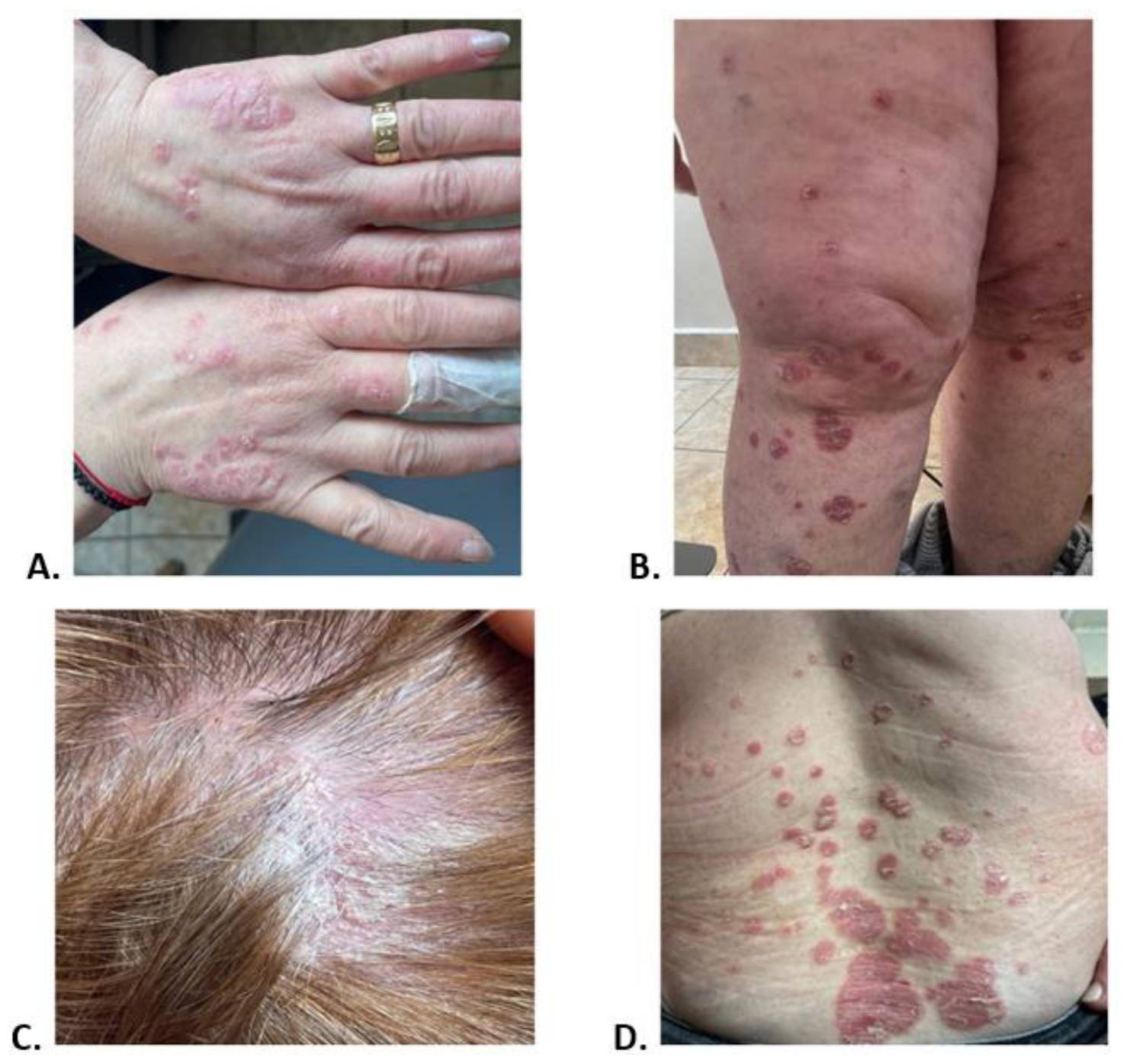
A 54-year-old woman presented in November 2024 with progressive worsening of a cutaneous eruption that began approximately four months prior. The onset was subtle, with a few erythematous plaques on the trunk initially interpreted as contact dermatitis. Over time, lesions expanded, became infiltrated, markedly scaly and pruritic, and progressively involved the scalp and limbs (Figure 3). The patient reported mild pruritus, skin tightness, and scaling that significantly impacted daily comfort. Psychosocial burden was evident: she avoided dark clothing due to visible scaling and described anxiety related to body image.
Her medical history was notable for invasive mucinous breast carcinoma, surgically treated in November 2023. Postoperative oncologic evaluation documented remission with no evidence of metastasis or recurrence. At presentation, she was receiving adjuvant endocrine therapy with anastrozole (1mg/day), and periodic oncologic follow-up remained negative.
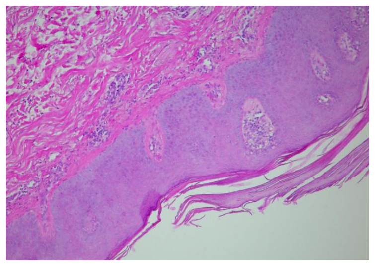
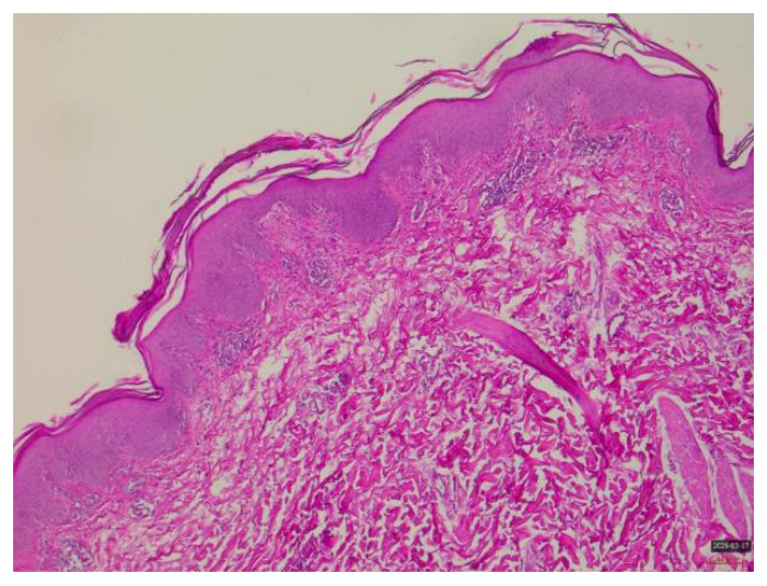
Dermatologic examination showed well-demarcated erythematous infiltrated plaques covered with adherent silvery scale involving the trunk, upper and lower limbs, and scalp. There were no clinical signs of psoriatic arthritis and no significant nail involvement. PASI score was 16.8 and DLQI score was 18, consistent with moderate-to-severe disease and major quality-of-life impairment. The diagnosis of plaque psoriasis was confirmed histopathologically (Figure 4).
The key challenge was not the psoriasis severity alone, but the recent oncologic context. Patients with recent solid malignancies are another type of patient frequently excluded from trials of systemic/biologic therapies, and available evidence is largely observational (24,25). The main clinical dilemma was potential recurrence of neoplasia under systemic immunomodulatory therapies, particularly the conventional immunosuppressants such as methotrexate, as well as the biologic agents. Exerting various mechanisms of action, methotrexate is an effective immunomodulator through disruption of adenosine metabolism, which leads to repressed T-cell activation and B-cell down-regulation (26). Although its immunosuppressive effect is less targeted compared to biologic therapies, caution is advised in patients with recent neoplasia. Regarding a possible therapy with methotrexate, there is no robust evidence that using low doses increases recurrence risk in solid tumors, but caution is warranted, particularly within the first years after cancer treatment.
Because of these aspects, therapeutic decisions were discussed in an interdisciplinary manner with the treating oncologist. Considerations included: approximately one year since surgery, absence of clinical/imaging recurrence, mucinous histology, which has a relatively favorable prognosis, and the significant psoriasis burden. A conservative systemic strategy was chosen: methotrexate 10 mg/week with folic acid supplementation, under strict hematologic and hepatic monitoring, combined with phototherapy to accelerate control. Clinical response was progressive. After approximately eight weeks, erythema and plaque infiltration decreased. At four months, the PASI score decreased to three with marked DLQI score improvement. By August 2025, complete remission was achieved (PASI score 0; DLQI score 0), without significant laboratory abnormalities and without oncologic recurrence during follow-up.
Given remission and the patient’s preference to minimize prolonged systemic exposure, methotrexate was temporarily discontinued with close monitoring. However, relapse occurred in January 2026, initially on the scalp and later on the trunk (PASI score 9; DLQI score 11). Concomitant oncologic assessment remained negative. Following renewed interdisciplinary discussion, methotrexate was restarted at the same dose, with plans to consider biologic transition if long-term control becomes necessary, guided by safety profile and available data in post-malignancy populations.
This case underscores the importance of individualized treatment and sustained dermatology–oncology collaboration in patients with recent malignancy.
According to current guidelines, screening for latent tuberculosis infection is an essential step before initiating biologic therapy in patients with psoriasis. In this context, isoniazid prophylaxis is frequently used as an effective bactericidal agent against Mycobacterium tuberculosis. On the other hand, it is associated with a well-known risk of hepatotoxicity, ranging from asymptomatic transient elevations of transaminases to severe, potentially fatal acute hepatitis (27,28).
Interestingly, data from the literature indicate a wide variability in the frequency of isoniazid-induced hepatotoxicity. A meta-analysis that included over 22,000 patients treated for latent tuberculosis infection reported a frequency of approximately 2.6% for isoniazid-induced liver damage, with a very low mortality (0.02%) (29). However, real-world data suggest higher rates in certain subgroups of patients. A recent study reported hepatotoxicity in approximately 20% of patients treated with isoniazid, with a significant association with factors such as dyslipidemia, systemic inflammatory diseases including psoriasis, and male gender (27).
Because of these aspects, the correct interpretation of hepatotoxicity in patients receiving biologic therapy and antituberculosis prophylaxis is mandatory, as differentiating between isoniazid-induced toxicity and a potential adverse effect of biologic therapy is essential to avoid unnecessary interruption of effective treatment. Next, we present the challenges associated with concomitant use of biologic therapy and isoniazid in a patient with psoriasis and hepatotoxicity.
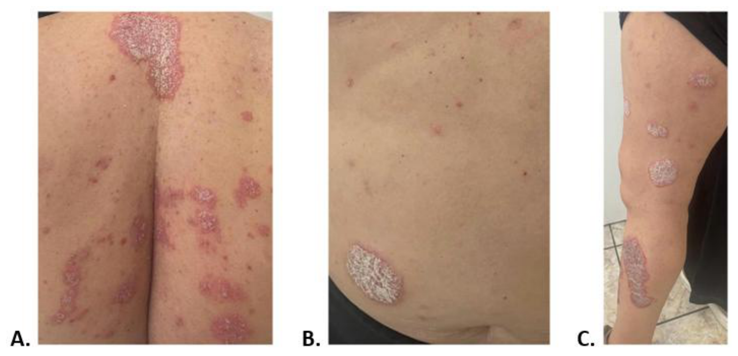
A 62-year-old woman with a 40-year history of plaque psoriasis was evaluated in April 2024 due to progressive worsening of cutaneous lesions. She had previously received multiple topical therapies and intermittent systemic courses (including methotrexate 15 mg/week), with partial responses and frequent relapses. In the months before presentation, plaques became thicker and more extensive on the trunk and limbs, accompanied by intense pruritus, sleep disturbance, and marked emotional distress (Figure 5). PASI score was 18.9, and DLQI score was 22, consistent with severe disease and major quality-of-life impact.
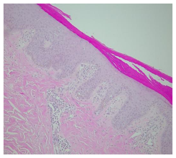
Given severity, long disease duration and insufficient response to prior treatments, biologic therapy with ixekizumab was initiated per approved regimen: 160 mg loading dose, followed by 80 mg every 2 weeks up to week 12, then 80 mg every 4 weeks. Ixekizumab is a selective IL-17A inhibitor with demonstrated efficacy and safety in pivotal phase three randomized trials UNCOVER-2 and UNCOVER-3 (30). Pre-treatment screening was performed according to current recommendations (31). A skin biopsy was obtained, confirming the diagnosis (Figure 6). Routine blood tests were within normal range and viral hepatitis screening was negative. On the other hand, QuantiFERON-TB Gold was positive, without respiratory symptoms or imaging findings suggestive of active tuberculosis, which made it consistent with latent tuberculosis.
In line with guidelines, isoniazid prophylaxis was initiated alongside biologic therapy, with regular hepatic monitoring (31). Dermatologic response was rapid – after three months, complete cutaneous response was achieved (PASI score 0; DLQI score 1). However, during routine monitoring, progressive transaminase elevation was observed:
The patient remained asymptomatic (no jaundice, significant asthenia or abdominal pain). No other hepatotoxic medications were used and no additional toxic exposures were identified.
A key clinical dilemma emerged: isoniazid-induced hepatotoxicity versus a possible biologic therapy-related adverse event. Isoniazid has a well-documented hepatotoxic potential, with increased risk in older patients, while IL-17 inhibitors generally have a favorable hepatic profile (32). However, attribution can be challenging in patients taking multiple medicines. Based on the temporal pattern and progressive rise under isoniazid exposure, a diagnosis of probable isoniazid-induced hepatocellular injury was established. For safety, biologic therapy was temporarily withheld and the patient underwent a gastroenterological evaluation.
Transaminases gradually decreased after discontinuation of anti-tuberculous prophylaxis. On the other hand, in January 2025, a partial psoriasis relapse occurred (PASI score 6; DLQI score 8) during biologic therapy cessation. In March 2025, because the liver enzymes were almost normalized (AST 46 U/L; ALT 51 U/L), biologic therapy with ixekizumab was resumed without isoniazid, under strict monitoring. The subsequent course was very good, with a complete response that was achieved by May 2025 (PASI score 0; DLQI score 1) and maintained through November 2025 without recurrent cytolysis.
The fact that the liver enzymes increased under isoniazid, normalized after discontinuation and did not increase again after reintroduction of biologic therapy strongly supports that isoniazid was the causative agent, rather than IL-17 inhibition. This case highlights the importance of systematic tuberculous screening, awareness of isoniazid hepatotoxicity, careful causality assessment if hepatotoxicity appears, and controlled re-challenge to preserve an effective biologic therapy.
Hepatic cirrhosis is a major cause of morbidity and mortality worldwide, frequently caused by hepatic steatosis, chronic alcohol consumption and viral infections (33). The disease progression is characterized by two distinct stages — compensated and decompensated cirrhosis, differentiated by the presence of complications such as ascites, hepatic encephalopathy, or gastrointestinal hemorrhage (33). Cirrhosis is the result of a complex process characterized by hepatocyte injury, stellate cell activation, and progressive fibrosis, in the context of persistent chronic inflammation (35). Metabolic imbalances, oxidative stress and immunological disorders contribute to disease progression and impaired liver function.
Chronic liver disease is frequently encountered in patients with psoriasis due to common systemic inflammatory mechanisms and metabolic dysfunction. Recent data suggest a causal relationship between certain autoimmune liver pathologies, such as primary biliary cirrhosis and psoriasis, supporting the role of immune disorders (34). However, the management of psoriasis in patients with cirrhosis is a major challenge, as many of the conventional systemic therapies are hepatotoxic or metabolized by the liver. Methotrexate, one of the most widely used systemic therapies in psoriasis, is contraindicated in the setting of advanced liver disease due to the risk of fibrosis and worsening liver dysfunction (34). In recent years, biological therapies have become an important therapeutic option, due to their targeted mechanism of action and more favorable safety profile compared to classic systemic therapies. Current data suggest that IL-17 inhibitors, IL-23 inhibitors, and IL-12/23 inhibitors are not associated with an increased risk of severe hepatotoxicity, making them a therapeutic option in patients with compensated cirrhosis, with careful selection and rigorous monitoring (35, 36).
Therefore, the treatment of psoriasis in patients with liver cirrhosis requires a careful assessment of the risk-benefit ratio. In the absence of completely safe therapeutic options, biological therapies may represent an effective alternative in selected patients with compensated cirrhosis. Because of these aspects, a multidisciplinary approach and rigorous monitoring are essential to optimize therapeutic outcomes and prevent complications (36). It is worth mentioning that the evaluation of patients with cirrhosis requires a complex approach, including imaging methods and biological parameters. Abdominal ultrasound is an accessible and noninvasive method for the assessment of liver fibrosis and portal hypertension, and is frequently used in the monitoring of these patients (37).
Lastly, we present a case that highlights the challenges encountered in this type of patient.
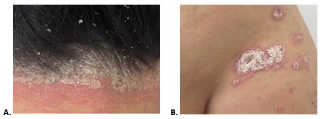
A 46-year-old woman with plaque psoriasis diagnosed in 2018 presented in July 2025 due to progressive worsening of cutaneous lesions over recent months. Lesions initially reappeared on elbows and knees, and then extended to limbs and scalp, with prominent scaling (Figure 7). Quality-of-life impairment was severe: DLQI score was 24, while PASI score was 12.6, indicating moderate disease with substantial psychosocial burden.
The complexity of this case was given by the hepatic comorbidity. The patient was previously diagnosed with compensated cirrhosis of unclear etiology (Child–Pugh score A, five points). Recent imaging and laboratory data showed advanced fibrosis, third-grade steatosis, and persistent cytolysis: ALT 159 U/L, AST 42 U/L, GGT 650 U/L. There were no clinical signs of decompensation (ascites, encephalopathy, or gastrointestinal bleeding).
Conventional systemic treatment options were severely limited. Methotrexate — commonly used in moderate-to-severe psoriasis — was considered contraindicated due to the risk of worsening liver injury and fibrosis progression (6). Other conventional systemic therapies also raised safety concerns. A careful risk–benefit assessment was performed. Even if psoriasis had a major impact on quality of life and also risk of progression, systemic intervention was needed to avoid further alteration of hepatic function.
The case was discussed with her gastroenterologist at the Carol Davila Central Military University Hospital, Bucharest. Factors supporting biologic therapy included: compensated cirrhosis, absence of severe portal hypertension/decompensation, regular liver monitoring capacity, and lack of safer alternatives. Therefore, biologic therapy with an IL-12/23 inhibitor (ustekinumab biosimilar) was initiated, given a relatively favorable hepatic safety profile in available data and no direct known association with severe hepatotoxicity. Baseline evaluation included viral hepatitis tests, infectious screening, complete blood count, and full liver panel. A strict monitoring protocol for transaminases and liver function markers was implemented.
Under treatment, improvement was observed: by four weeks, pruritus decreased and erythema diminished. At three months, the PASI score decreased to 8.7, and the DLQI score decreased to 12. Although complete remission was not achieved, the response was clinically meaningful given the disease complexity. Importantly, liver parameters remained stable, with no signs of clinical or biological decompensation. The patient received detailed counseling on monitoring and hepatic warning signs, and dermatology – gastroenterology collaboration continued.
This case illustrates that, in compensated cirrhosis, biologic therapy may be a viable alternative to hepatotoxic conventional agents, provided careful patient selection and rigorous interdisciplinary monitoring.
The management of psoriasis in the context of severe systemic comorbidities represents a major challenge in current dermatological practice, especially due to the lack of robust evidence from randomized clinical trials. Patients with HIV infection, a history of cancer, latent tuberculosis infection, or advanced liver disease are frequently excluded from these trials, which limits the direct applicability of therapeutic guidelines (5,6). In this context, data from real-world practice and clinical experience become essential to guide therapeutic decisions. The cases analyzed illustrate the significant variability of clinical presentation and the challenges of choosing treatment according to each patient’s profile.
In HIV-associated psoriasis, the literature suggests that the severity of the disease is not always directly correlated with the degree of immunosuppression, with complex mechanisms of immunological dysregulation being involved, especially at the level of the Th1/Th17 axis (8,16). In these situations, a stepwise therapeutic strategy is justified, starting with topical therapies and phototherapy, especially in localized forms (7). Clinical experience supports that avoiding systemic therapy when not absolutely necessary can reduce the risk of complications without compromising disease control.
In patients with a history of neoplasia, the main concern is the risk of tumor recurrence under immunomodulatory therapy. Although current data suggest only a modest increase in the overall oncological risk in psoriasis and do not clearly demonstrate a significant increase in recurrence under biological therapies, the level of evidence remains limited and heterogeneous (24,25). In this context, the time interval since oncological diagnosis, the type of tumor and the current status of the disease become critical factors in the choice of treatment. Close collaboration with the oncologist is essential to assess the individual risk and optimize the therapeutic strategy.
For latent tuberculosis infection, isoniazid prophylaxis is standard before initiating biologic therapy, but associated hepatotoxicity remains a relevant concern. Current guidelines recommend screening and appropriate treatment of latent tuberculosis infection before biologic therapy (31). Although the overall frequency of liver involvement is relatively low, it may become significant in certain subgroups of patients, particularly in the elderly or in the context of comorbidities (32). Careful analysis of the chronology of biological changes and response after treatment discontinuation is essential to establish causality and to avoid unnecessary discontinuation of effective biologic therapy.
In the context of chronic liver disease, especially cirrhosis, therapeutic options are limited by the risk of hepatotoxicity of conventional systemic therapies, especially methotrexate (5,38). Literature data suggest that biologic therapies, particularly IL-12/23 inhibitors, may have a favorable safety profile in patients with compensated liver disease (39).
Therefore, interdisciplinary collaboration between dermatologists, infectious disease specialists, oncologists, and gastroenterologists is absolutely essential in the management of these patients. Monitoring of clinical and biological parameters allows for rapid adjustment of treatment and prevention of complications. In the absence of clear evidence from randomized trials, the integration of real-world data provides valuable insight into the management of psoriasis associated with severe systemic comorbidities.
The management of moderate-to-severe psoriasis in the presence of major systemic comorbidities represents a complex therapeutic challenge in which decisions cannot be guided solely by cutaneous severity. Well-controlled HIV infection, recent malignancy (40-42), latent tuberculosis prophylaxis requirements, or compensated cirrhosis (43-47) should not be considered absolute contraindications to systemic or biologic therapy, but rather risk modifiers requiring careful stratification and dynamic monitoring.
The present case series illustrates that therapeutic strategies, including topical, phototherapeutic, and, in selected situations, systemic or biologic therapies, can be safely used in high-risk contexts when patient selection is rigorous, interdisciplinary collaboration is active, and surveillance is tailored to the clinical scenario. Sequential assessment of adverse events and ongoing re-evaluation of the risk–benefit balance is essential to avoid both undertreatment and unnecessary risk exposure. Also, three core principles emerge: individualized decision-making – PASI and DLQI scores are essential, but insufficient without a global patient risk assessment; interdisciplinary collaboration – dermatology, oncology, infectious diseases, gastroenterology and rigorous and dynamic safety monitoring, particularly in the setting of polypharmacy and complex systemic risk.
Taking all of these aspects into consideration, and in the absence of robust randomized evidence for these special populations, real-world experience contributes significantly to a risk-adapted dermatology approach, emphasizing structured vigilance over therapeutic avoidance.
The authors declare no conflict of interest. This research received no external funding.
Conceptualization, AFG, DOC, and CC; methodology, ADG, and CC; validation, DOC, CC, and CS; formal analysis, ADG, and CS; investigation, ADG, and HB; resources, ADG, AE, and HB; data curation, DOC, CS, and CC; writing—original draft preparation, ADG, and HB; writing—review and editing, ADG, DOC, and CC; visualization, AE; supervision, CC; project administration, CS. All authors have read and agreed to the published version of the manuscript.
Written informed consent has been obtained from the patients to publish this paper. The study was conducted in accordance with the World Medical Association Declaration of Helsinki (1964) and its later amendments (most recently revised in 2013). The study was approved by the Local Ethics Committee of the Dr. Carol Davila University Emergency Central Military Hospital, Bucharest (registration number 750, 22 January 2025).