
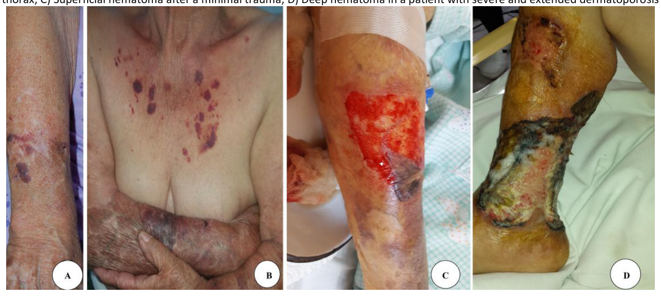
With the increase of the medium lifespan in developing countries, a new type of dermatosis was described in 2007 by Kaya and Saurat under the name of ‘dermatoporosis’ of chronic cutaneous insufficiency/fragility syndrome [1,2,3]. Clinical aspects comprise atrophy, purpura, and pseudo-stellate scars (Figure 1). Firstly, the British dermatologist Thomas Bateman described senile purpura in 1818 as purpuric patches arising in elderly people after minimal trauma, especially on the dorsum of the hands [4]. Later, in 1967 Colomb reported pseudo-stellate scars as fibrous scars with a stellate aspect arising on the forearms of the elderly [5]. One year after this description, Frederik and Zak made the connection between stellate pseudoscars and senile purpura, describing that both arise on the forearms of the elderly after minimal traumas. They also report that the shape of these scars can be round or linear, not only stellate [6]. In 1972, Colomb described the trade consisting of atrophy, Bateman’s purpura, and spontaneous pseudo-scars The term pseudoscars seems to be related to the fact that there is no solution of continuity with the exterior, the lesions are produced by a compression trauma without true lesions in the extreme external part of the skin, the epidermis [5,7]. Furthermore, there is a lack of reports between 1970 and the full documentation of Kaya and Saurat in 2007. Starting from this historical data and personal clinical cases, they bring valuable information according to clinical aspects, histopathology, ultrasonographic aspects, pathophysiology, and therapeutics [3]. The prevalence of dermatoporosis is about 30% in patients older than 65 years, but this aspect can vary according to the recruitment group: non-hospitalized patients, inpatients, outdoor patients, and patients for rehabilitation centers. The aim of this paper is to analyze published existing data for improving the diagnosis and management of dermatoporosis.
Only the future will decide if dermatoporosis is the expression of the general process of senescence, chronic sun exposure or even both [3]. Firstly, some data indicate that patients could have a genetic predisposition according to the connection with atopic dermatitis [8]. Furthermore, analyzing some aspects of aquaporines and skin aging previous statement is increased [9]. Aging plays a major role, the aging process acts mainly through chronic sun exposure, especially the professional type [3].
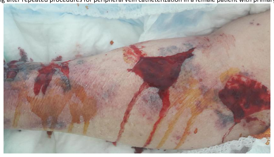
Intrinsic aging or endocrine aging plays a role, the disease is more frequent in postmenopausal women [10]. On the other way, seems that maternal age > 40 years at last pregnancy, breastfeeding for > 7 months per pregnancy and > 18 cumulative months, age < 20 years at first pregnancy, and menopause after 45 years play a protective role against dermatoporosis [11]. Iatrogenic factors like systemic steroid usage especially in patients with blistering diseases can induce dermatoporosis by decreasing the extracellular matrix leading to skin atrophy.
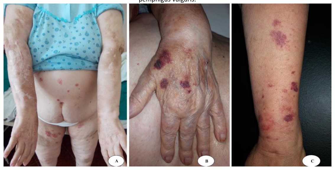
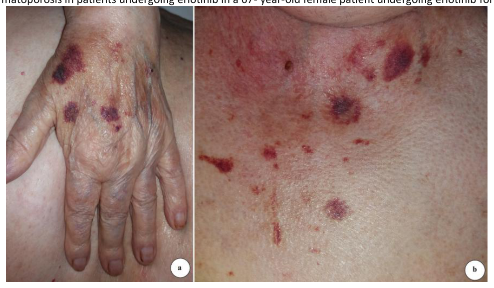
Regarding topical steroids seems that only clobetasol propionate 0.5% can induce dermatoporosis, can it be used for more than six months, this aspect can be visible on the forearms in patients with psoriasis (Figure 3) [12]. The author documented also an interesting case of a 42 female old patient suffering from a secreting cortisol tumor with a Cushing syndrome aspect and typical lesions of dermatoporosis on the forearms (Figure 4). Other medications could be anticoagulants, and epidermal growth factor inhibitors (EGFR) used in the treatment of different carcinomas (Figure 5). Associated comorbidities like osteoporosis, renal failure, and anemia were cited [13].
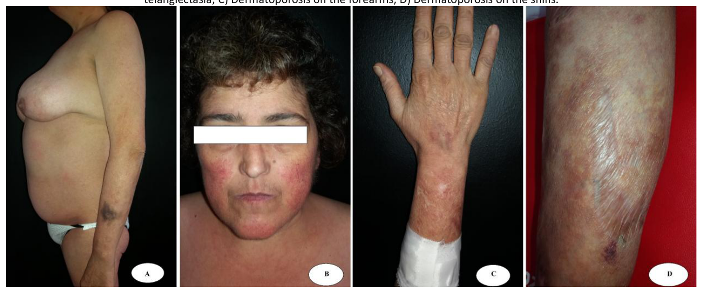
Moreover, there is a strong connection between the degree of osteoporosis and dermatoporosis [14,15]. It is not clear yet if osteoporosis has a direct influence on the onset of dermatoporosis or if both are the result of the general aging process having hormonal disturbances at the base. Additional information was brought by Kluger in 2016 describing the possible connection of dermatoporosis with the lack of vitamin C levels [16]. Laboratory data about skin expression or serum levels of vitamin C and dermatoporosis are lacking. This correlation is based only on the benefits of topical or systemic administration of vitamin C [16,17]. Recently, vitamin D deficiency was also proposed as a factor by Romano et al based on the connection between osteoporosis and dermatoporosis. Anyway, correlation data between decreased levels of vitamin D and dermatoporosis degree in the elderly are necessary for validation [18]. Finally, most of the patients present an association of these risk factors.
The clinical changes are skin atrophy, purpura, pseudoscars, hyperpigmentation, and ulcerations. Due to atrophy, there is an increased risk of the appearance of superficial or dissecting hematomas of the skin that form by the accumulation of blood in between the hypodermis and the muscular fascia due to local traumas [3,19]. Most lesions are located at the level of the forearms and shins [3]. Other sites are the chest, arms, and dorsal parts of the hands. It is a tendency for females to develop lesions on the chest and face compared with men. The appearance of lesions at the level of the face and scalp is reported in rare and isolated cases [3,10]. Skin atrophy is the main sign of dermatoporosis. It is essential for the development of the other changes, so that, on the background of exposure to trauma of the underlying structures, purpura and skin ulcers develop. These heal with hyperpigmentation and scars [3]. Senile purpura (Thomas Bateman, 1818) occurs in the elderly, especially on the backs of the hands, and is often found in the context of dermatoporosis [3.4]. It has a complex mechanism, being produced by both exposure of blood vessels due to atrophic skin, and by a decrease in the elastic fibers of the vascular endothelium. An increase in capillary permeability with age is also taken into account [4,20]. Colomb stellate pseudoscars are found on the forearms of the elderly and are produced by defective remodeling of the extracellular matrix due to functional disturbances of enzymes such as metalloproteinases [21]. Appearance on the shins and other sites is also possible. They can be stellate, linear, or plaque-like. The stellate ones are the most common [3,10]. They are formed by the rupture of collagen fibers but without clinical evidence of a break in continuity at the level of the epidermis. Repair is done by excessive proliferation of fibrous tissue, which leads to a clinically hypertrophic scar appearance [5,7]. Pseudoscars are found in approximately 30% of people over the age of 70. Hyperpigmentations appear as residual lesions and as a sign of photodamaged skin [3,10,21]. Cutaneous ulcerations appear based on the cutaneous atrophy after some unnoticeable frequent minimal traumas. They appear at the level of the extremities [3].
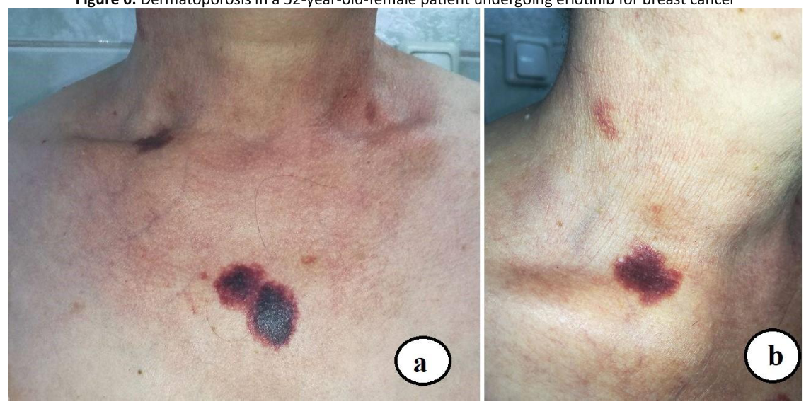
When the cause is obvious like usage of corticosteroids, local or oral, disruption of the treatment can improve greatly and stop the development of further purpura. Superficial hematomas are due to blood accumulation between the dermis and the subcutaneous fat. Clinically appears as a detachment of a skin region from the subjacent adipose tissue. They appear in the advanced stages of dermatoporosis and are found at the level of the shins as well as at the dorsal surface of the forearms [3]. Deep Dissecting hematomas of the skin (DDH) represents the main complication of dermatoporosis. They form as the accumulation of blood between subcutaneous fat and muscular fascia. It represents a surgical emergency due to the risk of block detachment of the cutaneous structures with exposure to infections of major muscular tissue areas. Due to the profound localization of blood collection, the detachment of the skin does not occur immediately. Thus, the lesions mimic cellulitis, presenting as painful erythematous-edematous plaques. People over 80 years with are affected, especially the females and they are located strictly on the calves. The positive diagnosis is done through magnetic resonance imaging (MRI) that shows a hypoechogenic band in the virtual space located between the hypodermis and the muscular fascia. DHH represents a major complication of the disease, the emergency surgical drainage being necessary. The repair of the residual defects is done slowly by the application of topical hyaluronic acid or with skin grafting [3,19] The development of non-melanocytic skin cancer is possible also on dermatoporotic skin, the author saw a case of actinic keratosis and one of cutaneous horn arising in the area of dermatoporosis (Figure 6).
Clinical aspects are usually enough for the diagnosis. More than that, Saurat developed a self-diagnosis tool (IDA: Index Dermatoporosis Assessment) consisting of a two-module questionnaire that is addressed to individuals aged ≥65 years. The technique was validated in France and Mexico. Skin ultrasonography quantifies the grade of atrophy showing a thickness of skin approx. 0.7-0.8 mm at the level of the regions with dermatoporosis (normal value = 1.3-1.5 mm). The usefulness of reflectance confocal microscopy (RCM) for the diagnosis of dermatoporosis and quantifying skin thickness was proven by Mezinger, but is expensive and not accessible for routine. Electronic microscopy highlights the disorganization of collagen with parallel exposure of the fibers on the surface of the epidermis (88). Anyway, skin biopsy is an invasive tool, so is recommended to be avoided in such patients and usually is unnecessary when the diagnosis is obvious but might be necessary to rule out other diagnoses in case of atypical location or isolated lesions. For these cases, a 3 mm punch biopsy can be performed on non-erosive/ulcerated lesions without any risk. A general blood count to exclude bleeding disorders, exploration of renal function, and osteoporosis assessment is strongly recommended [3,8]. The fragility of dermatoporotic areas with bleeding and delayed healing results in significantly limited skin biopsies and secondary histopathological and immunohistochemistry studies.
Depending on the main etiological factors, dermatoporosis can be classified as primary or secondary. Regardless of the form, the lesions are located on the same regions of election such as forearms, shins, anterior part of the thorax, and dorsum of hands [3,10]. Face and scalp location was also cited [10]. Primary dermatoporosis or actinic is due to chronic exposure to the sun, especially in patients over 60 years who are affected. Secondary dermatoporosis lies behind iatrogenic factors, especially the prolonged utilization of corticotherapy. It is characterized by the possibility of the lesions appearing under the age of 60 as well as by partial or complete resolution of the lesions after corticotherapy cessation [3]. The systemic usage as well as topic one can lead to the development of the lesions. Regardless of the way of administration or the cutaneous location in the case of local therapy, the lesions develop in s the same regions/ zones as well as in primary dermatoporosis [3,6]. The disease presents four evolutionary stages. The first stage is characterized by atrophies, purpura as well as pseudo scars, and adding the presence of cutaneous ulcerations leads to the second stage. In the third stage, the ulcerations are increased in size and number. Development of DDH is characteristic of the fourth stage [3,6,19].
The prevention consists firstly in avoidance of the triggers. Regarding this, there is the necessity to use the factors for photoprotection and the avoidance of trauma in patients who are already suffering from primary dermatoporosis. Careful organization at home with the furniture (« coffee table syndrome »), prevention of falling, also proper information of nursing staff the way they handle the patients, no adhesive directly on the skin, avoid excessive application of potent or highly potent corticosteroids. The first topical was proposed by Kaya in 2007, at the same time as the disease was described. The application of hyaluronic acid 1% leads to the improvement of the lesions [3]. In a recent study, topical or oral hydrolyzed collagen does not improve precocious lesions of dermatoporosis – stage I [23]. Topical retinoids are cited to be efficient in the treatment of dermatoporosis since their role is the re-establishment of the differential cellular process. The use of the combination of hyaluronic acid and retinyl-aldehyde is more effective than the separate application of these two [24]. Alpha-hydroxy-acids (AHA) are efficient by diminishing the grade of atrophy [25]. Humbert and colleagues sustain that dermatopororis, especially the Bateman senile purpura, is due to a deficit of vitamin C skin level and it shows an abatement of the lesions after applying topically vitamin C 5% [17]. There is limited data regarding the efficacy of topical application of topical human epidermal growth factor or dehydroepiandrosterone [26,27]. Systemic administration of vitamins C and D could bring some benefits [16,18]. An isolated case of dermatoporosis in the upper limbs treated with polymethylmethacrylate (PMMA) using the BioSculpt® technique was reported.
The authors declare no conflict of interest. No artificial intelligence automatically generated text was inserted in this manuscript, and no image was previously published in another journal or is under consideration of being published elsewhere. This research received no external funding.
Conceptualization, MAB; methodology, MAB, ACC; software, ACC; validation, DOC, ALT, NK; formal analysis, NK; investigation, MAB, ACC; resources, DOC, ALT, NK; data curation, ALT, NK; writing—original draft preparation, MAB, NK, ACC; writing—review and editing, DOC, ALT; visualization, ALT, NK; supervision, MAB, NK; project administration, DOC, ALT. All authors have read and agreed to the published version of the manuscript.
The study was conducted in accordance with the Declaration of Helsinki.
Consent was obtained from all subjects involved in the study. Written informed consent has been obtained from the patients to publish this paper.