Paneth Cells: A Comprehensive Review of Their Role, Prevalence and Molecular Mechanisms in Colorectal Neoplastic Lesions

1 - Pathology Department, Central Military Emergency University Hospital, Bucharest, Romania; florea.maria@drd.umfcd.ro

2 - Pathology Department, University of Medicine and Pharmacy “Carol Davila”, Bucharest, Romania

3 - Department of Special Motricity and Medical Recovery, The National University of Physical Education and Sports, Bucharest, Romania; lucicaeftimie@yahoo.com

4 - Pathology Department, Saint-Antoine Hospital, Paris, France; diana.enea21@yahoo.com

5 - Pathology Department, Fundeni Clinical Institute, Bucharest, Romania; gabriel.becheanu@umfcd.ro

6 - Pathology Department, Emergency University Hospital, Bucharest, Romania; mariana.costache@umfcd.ro

Correspondence: Lucian G. Eftimie, lucicaeftimie@yahoo.com

DOI: https://doi.org/10.55453/rjmm.2025.128.2.10

Received: 3 December 2024

Revised: 7 January 2025

Accepted: 12 January 2025

Abstract:

Paneth cells (PCs) are specialized epithelial cells located at the base of the crypts of Lieberkühn in the small intestine and colon, that have been occasionally identified in colorectal adenomas and adenocarcinomas. Their presence in these lesions has raised questions regarding their role in colorectal neoplasia and their potential association with adenoma burden. To understand their potential role in carcinogenesis, we conducted an extensive review of the available literature on PubMed, focusing on the occurrence, molecular mechanisms, and implications of Paneth cells in neoplastic lesions of the colorectum. This review synthesizes current findings and discusses the potential of Paneth cells as diagnostic biomarkers and therapeutic targets in colorectal cancer.

Keywords:
Citation:

Florea MA, Eftimie LG, Enea D, Becheanu G, Costache M. Paneth Cells: A Comprehensive Review of Their Role, Prevalence and Molecular Mechanisms in Colorectal Neoplastic Lesions. R. J. Mil. Med. 2025, 128(2): 166-170; https://doi.org/10.55453/ rjmm.2025.128.2.10

Article content:

Introduction

Paneth cells are highly secretory cells with a lifespan of approximately two months [1], crucial in maintaining gut health and innate immunity [2]. The best-known function of PCs is controlling the microbiome composition, but they have much broader functions. These cells secrete antimicrobial peptides, such as defensins and lysozymes, which help protect the intestinal lining from pathogens. PCs also support intestinal stem cells by providing essential growth factors, ensuring continuous renewal of the gut lining. Some authors propose that Paneth cells serve as a niche for stem cells [3-5], due to their location and expression of factors that support the niche (Notch, Wnt, growth factors).

Paneth cells are typically found at the base of the crypts, along with enteroendocrine cells and stem cells, and have distinct histological features: they have a triangular shape, bright red cytoplasmic granules, containing antimicrobial peptides, and basally located nuclei (Figure 1).

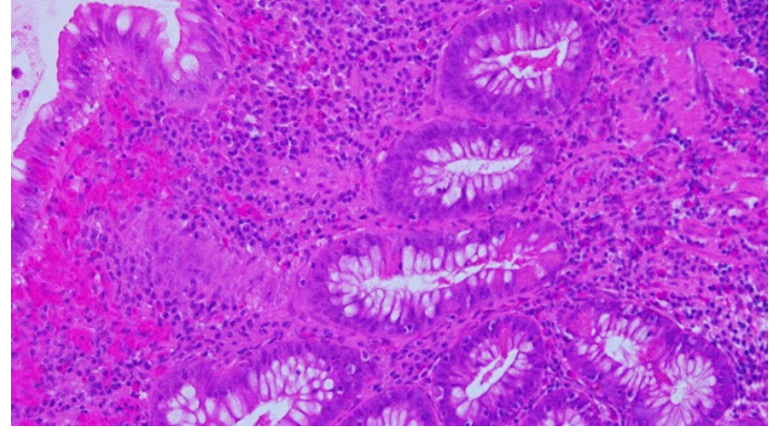
In the small intestine, the abundance of PCs increases progressively from the proximal to the distal regions, aligning with heightened bactericidal activity [6]. In contrast, in the colon, their numbers decline from the proximal to the distal areas and are absent in the left colon and rectum under normal conditions. Paneth cells in the left colon could be a sign of chronic mucosal injury (Figure 2). Their dysfunction has been linked to various gastrointestinal disorders, including inflammatory bowel disease (Crohn’s disease and ulcerative colitis) and graft-versus-host disease [7,8].

Human defensin 5 (HD-5) has been previously shown to be a specific marker of Paneth cells in the gastrointestinal tract and one of the target genes of the APC/β-catenin/Tcf pathway [9].

Methodology

A comprehensive search was conducted on PubMed using keywords such as “Paneth cells”, “colonic adenomas”, “serrated”, and “colorectal cancer” (CRC). Relevant studies published from 1965 to 2024 were selected, including original studies and case reports. A total of 49 studies were found, out of which 14 articles were considered relevant to our review, focusing on the role of Paneth cells in early neoplastic lesions and their potential as biomarkers in colorectal carcinogenesis.
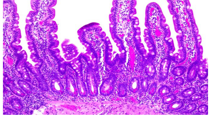
Photomicrograph of Paneth cells at the base of normal small bowel mucosa, HE stain, 100x magnification.
Figure 1: Paneth cells located at the base of a normal-looking small bowel mucosa (HE stain, 100x).
Photomicrograph of Paneth cell metaplasia in inflamed left colonic mucosa of a patient with ulcerative colitis, HE stain, 400x magnification.
Figure 2: Paneth cell metaplasia in the left colonic inflamed mucosa of a patient with ulcerative colitis (HE stain, 400x).

Results

A summary of our research is presented in Table 1, which includes the main findings regarding the presence of PCs in neoplastic lesions of the colorectum, based on original studies.

PCs have been described in colonic epithelial neoplasms, particularly in adenomas (Figures 3A and 3B). Based on the studies we reviewed from PubMed, Paneth cells were encountered in colorectal adenomas. As one would expect, due to their normal distribution in the colon, PC differentiation is more frequently observed in adenomas located in the proximal colon and in patients with familial adenomatous polyposis [9-11]. The reported prevalence of PC differentiation in adenomas ranges widely from 0.2% to 50% in our research [9,12-19].
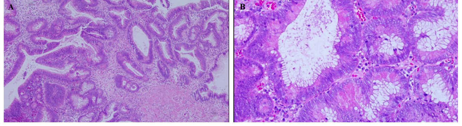
Photomicrographs, panels A and B, of Paneth cells within a tubulo-villous adenoma with low-grade dysplasia, HE stain, 100x and 400x magnification.
Figure 3 (A, B): Presence of Paneth cells in a tubulo-villous adenoma with low-grade dysplasia (HE stain, 100x and 400x).

In 1984, Bansal and Fenoglio identified PCs (described as “Paneth cell metaplasia”) in 0.2% of colonic adenomas among 3,215 patients, which was the largest number of patients included in the studies that we identified. They proposed that PCs in colonic adenomas represent the terminal differentiation of neoplastic cells. However, since their study, limited research has been conducted to evaluate the presence of PCs in relation to colonic neoplasms. As a result, it remains unclear whether PC differentiation is associated with specific types of colonic tumors.

Table 1: Main findings regarding the presence of PCs in neoplastic lesions of colorectum, based on original studies.
First Author/Publication year/Reference number Main findings, including the number of colorectal polyps examined/percent of lesions containing Paneth cells
Gibbs/1967/[11]
  • 69 solitary adenomas/27% PCs
  • 23 adenomas from 11 cases of polyposis coli/34% PCs
  • 140 CRC/3% PCs
Subbuswamy/1973/[12]
  • 5 adenomas and 26 CRC
  • Describes immature Paneth cells and intermediate cells (with intermediate features between Paneth cells and goblet cells)
Bansal/1984/[13]
  • 5770 adenomas/0,2% PCs
  • Presence of PCs is associated with a tubulovillous architecture
  • Paneth cell, squamous cell, and melanocyte differentiation can be seen in adenomas when epithelial cells undergo neoplastic alterations
Iwashita/1989/[14]
  • 212 adenomas/25% PCs
Rubio/2003/[15]
  • Intense lysozyme expression was found in 9.5% of the hyperplastic polyps, in about 90% of conventional adenomas and serrated adenomas and in 76.9% of the CRC
Mee Joo/2009/[9]
  • 39 adenomas/38,5% PCs; 40 CRC/1% PCs
  • None of the serrated polyps presented PCs
  • Coexpression of HD-5 and nuclear β-catenin staining was identified in most conventional adenomas (74.4%, P < .01)
  • Paneth cell differentiation was more frequent in right-sided adenomas
Pai/2013/[16]
  • 152 adenomas/17,1% PCs
  • Presence of Paneth cells is associated with male sex and adenoma burden
Mahon/2016/[17]
  • 1900 adenomas/18,8% PCs
  • PCs-containing adenomas at the distal colorectum were inversely associated with the presence of synchronous advanced adenomas or CRC
López-Arribillaga/2021/[18]
  • 38 adenomas/about 50% of PCs
  • Accumulation of PCs in early colorectal adenomas is associated with Beta-Catenin signalling and poor patient prognosis

Lysozyme immunohistochemical positive reaction, a sensitive PCs marker, was identified by Rubio, in 2003, in most colorectal adenomas, in many CRCs and in 9,5% of hyperplastic polyps that he examined, raising the question if intense lysozyme production might herald a possible dysplastic evolution in some hyperplastic polyps [15]. Later it was proven that lysozyme expression is less specific than HD-5 in gastrointestinal tissues [19].

In their study, Mee Joo et al [9] found histologic evidence of PCs differentiation in 38.5% of colonic adenomas and in 1 adenocarcinoma (2.5%) but not in any of the other types of polyps in this study. They also investigate the role of HD-5 and β-catenin in this process, as HD-5 has been previously shown to be a specific marker of Paneth cells [19-21] and is also one of the targeted genes of the APC/β-catenin/Tcf pathway of colon carcinogenesis. APC (adenomatous polyposis coli) is a key tumor suppressor gene, which helps control the Wnt signaling pathway. Its mutation plays a crucial role in the progression of colorectal cancer through the adenoma-carcinoma sequence, both in hereditary conditions like familial adenomatous polyposis and in sporadic cases. The loss of APC function causes β-catenin to accumulate in the cell, which promotes uncontrolled cell growth and the formation of conventional adenomas [22].

HD-5 was expressed in their study in neoplastic tissues, in cells with and without histologic evidence of Paneth cell differentiation, meanwhile in normal colonic mucosa HD-5 was expressed exclusively in normal and metaplastic Paneth cells [9]. They found a significant correlation between nuclear β-catenin and HD-5 immunohistochemical expression in adenomatous and cancerous areas since all neoplastic cells with histologic evidence of Paneth cell differentiation showed nuclear accumulation of β-catenin. This suggests that the activation of APC/β-catenin/Tcf pathway may play a role in Paneth cell differentiation in human colonic neoplasms [9].

Furthermore, in a more recent study, the authors indicated that Paneth cells contribute to the initial steps of cancer progression, and their accumulation in early colorectal adenoma is associated with β-catenin signaling and poor patient outcome [18]. Their immunohistochemical research proved also that PCs in adenomas show positive synaptophysin and membranous c-kit expression compared to non-dysplastic cells in the adjacent colonic mucosa.

Also, the presence of PCs containing adenomas had a strong association with male sex and adenoma burden [16]. No association was identified with the patient’s age, adenoma size, villous architecture, or grade of dysplasia. In their study, Mahon et al showed that there is a strong inverse association between PCs-containing adenomas in the distal colon and the presence of synchronous advanced adenomas or CRC [17].

Concerning colonic serrated polyps, Paneth cells have not been reported in this type of lesion, although available literature on the topic is very limited [9,23]. The absence of Paneth cells in all types of serrated polyps [9] supports the fact that these lesions are biologically heterogenous, characterized by aberrant cell proliferation and differentiation, leading to cancer through a distinct pathway, characterized by BRAF and KRAS mutations [24].

Although Paneth cell differentiation is relatively frequent in adenomas, it is rarely reported in adenocarcinomas of the gastrointestinal system [9,12,25,26].

Dysplasia with increased Paneth cell differentiation is a type of nonconventional dysplasia described in patients with inflammatory bowel disease. Also, Paneth cell differentiation was observed at a lower extent in goblet cell deficient dysplasia [27]. Changes in Paneth cell function have been shown to influence the gut microbiota, which in turn may impact the progression of colorectal cancer. Dysbiosis could exacerbate Paneth cell dysfunction, creating a feedback loop that drives tumor progression.

Conclusion

The presence of Paneth cells in the colon is often associated with pathological states such as inflammation and neoplasia. The appearance of Paneth cells in colonic neoplastic lesions reflects alterations in epithelial differentiation and could serve as a marker of early neoplastic transformation.

Molecular mechanisms governing Paneth cell differentiation, including the Wnt/β-catenin signaling pathway, significantly contribute to their presence, especially in colonic adenomas. Further research is needed to better understand the mechanisms underlying Paneth cell plasticity and their role in tumor progression. Given their potential to serve as early biomarkers for neoplastic changes, Paneth cells may offer significant clinical utility in the early detection, diagnosis, and monitoring of precancerous lesions and colorectal cancer.

Conflicts of interest and sources of funding

The authors declare no conflict of interest. No artificial intelligence automatically generated text was inserted in this manuscript, and no image was previously published in another journal or is under consideration of being published elsewhere.

Authors’ contribution

Conceptualization M.A.F and L.G.E.; methodology M.A.F.; software D.E.; validation G.B. and M.C.; formal analysis M.A.F.; investigation M.A.F.; resources L.G.E. and D.E.; data curation M.C.; writing—original draft preparation M.A.F.; writing—review and editing L.G.E.; visualization G.B.; supervision M.C.; project administration L.G.E.; funding acquisition M.A.F. All authors have read and agreed to the published version of the manuscript elaboration.

Ethics approval and consent to participate

Not applicable.

Patient consent for publication

Not applicable.

References

  1. Ireland H, Houghton C, Howard L, Winton DJ. Cellular inheritance of a Cre-activated reporter gene to determine Paneth cell longevity in the murine small intestine. Developmental Dynamics. 2005 Aug; 233(4):1332-6. doi: 10.1002/dvdy.20446.
  2. Wallaeys C, Garcia-Gonzalez N, Libert C. Paneth cells as the cornerstones of intestinal and organismal health: a primer. EMBO Molecular Medicine. 2023 Feb 8;15(2):e16427. doi: 10.15252/emmm.202216427.
  3. Sato, T., van Es, J. H., Snippert, H. J., Stange, D. E., Vries, R. G., van den Born, M., Barker, N., Shroyer, N. F., van de Wetering, M., & Clevers, H. (2011). Paneth cells constitute the niche for Lgr5 stem cells in intestinal crypts. Nature, 469(7330). https://doi.org/10.1038/nature09637.
  4. Quintero M, Samuelson LC, Paneth Cells: Dispensable yet Irreplaceable for the Intestinal Stem Cell Niche, Cellular and Molecular Gastroenterology and Hepatology (2025), doi: 10.1016/j.jcmgh.2024.101443.
  5. Zhu, G., Hu, J., & Xi, R. (2021). The cellular niche for intestinal stem cells: a team effort. In Cell Regeneration (Vol. 10, Issue 1). https://doi.org/10.1186/s13619-020-00061-5.
  6. Nakamura K, Yokoi Y, Fukaya R, Ohira S, Shinozaki R, Nishida T, Kikuchi M, Ayabe T. Expression and Localization of Paneth Cells and Their α-Defensins in the Small Intestine of Adult Mouse. Front Immunol. 2020 Oct 13:11:570296. doi: 10.3389/fimmu.2020.570296.
  7. Deuring JJ, Fuhler GM, Konstantinov SR, Peppelenbosch MP, Kuipers EJ, Haar C de, van der Woude C J. Genomic ATG16L1 risk allele-restricted Paneth cell ER stress in quiescent Crohn’s disease. Gut. 2014 Jul;63(7):1081-91. doi: 10.1136/gutjnl-2012-303527.
  8. Levine JE, Huber E, Hammer STG, Harris AC, Greenson JK, Braun TM, Ferrara JLM, Holler E. Low Paneth cell numbers at onset of gastrointestinal graft-versus-host disease identify patients at high risk for nonrelapse mortality. Blood. 2013 Aug 22;122(8):1505-9. doi: 10.1182/blood-2013-02-485813.
  9. Joo M, Shahsafaei A, Odze RD. Paneth cell differentiation in colonic epithelial neoplasms: evidence for the role of the Apc/β-catenin/Tcf pathway. Human Pathology. 2009 Jun;40(6):872-80. doi: 10.1016/j.humpath.2008.12.003.
  10. Odze, R., Gallinger, S., So, K., & Antonioli, D. (1994). Duodenal adenomas in familial adenomatous polyposis: relation of cell differentiation and mucin histochemical features to growth pattern. Modern Pathology: An Official Journal of the United States and Canadian Academy of Pathology, Inc, 7(3).
  11. Gibbs NM. Incidence and significance of argentaffin and paneth cells in some tumours of the large intestine. J Clin Pathol. 1967 Nov;20(6):826-31. doi: 10.1136/jcp.20.6.826.
  12. Subbuswamy SG. Paneth cells and goblet cells. J Pathol. 1973 Nov;111(3):181-9. doi: 10.1002/path.1711110306.
  13. Bansal M, Fenoglio CM, Robboy SJ, King DW. Are metaplasias in colorectal adenomas truly metaplasias? Am J Pathol 1984;115:253-65.
  14. Iwashita A, Watanabe H, Enjoji M. Argyrophil and argentaffin cells in adenomas of the colon and rectum. Fukuoka Acta Medica. 1989 Feb;80(2):114-24.
  15. Rubio CA. Colorectal Adenomas Produce Lysozyme. Anticancer Res. 2003 Nov-Dec;23(6D):5165-71.
  16. Pai RK, Rybicki LA, Goldblum JR, Shen B, Xiao SY, Liu X. Paneth cells in colonic adenomas: Association with male sex and adenoma burden. Am J Surg Pathol. 2013 Jan;37(1):98-103. doi: 10.1097/PAS.0b013e318267b02e.
  17. Mahon M, Xu J, Yi X, Liu X, Gao N, Zhang L. Paneth Cell in Adenomas of the Distal Colorectum Is Inversely Associated with Synchronous Advanced Adenoma and Carcinoma. Sci Rep. 2016 May 18:6:26129. doi: 10.1038/srep26129.
  18. López-Arribillaga E, Yan B, Lobo-Jarne T, Guillén Y, Menéndez S, Andreu M, Bigas A, Iglesias M, Espinosa L. Accumulation of paneth cells in early colorectal adenomas is associated with beta-catenin signaling and poor patient prognosis. Cells. 2021 Oct 28;10(11):2928. doi: 10.3390/cells10112928.
  19. Cunliffe RN. Alpha-defensins in the gastrointestinal tract. Mol Immunol. 2003 Nov;40(7):463-7. doi: 10.1016/s0161-5890(03)00157-3.
  20. Cunliffe RN, Rose FR, Keyte J, Abberley L, Chan WC, Mahida YR. Human defensin 5 is stored in precursor form in normal Paneth cells and is expressed by some villous epithelial cells and by metaplastic Paneth cells in the colon in inflammatory bowel disease. Gut. 2001 Feb;48(2):176-85. doi: 10.1136/gut.48.2.176.
  21. Shen B, Porter EM, Reynoso E, Shen C, Ghosh D, Connor JT, Drazba J, Rho HK, Gramlich TL, Li R, Ormsby AH, Sy M-S, Ganz T, Bevins CL. Human defensin 5 expression in intestinal metaplasia of the upper gastrointestinal tract. J Clin Pathol. 2005 Jul;58(7):687-94. doi: 10.1136/jcp.2004.022426.
  22. Fodde, R. The APC gene in colorectal cancer. Eur J Cancer. 2002 May;38(7):867-71. doi: 10.1016/s0959-8049(02)00040-0.
  23. Santiago RM, Pua GL. Paneth Cells in Colonic Adenomas: Association with Higher Adenoma Burden. PJP, 1(1), 10–14.
  24. Pai RK, Makinen MJ, Rosty O. Serrated lesion and polyps. WHO classification of tumours of the digestive system, International Agency for Research on Cancer, Lyon (2019), pp. 163-169.
  25. Holmes EJ. (1965). Neoplastic paneth cells. Their occurrence in 2 adenomas and one carcinoma of the colon. 1965 Nov;18(11):1416-22. doi: 10.1002/1097-0142(196511)18:11.
  26. Nihed A, Soumaya M, Atika B, Ilhem BJ, Atef BA, Fehmi H, Moncef M. Paneth cell adenocarcinoma of the colon: A rare entity. Int J Surg Case Rep. 2019:65:313-316. doi: 10.1016/j.ijscr.2019.10.071.
  27. Choi WT, Yozu M, Miller GC, Shih AR, Kumarasinghe P, Misdraji J, Harpaz N, Lauwers GY. Nonconventional dysplasia in patients with inflammatory bowel disease and colorectal carcinoma: a multicenter clinicopathologic study. Mod Pathol. 2020 May;33(5):933-943. doi: 10.1038/s41379-019-0419-1.

Paneth Cells: A Comprehensive Review of Their Role, Prevalence and Molecular Mechanisms in Colorectal Neoplastic Lesions

Cite this article

APA Style

Florea, M.A., Eftimie, L.G., Enea, D., Becheanu, G., & Costache, M. (2025). Paneth cells: a comprehensive review of their role, prevalence and molecular mechanisms in colorectal neoplastic lesions. Romanian Journal of Military Medicine, 128(2), 166-170. https://doi.org/10.55453/rjmm.2025.128.2.10

Vancouver Style

Florea MA, Eftimie LG, Enea D, Becheanu G, Costache M. Paneth Cells: A Comprehensive Review of Their Role, Prevalence and Molecular Mechanisms in Colorectal Neoplastic Lesions. Rom J Mil Med. 2025;128(2):166-170. doi:10.55453/rjmm.2025.128.2.10.

Harvard Style

Florea, M.A., Eftimie, L.G., Enea, D., Becheanu, G. & Costache, M. 2025, 'Paneth Cells: A Comprehensive Review of Their Role, Prevalence and Molecular Mechanisms in Colorectal Neoplastic Lesions', Romanian Journal of Military Medicine, vol. 128, no. 2, pp. 166-170, doi:10.55453/rjmm.2025.128.2.10.