Atypical Fibroxanthoma versus Hemosiderotic Dermatofibroma: Challenges in Differential Diagnosis – Literature Review and Case Report

1 - Department of General Surgery, CF2 Clinical Hospital, Bucharest, Romania; eclarer_mon_ame@yahoo.com (SP); kover_zoltan@yahoo.com (ZJK); dancostea2006@yahoo.com (DGC)

2 - 1st Department of Cardiovascular Anesthesiology and Intensive Care, Prof. Dr. C. C. Iliescu Emergency Institute for Cardiovascular Diseases, Bucharest, Romania; andrei.bodor@rez.umfcd.ro

3 - Department of General Surgery, Bagdasar-Arseni Emergency Clinical Hospital, Bucharest, Romania; serban_duțescu@yahoo.com

4 - Department of Dermatology, Clinical Hospital of Infectious and Tropical Diseases ”Prof. Dr. Victor Babeș”, Bucharest, Romania; mihaela_anca_popescu@yahoo.com

5 - Department of Histopathology, Bagdasar-Arseni Emergency Clinical Hospital, Bucharest, Romania; stefanbedereag@gmail.com

6 - Carol Davila University of Medicine and Pharmacy, Bucharest, Romania; horatiu.moldovan@umfcd.ro

7 - Academy of Romanian Scientists, Bucharest, Romania

8 - Titu Maiorescu University, Faculty of Medicine, Department of Anesthesia and Intensive Care, Bucharest, Romania; gabriel.gorecki@prof.utm.ro

9 - Department of Anesthesia and Intensive Care, CF2 Clinical Hospital, Bucharest, Romania

Correspondence: Andrei Bodor, andrei.bodor@rez.umfcd.ro

DOI: https://doi.org/10.55453/rjmm.2025.128.2.9

Received: 5 December 2024

Revised: 9 January 2025

Accepted: 15 January 2025

Abstract:

Atypical fibroxanthoma and hemosiderotic dermatofibroma are both dermal tumors that can raise significant challenges for differential diagnosis. The definitive diagnosis is established based on histopathological appearance and immunohistochemistry. The aim of this paper is to present the differential diagnosis challenges encountered in the case of a 67-year-old male patient who developed within a month, following trauma to the lower third of the right thigh, a large asymptomatic tumor with an irregular outline.

Keywords:
Citation:

Popescu S, Kövér ZJ, Costea DG, Bodor A, Duțescu S, Popescu MA, Bedereag S, Moldovan H, Gorecki GP. Atypical Fibroxanthoma versus Hemosiderotic Dermatofibroma: Challenges in Differential Diagnosis - Literature Review and Case Report. R. J. Mil. Med. 2025, 128(2): 156-165; https://doi.org/10.55453/rjmm.2025.128.2.9

Article content:

INTRODUCTION

Atypical fibroxanthoma, first described in the 1960s by EB Helwig, is a rare spindle-cell dermal tumor with histological features resembling a sarcoma, but with a predominantly benign evolution and an intermediate malignant potential, hence it is considered by some authors to be a form of undifferentiated pleomorphic sarcoma and it is also known as cutaneous pseudosarcoma or paradoxical fibrosarcoma [1-5]. This type of tumor usually occurs among elderly men, following exposure to ultraviolet radiation or radiotherapy, is mainly located at the cephalic extremity and has a rapid development over the course of several weeks or months [2-4].

Dermatofibroma represents a slowly progressing benign nodular dermal tumor associated with post-inflammatory tissue reaction and dermal fibrosis, with malignant potential, predominantly affecting young adult females [6-9]. The lesion is often asymptomatic, although some patients complain of pruritus, discomfort or pain and it is characteristically localized on the extremities – especially the lower limbs, although cases with osseous, periorbital, gastrointestinal, genitourinary or intracranial localization have also been reported [6,7].

The aim of this paper is to present the differential diagnosis challenges encountered in that category of cases, ilustrating the disscussion with the case of a 67-year-old male patient who developed within a month, following trauma to the lower third of the right thigh, a rather large asymptomatic firm tumor with an irregular outline.

The diagnosis of both atypical fibroxanthoma and dermatofibroma requires going through several steps, starting with the information provided by the patient’s history and physical examination, and complementing those with imaging investigations, which provide additional information regarding the location and involvement of adjacent tissues. Nevertheless, the definitive laboratory examinations are

histopathology and immunohistochemistry. In this case, histopathology showed changes suggestive for an atypical fibroxanthoma; the diagnosis was also supported by clinical criteria (macroscopic appearance, conditions of appearance, location, rapid progression to remarkable dimensions). Immunohistochemistry, however, established the diagnosis of a hemosiderotic dermatofibroma, thus changing the prognosis of the disease.

DISCUSSION

Definition

Atypical fibroxanthoma is defined as a dermal tumor with intermediate malignant potential, characteristically appearing among elderly men, following exposure to ultraviolet radiation (UV) or radiotherapy, predominantly located on the cephalic extremity, with rapid development over a period of several weeks or months [1-5]. However, cases have also been reported in young people without any associated risk factors [1,10].

Dermatofibroma, also known as superficial benign fibrous histiocytoma, represents a frequently asymptomatic, slowly progressing, benign, usually small nodular dermal tumor with malignant potential, predominantly affecting young adult females, with characteristic localization on the extremities – especially the lower limbs [6,7,8,11]. Although it most often appears as a hyperkeratotic solitary nodule with a diameter of approximately 0.3 – 1 cm, dermatofibroma is known for its polymorphous clinical and histopathological presentation, with various types of dermatofibromas having been described in specialized literature over the years [6,8].

Etiopathogenesis

In the case of atypical fibroxanthoma, there have been discovered UV-induced mutations of the p53 gene, which determine a decrease in the DNA ability to repair, but it has also been linked to trauma, radiotherapy, xeroderma pigmentosum and other diseases with defective DNA repair, as well as immunosuppressive disorders such as HIV and diabetes [1,2,3,12].

Regarding dermatofibroma, although its etiopathogenesis is incompletely elucidated, an association with trauma, especially with arthropod stings and tattoos, has been noted, although this hypothesis has been questioned following cytogenetic studies, which, along with the clinical evolution, recurrence frequency, and metastatic potential, represented arguments supporting the neoplastic origin of the lesion [6,7,13-15]. In the multiple eruptive variants, recorded in 0.3% of patients, immunological abnormalities are considered to play a role in the onset of the disease [6,7]. Thus, among patients with multiple lesions, a frequent association with HIV infection and systemic lupus erythematosus has been noted, or, more rarely, with immunosuppression following heart transplantation and conditions such as dermatomyositis, atopic dermatitis, myasthenia gravis, Hashimoto’s thyroiditis, Basedow-Graves disease, Down syndrome, cutaneous T-cell lymphoma, leukemias, multiple myeloma, and myelodysplastic syndrome [6-8,16-23]. Additionally, antiretroviral agents and biologic therapy with monoclonal anti-CD11 antibodies have been implicated in the onset of the disease [7,8,24].

Clinical Presentation

Atypical fibroxanthoma presents as an erythematous nodular lesion, often ulcerated, with a moist appearance, located in sun-exposed regions, with erythematous, atrophic and telangiectatic skin [1,2,25]. Although the preferred location is on the head and neck region, cases with tumors located on the trunk and limbs have also been reported [1-4]. The tumor rarely exceeds 3 cm in diameter, and due to its rapid evolution patients often present within up to 6 months of onset [1-4]. In exceptional cases, association with xeroderma pigmentosum has been reported [1].

Dermatofibroma presents itself as a “button-like” papular lesion of firm, elastic consistency, with a diameter of 0.5-1 cm, surrounded by a discrete halo, covered with fine scales, non-adherent to deep planes, predominantly located on the limbs [6,7,8,11,26,27]. Dermatofibroma is covered with variously-colored teguments, often brown, although they can vary from pink-gray to black, its color being often attributed to the presence of hemosiderin, although recent studies have found an increased expression of stem cell factors and increased melanocyte tyrosinase at the lesion site [6,7,11,26,27]. Lateral compression reveals the presence of a central dimple, which is considered a clinically useful sign in differential diagnosis [6,7,28,29].

A series of clinical-pathological variants of dermatofibroma have been described (common, cellular, aneurysmal, hemosiderotic, epithelioid, lipidized, atrophic, clear cell, balloon cell, signet-ring cell, granular cell, atypical, palisading, keloidal, lichenoid, myxoid), among which the variants presented below in table 1 are included [6-9,23,26,30,31].

Upon dermatoscopy, atypical fibroxanthoma does not present specific features – Bugatti and Filosa described regions of white color, specific for fibrosis and healing in the middle and deep dermis, as well as an atypical polymorphic vascular network, with a punctate, linear, hairpin or arborescent vascular pattern diffusely spread over the tumor surface [1-4,32].

Dermatofibroma has a reticular-circular dermatoscopic appearance, with a regular pigmentary network, rare black dots and brown globules, with an imprecisely defined central white patch [6,7,8,33,34]. Occasionally, multiple white spots can be seen, resembling a galactic image according to some authors [6-9,23].

Histopathological examination

Atypical fibroxanthoma presents as an exophytic, non-encapsulated, relatively well-defined dermal tumor, surrounded by an epidermal collar, composed of large spindle cells, anaplastic or with a pleomorphic appearance similar to histiocytes, often appearing

multinucleated, chaotically spread or occasionally organized in bundles, with an increased number of mitotic figures and cellular atypia [1-4,32]. Histiocyte-like cells often contain lipids and hemosiderin [1,3]. The lesion may extend to the adipose tissue, deforming adjacent structures, but without invasive characteristic [1-4]. Rarely, atypical granular or clear round-oval cell variants without mitotic figures have been described, features that, along with the presence of hemosiderin, may lead to difficulties in diagnosis, with atypical fibroxanthoma potentially being confused with a high-grade sarcoma or a malignant melanoma [1-4]. Electron microscopy demonstrates the fibrohistiocytic nature of the tumor [2,35].

Table 1: Clinical-histopathological variants of dermatofibroma [6,7,8,9,23,26,30,31].
Type of dermatofibroma Characteristics
Cellular dermatofibroma Less than 5% of cases More often among young males Cases have been reported in children Preferential localization on the limbs Present in other regions (head, neck, fingers) Of larger size (usually up to 2 cm, but lesions of more than 5 cm have been reported) Recurrence rate of circa 25% Potential for metastasis reported in a few cases
Aneurysmal dermatofibroma Can be confused with a tumor of vascular origin Exhibits the particularity of rapid growth Reaches considerable dimensions Recurrence rate of circa 19%
Atypical dermatofibroma More frequently among young males A papule, nodule or plaque of up to 1.5 cm Preferentially localized on the lower limbs Recurrence rate of about 14% Potential to metastasize
Epithelioid dermatofibroma Found especially among young women Polypoid highly vascularized lesions Can resemble an ulcerated pyogenic granuloma Usually located on the limbs
Hemosiderotic dermatofibroma Bluish or pink tumor Can be confused with melanoma or vascular tumors More frequent among young males Possibly an early variant of aneurysmal dermatofibroma Highly vascularized, small vessels Extravasated erythrocytes Intra/extracellular hemosiderin deposits

Dermatofibroma is a non-encapsulated tumor located in the mid dermis, composed of spindle cells organized in bundles surrounded by abundant collagenous deposits around which they seem to form a peripheral mold [6-9,23]. Moreover, the lesion is characterized by epidermal acanthosis, pseudoepitheliomatous hyperplasia, basaloid proliferation, rich pigmentation due to increased iron or melanin concentration, and the presence of a Grenz zone demarcating the normal papillary dermis from the overlying tumor [69,23,36]. Also, a proliferation of histiocyte-like or fibroblast-like cells associated with an abundant inflammatory infiltrate can be noted, as well as the presence of foamy macrophages, siderophages, and giant multinucleated cells [6-9,23,36]. Usually, neighboring structures are not affected, but in cases with storiform disposition, these may be invaded, suggesting a dermatofibrosarcoma protuberans [6,37].

Immunohistochemistry

Atypical fibroxanthoma is positive for vimentin, CD74, CD99, CD10, pro-collagen 1 and p53 and does not react with cytokeratin, MNF116 and AE1/AE3, S-100, desmin and smooth muscle actin, allowing differentiation from squamous cell carcinoma, malignant melanoma and leiomyosarcoma [1-4,38].

Dermatofibroma exhibits variable expression of factor XIIIa, CD34, stromelysin 3, transforming growth factor beta (TGF-β) I and II receptors, insulin-like growth factor-binding protein 7 (IGFBP7) binding protein 7, D2-40, cathepsin K and the CXCR4 cytokine receptor. Nestin is positive in only 13% of cases, and Cthrc1 (collagen triple helix repeat containing-1) is rarely present, aiding in differentiation from dermatofibrosarcoma protuberans [6-9,23,39-42].

The immunohistopathological features of various types of dermatofibroma are illustrated in Table 2, below [6-9,23,39-42].

The diagnosis of atypical fibroxanthoma is one of exclusion, primarily requiring differentiation from entities such as dermatofibrosarcoma protuberans, malignant melanoma and squamous cell carcinoma, which is mainly achieved through histopathological exam and immunohistochemistry [1-4].

In the case of dermatofibroma, the differential diagnosis is made with numerous conditions, including benign or malignant tumors

(leiomyoma, lipoma, leiomyosarcoma, dermatofibrosarcoma protuberans, carcinomas, malignant melanoma, cutaneous T-cell lymphoma, keratoacanthoma, angiohistiocytoma with multinucleated cells, hemosiderotic hemangioma), cutaneous metastases, various forms of cutaneous nevi (Spitz, blue nevus, dysplastic nevus, melanocytic nevus), atypical fibroxanthoma, cutaneous manifestations of various conditions such as HIV infection, reticulohistiocytosis, mastocytosis, prurigo nodularis, etc [6-9,23,39]. Although the clinical appearance is suggestive in typical cases, the definitive diagnosis is established based on histopathological appearance and immunohistochemistry.

Table 2: Immunohistopathological features of dermatofibroma [6-9,23,39-42]
Type of dermatofibroma Histopathology and immunohistochemistry
Cellular dermatofibroma Epidermal hyperplasia Highly cellular Lower polymorphism Composed of spindle cells with eosinophilic cytoplasm and storiform disposition Variable mitotic index Necrosis in 12% of cases Frequent focal invasion of subcutaneous tissue, with a lacy pattern Often positive for CD34, factor XIIIa and actin
Aneurysmal dermatofibroma Extensive hemorrhage Pseudovascular spaces similar to prominent caverns, without being lined with endothelial cells Mitotic index varies Often positive for CD34, factor XIIIa and actin
Atypical dermatofibroma Mono- or multinucleated spindle-shaped or histiocyte-like cells Pleomorphic, with mitotic figures and cellular atypia
Epithelioid dermatofibroma Cells with abundant eosinophilic cytoplasm and vesicular nuclei Often with a prominent vascular component Can be confused with Spitz nevus (unlike the latter, involvement of the dermo-epidermal junction cannot be observed) Positive for ALK (anaplastic lymphoma receptor tyrosine kinase) Negative for S-100
Hemosiderotic dermatofibroma Abundant hemosiderotic deposits Often positive for factor XIIIa Variably positive for CD34, usually negative tumor cells and positive endothelial cells Negative for ALK

The treatment of choice is surgical for both conditions. In the case of atypical fibroxanthoma, Mohs micrographic technique can be used if the lesion is not well delimited, and given the risk of local recurrence and lymphatic dissemination, this procedure is increasingly recommended for macroscopically well-defined tumors [1-4,43-46].

In the case of dermatofibroma, special attention is given to cellular, atypical and aneurysmal variants, due to the risk of local recurrence, as well as metastasis in the first two variants [6-9,23,47,48]. Conventional excisional biopsy is the most commonly used technique, although cases successfully treated with cryosurgery, CO2 or pulsed-dye lasers have been reported [6-9,49,50]. Intralesional corticosteroid injection has also been reported, but the results have been unsatisfactory [6-9,23,39].

Evolution and prognosis

Atypical fibroxanthoma mostly presents a benign course, but the risk of an unfavorable outcome requires careful patient monitoring [1-5]. Last but not least, sun exposure should be avoided, and it is recommended to use photoprotection [1-4].

For the same reasons as with atypical fibroxanthoma, surveillance of patients with dermatofibromas is recommended when there is suspicion of an aggressive clinical-histological variant. On average, the rate of local recurrences was estimated at about 20%, and in the specialized literature, metastases of over 5 cm in size with various locations, including pulmonary, have been reported in variants with increased malignant potential [6-9,23,51]. However, rare cases of spontaneous regression have also been reported, with lesions leaving behind lasting post-inflammatory hypopigmentation [6-9,23]. In the case of multiple eruptive variants, screening for possible associated autoimmune conditions is recommended [6-9,23,52].

CASE REPORT

We present the case of a 67-year-old patient of urban origin, with no significant hereditary or personal pathological history, who requested dermatological consultation after having developed a post-traumatic tumor in the lower third of the right thigh over the course of one month.

Upon clinical examination, a seemingly subcutaneous, round-oval mass with irregular contours, with a diameter of about 8/9 cm, was noted. The lesion was painless, firm and immobile upon palpation, with overlying violaceous-colored skin (Figure 1). No locoregional adenopathy was detected and there were no subjective complaints.
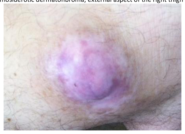
Hemosiderotic dermatofibroma, external aspect of the right thigh's distal third
Figure 1: Hemosiderotic dermatofibroma, external aspect of the right thigh’s distal third
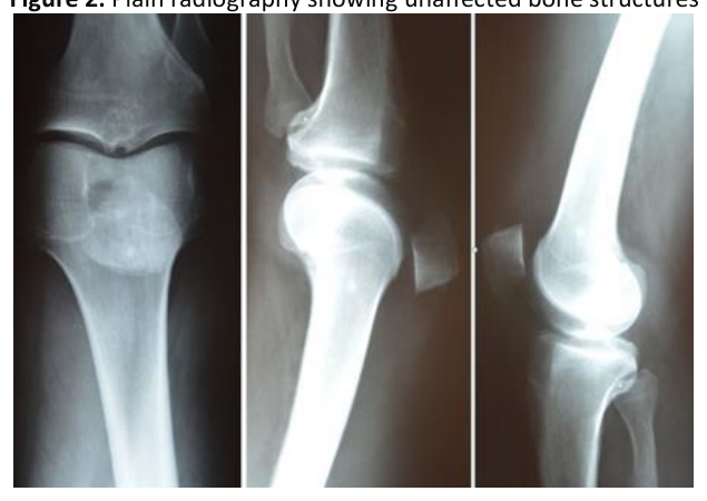
Plain radiography showing unaffected bone structures
Figure 2: Plain radiography showing unaffected bone structures
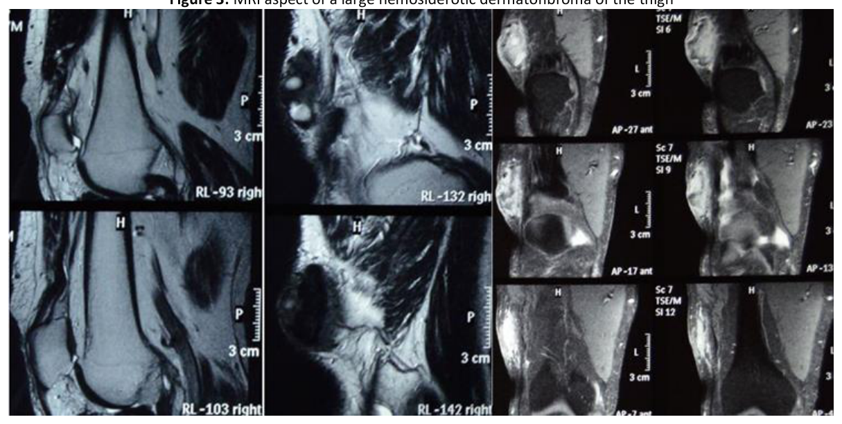
MRI aspect of a large hemosiderotic dermatofibroma of the thigh
Figure 3: MRI aspect of a large hemosiderotic dermatofibroma of the thigh
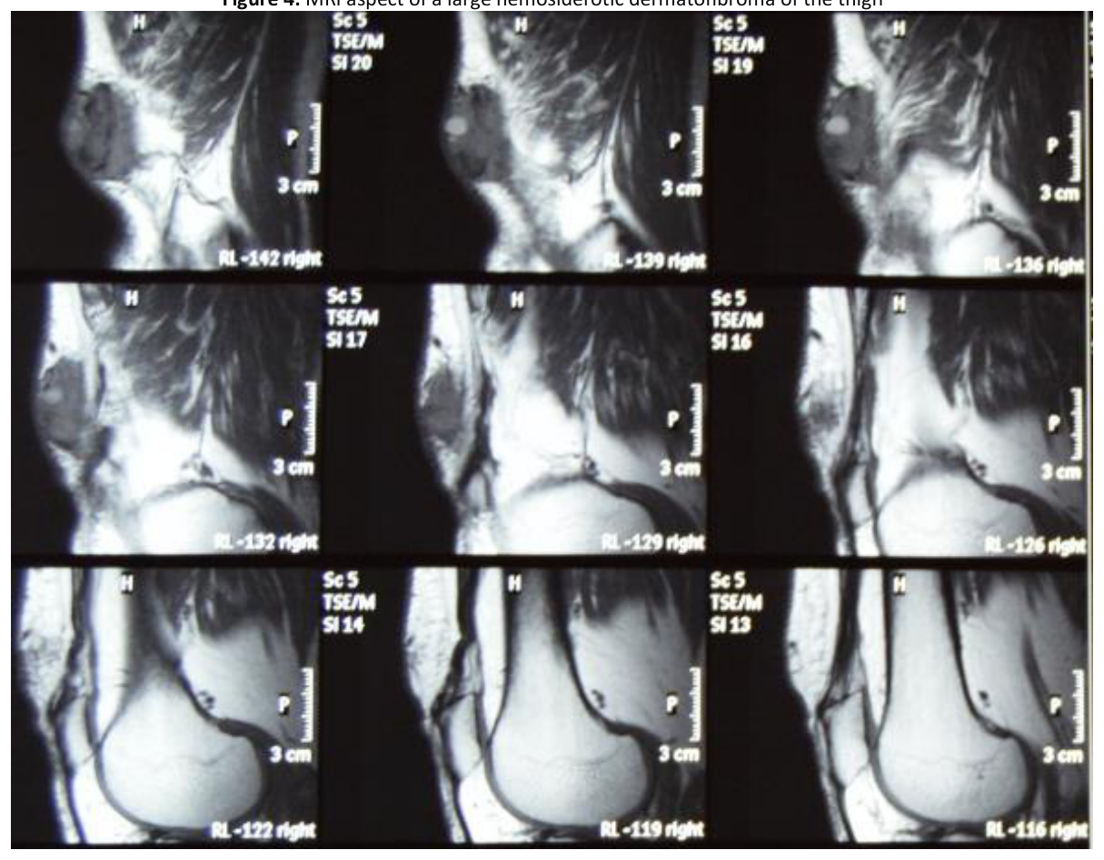
MRI aspect of a large hemosiderotic dermatofibroma of the thigh
Figure 4: MRI aspect of a large hemosiderotic dermatofibroma of the thigh
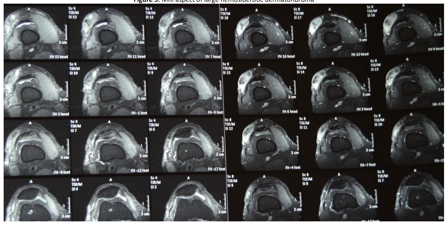
MRI aspect of large hemosiderotic dermatofibroma
Figure 5: MRI aspect of large hemosiderotic dermatofibroma
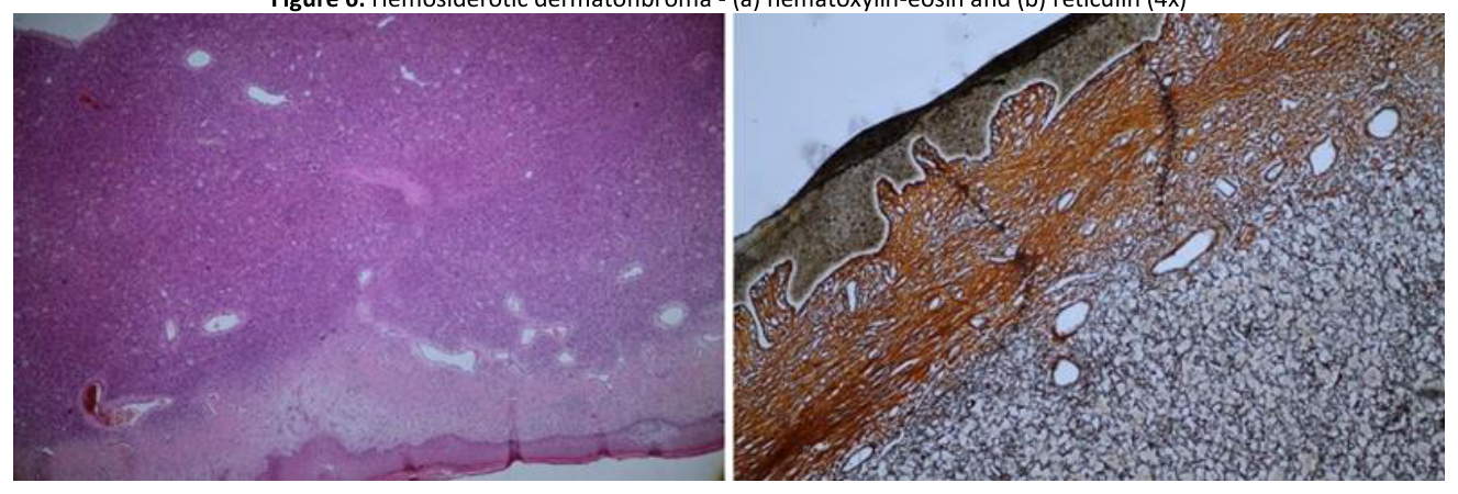
Hemosiderotic dermatofibroma - (a) hematoxylin-eosin and (b) reticulin (4x)
Figure 6: Hemosiderotic dermatofibroma – (a) hematoxylin-eosin and (b) reticulin (4x)
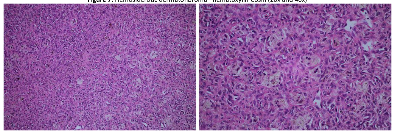
Hemosiderotic dermatofibroma - hematoxylin-eosin (20x and 40x)
Figure 7: Hemosiderotic dermatofibroma – hematoxylin-eosin (20x and 40x)
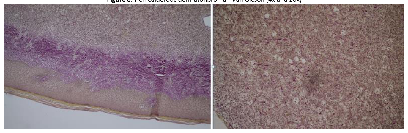
Hemosiderotic dermatofibroma - Van Gieson (4x and 20x)
Figure 8: Hemosiderotic dermatofibroma – Van Gieson (4x and 20x)
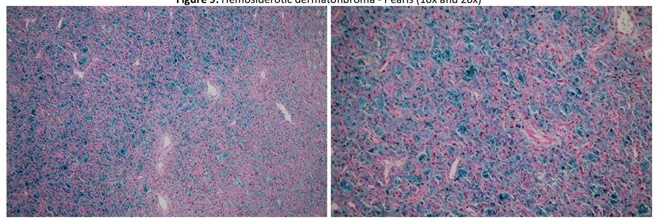
Hemosiderotic dermatofibroma - Pearls (10x and 20x)
Figure 9: Hemosiderotic dermatofibroma – Pearls (10x and 20x)

Immunohistochemical examination revealed diffuse positive staining for factor XIIIa in tumor cells, negative staining for CD34 in tumor cells and positive staining in vessels, positive staining for ACT in vascular walls, negative staining for CK 19 in tumor cells and positive

staining for Ki67 in 3-5% of tumor cells. Thus, the immunohistochemical examination, in association with the histopathological aspect, supported the diagnosis of hemosiderotic dermatofibroma.

The evolution of the presented patient was favorable, with no signs of local or regional recurrence that could be observed during a 1- year follow-up period. Upon discharge, the patient received recommendations to avoid local trauma, exposure to harsh weather conditions and ultraviolet radiation, as well as careful monitoring, with the indication to seek specialized medical service in the event of any worrisome changes being noted.

CONCLUSION

Although establishing a clinical diagnosis of dermatofibroma is facile in typical cases, benign fibrous histiocytomas are tricky lesions, with plenty of surprising cases that raised differential diagnosis problems having been reported in specialized literature. The most reliable diagnosis is made by correlating histopathology with immunohistochemistry findings.

The particularity of the case consisted of the clinical appearance of the lesion, especially in terms of its size and rapidly progressive evolution, as well as the relatively rare clinicopathological variant. Also, given that the literature specifies a predominance of young adults, the age of the presented patient can be considered an atypical feature.

Conflicts of interest and sources of funding

The authors declare no conflict of interest. No artificial intelligence automatically generated text was inserted in this manuscript, and no image was previously published in another journal or is under consideration of being published elsewhere.

Authors’ contribution

Conceptualization, S.P. and A.B.; methodology, Ș.D. and Z.J.K.; software, S.P. and D.G.C.; validation, Ș.D. and Ș.B.; investigation, S.P., Ș.D. and Ș.B; resources, H.M. and D.G.C.; data curation Z.J.K. and M.A.P.; writing—original draft preparation, S.P.; writing—review and editing, M.A.P. and A.B.; visualization, H.M. and G.P.G; supervision G.P.G and S.P.; project administration, A.B. and G.P.G. All authors have read and agreed to the published version of the manuscript.

Ethics approval and consent to participate

Not applicable.

Patient consent for publication

Written informed consent has been obtained from the patient to publish this paper.

References:

  1. Sakamoto A. Atypical Fibroxanthoma. Clin Med Oncol 2008; 2:117-27. Doi: 10.4137/cmo.s506. PMCID: PMC3161641. PMID: 21892274. [PubMed].
  2. Brown FC. Atypical Fibroxanthoma. Medscape, 2020. https://emedicine.medscape.com/article/1056204-overview#a5.
  3. Kolb L, Schmieder GJ. Atypical Fibroxanthoma. [Updated 2022 Sep 18]. In: StatPearls [Internet]. Treasure Island (FL): StatPearls Publishing; 2024 Jan-. Available from: https://www.ncbi.nlm.nih. gov/ books/ NBK459342/
  4. Dermal and Subcutaneous Tumors. Atypical Fibroxanthoma. In: Andrews’ Disease of the Skin. Clinical Dermatology, 8th ed.. W.B. Saunders Company, Philadelphia 1990; 28:613.
  5. Merati M, Scott J, Honda K, Bordeaux J. Atypical Fibroxanthoma Invading Parietal Bone. Dermatol Surg. 2018 Dec;44(12):1644-1646. [PubMed].
  6. Pierson J. Tam CC. Dermatofibroma. Medscape, 2020. https://emedicine.medscape.com/article/1056742-overview
  7. Myers DJ, Fillman EP. Dermatofibroma. [Updated 2022 Oct 24]. In: StatPearls [Internet]. Treasure Island (FL): StatPearls Publishing; 2024 Jan-. Available from: https://www.ncbi.nlm.nih.gov/books/NBK470538/.
  8. Orzan OA, Dorobanțu AM, Gurău CD, Ali S, Mihai MM, Popa LG, Giurcăneanu C, Tudose I, Bălăceanu B. Challenging Patterns of Atypical Dermatofibromas and Promising Diagnostic Tools for Differential Diagnosis of Malignant Lesions. Diagnostics (Basel) 2023 Feb; 13(4):671. Doi: 10.3390/diagnostics13040671. PMCID: PMC9955442. PMID: 36832159.
  9. Alves JVP, Matos DM, Barreiros HF, Bártolo EAFLF. Variants of Dermatofibroma – A Histopathological Study. An Bras. Dermatol. 2014; 89:472– 477. doi: 10.1590/abd1806-4841. 20142629. [PubMed] https://www.ncbi.nlm.nih.gov/pmc/articles/ PMC9955442/.
  10. Dahl I. Atypical fibroxanthoma of the skin. A clinico-pathological study of 57 cases. Acta Pathol Microbiol Scand A. 1976 Mar; 84(2):183-97. PMID: 1258644. [PubMed]. https://pubmed.ncbi. nlm.nih.gov/1258644/.
  11. Gaufin M, Michaelis T, Duffy K. Cellular Dermatofibroma: Clinicopathologic Review of 218 Cases of Cellular Dermatofibroma to Determine the Clinical Recurrence Rate. Dermatol Surg. 2019 Nov;45(11):1359-1364. doi: 10.1097/DSS.0000000000001833. PMID: 30741794. https://pubmed.ncbi. nlm.nih.gov/30741794/.
  12. Tolkachjov SN, Kelley BF, Alahdab F, Erwin PJ, Brewer JD. Atypical fibroxanthoma: Systematic review and meta-analysis of treatment with Mohs micrographic surgery or excision. J Am Acad Dermatol. 2018 Nov;79(5):929-934.e6. doi: 10.1016/j.jaad.2018.06.048. Epub 2018 Jul 5. PMID: 29981390. [PubMed]. https://pubmed.ncbi.nlm.nih.gov/29981390/.
  13. Chen TC, Kuo T, Chan HL. Dermatofibroma is a clonal proliferative disease. J Cutan Pathol. 2000 Jan. 27(1):36-9. https://read.qxmd.com/ read/10660130/dermatofibroma-is-a-clonal-proliferative-disease?redirected=slug.
  14. Evans J, Clarke T, Mattacks CA, Pond CM. Dermatofibromas and arthropod bites: is there any evidence to link the two?. Lancet. 1989 Jul 1. 2(8653):36-7. https://read.qxmd.com/read/2567804/dermatofibromas-and-arthropod-bites-is-there-any-evidence -to-link-the-two?redirected =slug.
  15. Lobato-Berezo A, Churruca-Grijelmo M, Martínez-Pérez M, Imbernón-Moya A, Vargas-Laguna ME, Fernández-Cogolludo E, et al. Dermatofi-broma Arising within a Black Tattoo. Case Rep Dermatol Med. 2014. 2014:745304. https://read.qxmd.com/read/25328722/dermatofibroma-arising-withina-black-tattoo?redirected=slug.
  16. García-Millán C, Aldanondo I, Fernández-Lorente M, Carrillo R, Jaén P. Multiple Eruptive Dermatofibromas in 2 Patients Infected with the Human Immunodeficiency Virus. Actas Dermosifiliogr. 2007; 98:702–706. doi: 10.1016/S0001-7310(07)70163-1. [PubMed]. https://pubmed.ncbi.nlm.nih.gov/ 18035028/. 17. Marque M, Pallure V, Huet P, Bessis D, Guillot B. Multiple familial “eruptive” dermatofibromas. Ann. Dermatol. Venereol. 2013; 140:452–454. doi: 10.1016/j.annder. 2013.02.018. [PubMed]. https://pubmed.ncbi.nlm.nih.gov/23773744/. 18. Huang PY, Chu CY, Hsiao CH. Multiple eruptive dermatofibromas in a patient with dermatomyositis taking prednisolone and methotrexate. J. Am. Acad. Dermatol. 2007; 57:S81–S84. doi: 10.1016/j.jaad.2006.05.070. [PubMed]. https://pubmed.ncbi. nlm.nih.gov/17097372/. 19. López N., Fernández A., Bosch R.J., Herrera E. Multiple eruptive dermatofibromas in a patient with Graves-Basedow disease. J. Eur. Acad. Dermatol. Venereol. 2008; 22:402–403. doi: 10.1111/j.1468-3083.2007.02355.x. https://pubmed.ncbi.nlm.nih.gov/ 18269629/. 20. Kimura Y., Kaneko T., Akasaka E., Nakajima K., Aizu T., Nakano H., Sawamura D. Multiple eruptive dermatofibromas associated with Hash-imoto’s thyroiditis and myasthenia gravis. Eur. J. Dermatol. 2010; 20:538–539. doi: 10.1684/ejd.2010.0981. [PubMed]. https://pubmed.ncbi.nlm. nih.gov/20406729/. 21. Monteagudo B., Suarez-Amor O., Cabanillas M., Leon-Mateos A., Perez-Valcarcel J., de las Heras C. Down syndrome: Another cause of immunosuppression associated with multiple eruptive dermatofibromas? Dermatol. Online J. 2009; 15:15. doi: 10.5070/D36VG9X727. [PubMed]. https://pubmed.ncbi.nlm.nih.gov/19931002/. 22. Alexandrescu D.T., Wiernik P.H. Multiple Eruptive Dermatofibromas Occurring in a Patient with Chronic Myelogenous Leukemia. Arch. Derma-tol. 2005; 141:397–398. doi: 10.1001/ archderm.141.3.397. [PubMed]. https://pubmed.ncbi.nlm.nih.gov/ 15781688/. 23. Soft-Tissue Tumours and Tumour-Like Conditions. Fibrous histiocytoma (dermatofibroma). In: Rook’s Textbook of Dermatology, 8th ed.WileyBlackwell Publishing, Oxford (2010); III:56.16-18. 24. Bachmeyer C., Cordier F., Blum L., Cazier A., Vérola O., Aractingi S. Multiple eruptive dermatofibromas after highly active antiretroviral thera-py. Br. J. Dermatol. 2000; 143:1336–1337. doi: 10.1046/j.1365-2133.2000.03924.x. [PubMed]. https://pubmed. ncbi.nlm.nih.gov/11122056/. 25. Mirza B, Weedon D. Atypical fibroxanthoma: a clinicopathological study of 89 cases. Australas J. Dermatol. 2005; 46(4):235–238. [PubMed]. https://pubmed.ncbi.nlm.nih.gov/16197421/. 26. Han TY, Chang HS, Lee JH, Lee WM, Son SJ. A clinical and histopathological study of 122 cases of dermatofibroma (benign fibrous histiocyto-ma). Ann Dermatol. 2011 May; 23(2):185-92. https://read.qxmd.com/read/21747617/a-clinical-andhistopathologi-cal-study-of-122-cases-ofdermatofibroma-benign-fibrous-histiocytoma?redirected=slug. 27. Şenel E, Yuyucu Karabulut Y, Doğruer Şenel S. Clinical, histopathological, dermatoscopic and digital microscopic features of dermatofibroma: a retrospective analysis of 200 lesions. J Eur Acad Dermatol Venereol. 2015 Oct. 29 (10):1958-66. https://read.qxmd.com/read/25786895/clinicalhistopathological-dermatoscopic-and-digital-microscopic-features-of-dermatofibroma-a-retrospective-analysis-of-200-lesions?redirected=slug. 28. Lee WJ, Jung JM, Won CH, Chang SE, Choi JH, Moon KC, Lee MW. Clinical and histological patterns of dermatofibroma without gross skin surface change: A comparative study with conventional dermatofibroma. Indian J Dermatol Venereol Leprol. 2015 May-Jun;81(3):263-9. [PubMed]. https://www.ncbi.nlm.nih.gov/books/NBK470538/. 29. Mentzel T, Wiesner T, Cerroni L, Hantschke M, Kutzner H, Rütten A, Häberle M, Bisceglia M, Chibon F, Coindre JM. Malignant dermatofibro-ma: clinicopathological, immunohistochemical, and molecular analysis of seven cases. Mod Pathol. 2013 Feb;26(2):256-67. [PubMed]. https://pubmed.ncbi.nlm.nih.gov/22996372/. 30. Luzar B, Calonje E. Cutaneous fibrohistiocytic tumours – an update. Histopathology. 2010 Jan; 56(1):148-65. doi: 10.1111/j.13652559.2009.03447.x. PMID: 20055912. [PubMed]. https://pubmed.ncbi.nlm.nih.gov/20055912/. 31. Lagziel T, Sylvester S, Hultman CS, Asif M. Hemosiderotic Dermatofibroma: A Rare and Atypical Variant Capable of Clinically Resembling Melanoma. Cureus. 2020 Jan 22; 12(1):e6736. doi: 10.7759/cureus.6736. PMID: 32133258; PMCID: PMC7034752. https://www.ncbi.nlm.nih. gov/pmc/articles/PMC7034752/.
  17. Bugatti L, Filosa G. Dermatoscopic features of cutaneous atypical fibroxanthoma: three cases. Clin Exp Dermatol. 2009 Dec. 34(8):e898-900. https://read.qxmd.com/read/20055861/dermatoscopic-features-of-cutaneous-atypical-fibroxanthoma-three- cases?redirected=slug. 33. Zaballos P, Puig S, Llambrich A, Malvehy J. Dermoscopy of dermatofibromas: a prospective morphological study of 412 cases. Arch Dermatol. 2008 Jan. 144(1):75-83. https://read.qxmd.com/read/18209171/dermoscopy-of-dermatofibromas -a-prospective-morphological-study-of-412cases?redirected=slug. 34. Aytekin S., Kaynak E., Ayhan E. Dermoscopy of dermatofibromas: A new perspective. Int. J. Clin. Pract. 2021; 75:e14547. doi: 10.1111/ijcp.14547. https://pubmed.ncbi.nlm.nih.gov/34137132/. 35. Starink TH, Hausman R, Van Delden L, Neering H. Atypical fibroxanthoma of the skin. Presentation of 5 cases and a review of the literature. Br J Dermatol. 1977 Aug. 97(2):167-77. https://read.qxmd.com/read/334235/atypical-fibroxanthoma-of- the-skin-presentation-of-5-cases-and-a-reviewof-the-literature?redirected=slug.
  18. Calonje E., Brenn T., Lazar A., Mckee P. Dermal tumors. In: McKee’s Pathology of the Skin with Clinical Correlations. 4th ed. Elsevier Saun-ders; Edinburgh, UK: 2012. pp. 1643–1662. https://books.google.ro/books?hl=en&lr=&id= pMN1DwAAQBAJ&oi=fnd&pg=PP1&ots=OGZDaU1b_i&sig=HT6Ju_PF1A5fT9XKDafkkZxEAH4&redir_esc=y#v=onepage&q&f=false.
  19. Wick MR, Ritter JH, Lind AC, Swanson PE. The pathological distinction between “deep penetrating” dermatofibroma and dermatofibrosar-coma protuberans. Semin Cutan Med Surg. 1999 Mar. 18(1):91-8. https://read.qxmd.com/read/10188848/ the-pathological-distinction-between-deeppenetrating-dermatofibroma-and-dermatofibrosarcoma-protuberans?redirected=slug.
  20. Hartel PH, Jackson J, Ducatman BS, Zhang P. CD99 immunoreactivity in atypical fibroxanthoma and pleomorphic malignant fibrous histio-cytoma: a useful diagnostic marker. J. Cutan Pathol. 2006; 33(Suppl 2):24–8. [PubMed]. https://pubmed.ncbi.nlm.nih.gov/16972949/.
  21. Kim HJ, Lee JY, Kim SH, et al. Stromelysin-3 expression in the differential diagnosis of dermatofibroma and dermatofibrosarcoma protuber-ans: comparison with factor XIIIa and CD34. Br J Dermatol. 2007 Aug. 157(2):319-24. https://read.qxmd.com/read/17596171/stromelysin-3-expression-in- the-differential-diagnosis-of-dermatofibroma-and-dermatofibrosarcoma-protuberans-comparison-with-factor-xiiia-and-cd34?redirected=slug.
  22. Maekawa T, Jinnin M, Ihn H. The expression levels of thrombospondin-1 in dermatofibroma and dermatofibrosarcoma protuberans. Eur J Dermatol. 2011 Jul-Aug. 21(4):534-8. https://read.qxmd.com/read/21697041/the-expression-levels-of- thrombospon-din-1-in-dermatofibroma-anddermatofibrosarcoma-protuberans?redirected=slug.
  23. Li J, Yu Y, Yang Y, Wang L, Cao J, Liang X, et al. IGFBP7, a novel immunohistochemical marker in differentiating dermatofibroma from dermatofibrosarcoma protuberans. J Eur Acad Dermatol Venereol. 2012 Mar. 26(3):382-5. https://read.qxmd.com/read/21492256/igfbp7-a-novelimmunohistochemical-marker-in-differentiating-dermatofibroma-from-dermatofibrosarcoma-protuberans?redirected=slug.
  24. Yan X, Takahara M, Xie L, Tu Y, Furue M. Cathepsin K expression: a useful marker for the differential diagnosis of dermatofibroma and dermatofibrosarcoma protuberans. Histopathology. 2010 Sep. 57(3):486-8. https://read.qxmd.com/ read/20727023/cathepsin-k-expression-a-usefulmarker-for-the-differential-diagnosis-of-dermatofibroma-and-dermatofibrosarcoma-protuberans?redirected=slug.
  25. Giuffrida TJ, Kligora CJ, Goldstein GD. Localized cutaneous metastases from an atypical fibroxanthoma. Dermatol Surg. 2004 Dec. 30(12 Pt 2):15614. https://read.qxmd.com /read/15606841/localized-cutaneous-metastases-from-an-atypical- fibroxanthoma?redirected=slug.
  26. Grosso M, Lentini M, Carrozza G, Catalano A. Metastatic atypical fibroxanthoma of skin. Pathol Res Pract. 1987 Jun. 182(3):443-7. https://read.qxmd.com/read/3306632/metastatic-atypical-fibroxanthoma-of-skin?redirected=slug.
  27. Polcz MM, Sebaratnam DF, Fernández-Peñas P. Atypical fibroxanthoma management: Recurrence, metastasis and disease-specific death. Australas J Dermatol. 2017 Aug 17. https://read.qxmd.com/read/28815551/atypical-fibroxanthoma -management-recurrence-metastasis-anddisease-specific-death?redirected=slug.
  28. Zalla MJ, Randle HW, Brodland DG, et al. Mohs surgery vs wide excision for atypical fibroxanthoma: follow-up. Dermatol Surg. 1997 Dec. 23(12):1223-4. https://read.qxmd.com/ read/9426678/mohs-surgery-vs-wide-excision-for-atypical-fibroxanthoma -follow-up?redirected=slug.
  29. Doyle L.A., Fletcher C.D. Metastasizing “Benign” Cutaneous Fibrous Histiocytoma: A clinicopathologic analysis of 16 cases. Am. J. Surg. Pathol. 2013; 37:484–495. doi: 10.1097/PAS.0b013e31827070d4. [PubMed]. https://pubmed.ncbi.nlm.nih. gov/23426120/.
  30. Gershtenson P.C., Krunic A.L., Chen H.M. Multiple clustered dermatofibroma: Case report and review of the literature. J Cutan Pathol 2010; 37:e42–e45. doi: 10.1111/j.1600-0560.2009. 01325.x. [PubMed]. https://pubmed.ncbi.nlm.nih.gov/19614987/.
  31. Krupa Shankar DS, Kushalappa AA, Suma KS, Pai SA. Multiple dermatofibromas on face treated with carbon dioxide laser. Indian J Dermatol Venereol Leprol. 2007 May-Jun. 73(3):194-5. https://read.qxmd.com/read/17558056/multiple-dermatofibro-mas-on-face-treated-with-carbondioxide-laser?redirected=slug.
  32. Alonso-Castro L, Boixeda P, Segura-Palacios JM, de Daniel-Rodríguez C, Jiménez-Gómez N, Ballester-Martínez A. Dermatofibromas treated with pulsed dye laser: Clinical and dermoscopic outcomes. J Cosmet Laser Ther. 2012 Apr. 14(2):98-101.https://read.qxmd.com/ read/22384809/dermatofibromas-treated-with-pulsed-dye-laser-clinical-and -dermoscopic-outcomes?redirected=slug. 51. Parish LC, Yazdanian S, Lambert WC, Lambert PC. Dermatofibroma: a curious tumor. Skinmed. 2012 Sep-Oct; 10(5):268-70. [PubMed]. https://pubmed.ncbi.nlm.nih.gov/ 23163067/.
  33. Costache, D.O.; Blejan, H.; Poenaru, M.; Costache, R.S. Skin Cancer Correlations in Psoriatic Patients. Cancers 2023, 15, 2451. https://doi.org/10.3390/cancers15092451

Atypical Fibroxanthoma versus Hemosiderotic Dermatofibroma: Challenges in Differential Diagnosis – Literature Review and Case Report

Cite this article

APA Style

Popescu, S., Kövér, Z.J., Costea, D.G., Bodor, A., Duțescu, Ș., Popescu, M.A., Bedereag, Ș., Moldovan, H., & Gorecki, G.P. (2025). Atypical fibroxanthoma versus hemosiderotic dermatofibroma: challenges in differential diagnosis – literature review and case report. Romanian Journal of Military Medicine, 128(2), 156-165. https://doi.org/10.55453/rjmm.2025.128.2.9

Vancouver Style

Popescu S, Kövér ZJ, Costea DG, Bodor A, Duțescu Ș, Popescu MA, et al. Atypical Fibroxanthoma versus Hemosiderotic Dermatofibroma: Challenges in Differential Diagnosis – Literature Review and Case Report. Rom J Mil Med. 2025;128(2):156-165. doi:10.55453/rjmm.2025.128.2.9.

Harvard Style

Popescu, S., Kövér, Z.J., Costea, D.G., Bodor, A., Duțescu, Ș., Popescu, M.A., Bedereag, Ș., Moldovan, H. & Gorecki, G.P. 2025, 'Atypical Fibroxanthoma versus Hemosiderotic Dermatofibroma: Challenges in Differential Diagnosis – Literature Review and Case Report', Romanian Journal of Military Medicine, vol. 128, no. 2, pp. 156-165, doi:10.55453/rjmm.2025.128.2.9.