
Heart failure with preserved ejection fraction (HFpEF) is a challenging syndrome in contemporary cardiology, due to the heterogeneous nature of the disease. Compared to heart failure with reduced ejection fraction (HFrEF), it is more difficult to diagnose. It requires integration of clinical, echocardiographic and biochemical data in order to suggest the diagnosis. Due to its heterogeneity, treatment options have not been as thoroughly studied as in HFrEF. However, new trials are starting to refine the treatment protocols of HFpEF, and it remains to be seen how the conclusions of these trials will be integrated into the new guidelines for management of HFpEF developed by international cardiology societies and international heart failure societies [1].
The definition of HFpEF has been refined with the accumulation of increasing evidence. It is currently defined as a clinical syndrome manifesting with heart failure symptoms (exertional dyspnea, fatigue, reduced exercise capacity, peripheral edema of no other cause) associated with hemodynamic changes (spontaneous or provokable increased left ventricular filling pressures, alongside elevated natriuretic peptides) in the presence of a normal left ventricular ejection fraction (≥50%) [2,3].
There are multiple risk factors for developing HFpEF. Out of these, the most frequent are [4]:
The clinical examination usually shows signs and symptoms of heart failure (dyspnea, peripheral edema, reduced exercise capacity of no other cause). The ECG shows no pathognomonic findings, although it can reveal the pathophysiology of the disease in some situations, showing left ventricular hypertrophy criteria (such as a positive Sokolow-Lyon index or Cornell Index), atrial fibrillation or low QRS voltage in limb leads suggestive of cardiac amyloidosis. Moreover, diffuse low QRS voltage may be seen in obese patients. These changes can already hint towards a phenotype of HFpEF [5,6].
Whereas laboratory assessment can reveal multiple risk factors, such as kidney disease or diabetes mellitus, the most important biomarkers in HFpEF are natriuretic peptides (BNP/B-type natriuretic peptide and NT-proBNP/N-terminal-proBNP). However, compared to HFrEF, the importance of natriuretic peptides is more nuanced, as there are no clear cutoff criteria. The ESC guidelines recommend low cutoff values for screening – NT-proBNP of 125 pg/mL or BNP of 35 pg/mL; these cutoffs have modest specificity in chronic heart failure, but have a very high negative predictive value in the acute setting. Moreover, these cutoffs have less negative predictive value in the chronic setting, especially with the increasing age of the patient. Thus, some authors suggest different cut-off values for young (<50 years, an NT-proBNP cut-off of 50 pg/mL), middle-aged (50-75 years, an NT-proBNP cut-off of 75 pg/mL), and elderly patients (>75 years, an NT-proBNP cut-off of 250 pg/mL). Subsequently, other factors increasing natriuretic peptide levels independent of heart failure should be taken into consideration. Some of these are presented in Table 1 [7–10].
| Cardiac causes of increased natriuretic peptides | Noncardiac causes of increased natriuretic peptides |
|---|---|
| Acute coronary syndromes | Age |
| Atrial fibrillation | Female gender |
| Valvular heart disease | Renal impairment |
| Cardiomyopathies | Pulmonary embolism |
| Myocarditis | Obstructive sleep apnea |
| Cardioversion | Systemic bacterial infections |
| Chemotherapy; toxins; critical illness; burns |
Echocardiographic assessment is the cornerstone of HFpEF diagnosis, as it offers a non-invasive means of identifying structural and functional cardiac abnormalities, while also confirming the preserved left ventricular ejection fraction. The definition of HFpEF includes objective evidence of cardiac structural and/or functional abnormalities consistent with the presence of either LV diastolic dysfunction and/or raised LV filling pressure. Left ventricular volumes and diameters should be recorded to accurately assess systolic function and ventricular geometry. Furthermore, both concentric and eccentric left ventricular myocardial hypertrophy may also be identified (by measuring left ventricular mass index and relative wall thickness), and in that case, screening for cardiac amyloidosis might be taken into consideration. Other structural parameters assessed are left atrial volumes and left atrial volume index, measured at end-systole. Diastolic function parameters are very important for highlighting increased left ventricular filling pressures. The most frequent functional parameters assessed are: early mitral inflow velocity (E), E/A ratio in sinus rhythm, E/average e’ ratio, and tricuspid regurgitation velocity. However, newer algorithms for assessing LV filling pressures are under development, taking into account other echocardiographic parameters, such as pulmonary S/D ratio or left atrial strain [4,11–14].
There are 2 scores mentioned in the 2021 ESC Guidelines for the diagnosis and treatment of acute and chronic heart failure, although a recommendation for their usage is not offered. These scores are HFA-PEFF and H2-FPEF.
HFA-PEFF is recommended by the Heart Failure Association and includes a 4-step algorithm
1. P – Pretest assessment – assessing signs and symptoms of HF, risk factors, ECG, standard echocardiography (to confirm normal ejection fraction and to exclude other causes of heart failure)
2. E – Echocardiographic (comprehensive) and natriuretic peptide assessment (if not measured in step 1)
3. F1 – Functional testing in case of uncertainty – exercise stress echocardiography to assess stress diastology and/or invasive hemodynamic measurements to assess pulmonary capillary wedge pressure (PCWP)
4. F2 – Final etiology
Step 2 includes 3 measurement domains, with Major (2 points) and Minor (1 point) criteria. These domains are functional, morphological and natriuretic peptide, as presented in Table 2.
A score of at least 5 in step 2 suggests the diagnosis of HFpEF, whereas a score between 2 and 4 recommends a diastolic stress test or invasive hemodynamic measurements. It is important to note that the score in each domain is not cumulative; the maximum score in each domain is taken into account.
Step 3 requires functional testing to assess elevated LV filling pressure during stress or invasive hemodynamic tests at rest or at stress. An average E/e’ ≥ 15 (adding 2 points) or tricuspid regurgitation velocity > 3.4 m/s (adding 3 points) during non-invasive exercise stress echocardiography is considered positive. The score obtained in step 3 after exercise stress echocardiography is added to the score from step 2. A total score of at least 5 points confirms HFpEF.
A pulmonary capillary wedge pressure ≥ 15 mmHg at rest or ≥ 25 mmHg during exercise by invasive hemodynamic measurement is considered positive, and the diagnosis of HFpEF is confirmed.
| 1. Functional measurement domain | ||
|---|---|---|
| e’ – peak early diastolic velocity of mitral annular motion (cm/s) | Major (2 points) | Septal e’< 7 or lateral e’<10 for <75 years Septal e’<5 or lateral e’ < 7 for ≥ 75 years |
| Minor (1 point) | N/a | |
| E/E’ – peak early diastolic velocity of mitral inflow divided by the mean value of e’ recorded at the septal and lateral mitral annulus | Major (2 points) | E/e’ ≥ 15 |
| Minor (1 point) | E/e’ ratio between 9 and 14 | |
| Tricuspid regurgitation velocity (m/s) or Pulmonary arterial systolic pressure (mmHg) | Minor (1 point) | Peak velocity > 2.8 PAPs > 35 mmHg |
| LV GLS (% as positive value) | Minor (1 point) | GLS < 16 |
| 2. Morphological measurement domain | ||
| LAVI – left atrial volume index (ml/m2) | Major (2 points) | Sinus rhythm: > 34/ atrial fibrillation: > 40 |
| Minor (1 point) | Sinus rhythm: 29-34/ atrial fibrillation > 34-40 | |
| LVH – left ventricular hypertrophy LV max index – LVMI (g/m2) | Major (2 points) | Male: LVMI ≥ 149 and RWT > 0.42; Female: LVMI ≥ 122 and RWT > 0.42 |
| LV relative wall thickness (RWT) | Minor (1 point) – at least 1 positive criterion | Male: LVMI ≥ 115/RWT >0.42/LV wall thickness ≥ 12 mm Female: LVMI ≥ 95/RWT >0.42/LV wall thickness ≥ 12 mm |
| 3. Natriuretric peptide domain | ||
| Serum concentration of BNP OR Serum concentration of NT-proBNP (pg/mL) | Major (2 points) | Sinus rhythm: BNP>80/ atrial fibrillation: >240 OR Sinus rhythm: NT-proBNP>220/ atrial fibrillation: >660 |
| Minor (1 point) | Sinus rhythm: BNP 35-80/ atrial fibrillation: 105-240 OR Sinus rhythm: NT-proBNP>125-2250/ atrial fibrillation: >375-660 |
|
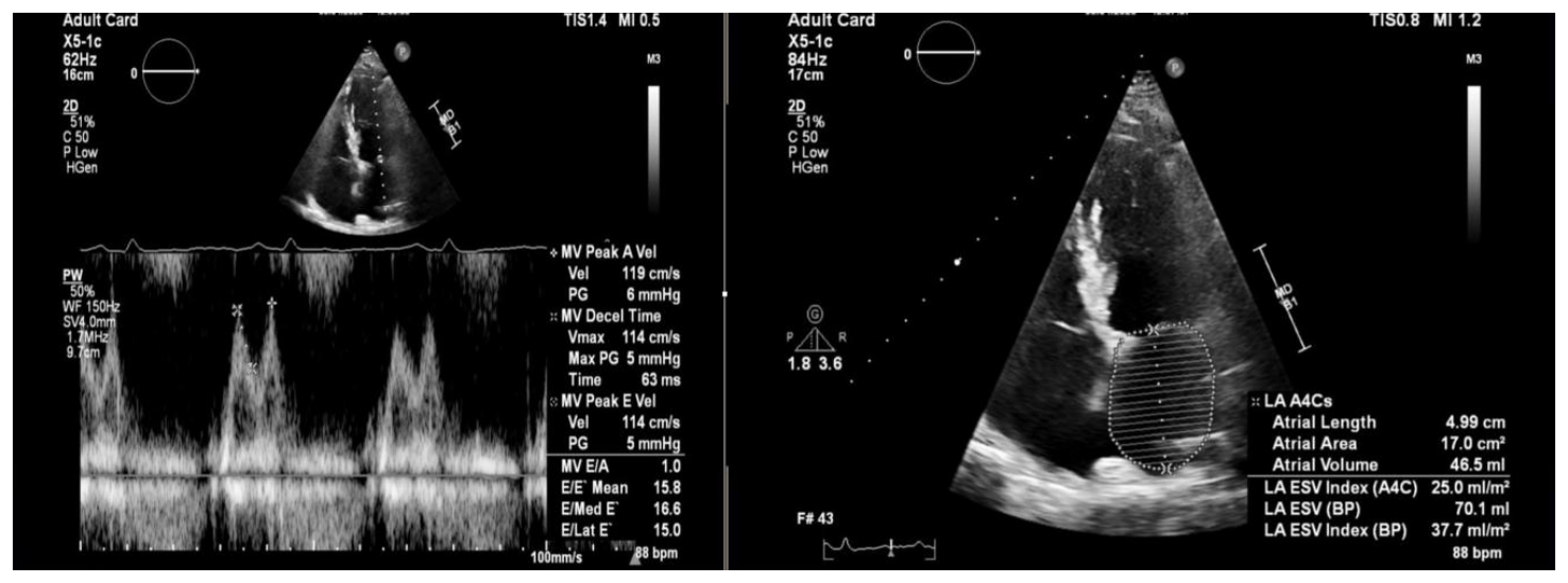
An example of elevated LV filling pressures estimated by transthoracic echocardiography from the personal collection of the authors is presented in Figure 1.
The other score mentioned in the 2021 ESC guidelines for heart failure is the H2FPEF score, which is more clinically accessible. The focus of this score is the clinical probability of HFpEF based on associated diseases and characteristics of the patient, with less focus on echocardiographic parameters. It must also be noted that the H2FPEF score does not include plasmatic natriuretic peptide dosing. The score incorporates and rates the following items:
Regarding the concordance of these scores, there are multiple studies comparing them. In a meta-analysis published by Estes-Schmatzl et al. in 2025, the 2 diagnostic algorithms produced discordant results in the same patient population in 28-41% of cases. The H2FPEF score had a higher sensitivity, identifying patients with obesity or atrial fibrillation HFpEF phenotypes, whereas the HFA-PEFF algorithms had a higher specificity, as they required more complex and comprehensive imaging assessment. Moreover, in another article published in 2022 by Amanai et al, the only H2FPEF score could be used to discriminate between patients with dyspnea and those with poor exercise capacity. These practical insights suggest complementary roles, rather than competitive ones, for these 2 scores in clinical practice, as H2FPEF is more suitable for primary care and general cardiology, whereas HFA-PEFF could be used in specialized heart failure clinics [15–20].
Furthermore, as machine learning and AI become more accessible in medicine, the implementation of AI algorithms for HFpEF might become widespread. There are multiple proposed models being developed. One model is the EchoGo Heart Failure model, which uses neural network analysis of an apical 4-chamber transthoracic echocardiogram video to predict HFpEF with a sensitivity of 82% and specificity of 81%. Another model, called AIM-HFpEF, integrates data from electronic health records and analyses them using natural language processing, with a sensitivity of 89% and a specificity of 62% for the diagnosis of HFpEF [21,22].
After 2021, there was a paradigm shift in the treatment for HFpEF, with the validation of SGLT2 inhibitors in patients with chronic heart failure with preserved ejection fraction. In 2021, EMPEROR-PRESERVED showed a reduction in cardiovascular death or heart failure hospitalization in patients treated with empagliflozin, while also improving health-related quality of life. The primary outcomes were reached by reducing heart failure hospitalization, as the reduction of cardiovascular deaths did not reach statistical significance alone, only in association with HF hospitalization [23–25].
After the validation of empagliflozin in 2021, the results of the DELIVER trial showed benefits in reducing cardiovascular death or worsening heart failure, while maintaining efficacy on the whole spectrum of ejection fraction by using another SGLT2 inhibitor, dapagliflozin [26–29].
These two trials led to ESC publishing a focused update of the heart failure guidelines in 2023, recommending SGLT2 inhibitors with a class I recommendation, with an A level of evidence [1].
Furthermore, 2024 marked the publication of 2 trials assessing GLP-1 agonists in HFpEF patients with obesity. The STEP-HFpEF trial followed semaglutide in patients with obesity-related HFpEF. The primary endpoints reached were a reduction in body weight and an increase in perceived health-related quality of life, as assessed by KCCQ-CSS. In a subgroup analysis, the body weight reduction in women was higher. However, it must be noted that only 3.6% of patients received SGLT2 inhibitors for HFpEF, and it is difficult to assess whether the benefits were driven by weight loss or improvement in HF symptoms. This study highlights the benefit of semaglutide in improving HF-related symptoms and exercise function, regardless of baseline health status or sex, but requires a larger study in order to follow cardiovascular events, as STEP-HFpEF was underpowered for clinical events [30–32].
Another GLP-1 agonist trial was the SUMMIT trial, published in 2024, which compared the safety and efficacy of tirzepatide among patients with HFpEF and obesity. Tirzepatide is a GLP-1 and GIP agonist, and it was shown in this trial that it can reduce cardiovascular death and a worsening HF event compared to placebo, while also increasing patient-oriented quality of life and functional capacity in obese patients with HFpEF. This molecule has also been shown to reverse left ventricular remodeling by reducing left ventricular mass and paracardiac adipose tissue on cardiac magnetic resonance studies. Moreover, the reduction of high-sensitivity C-reactive protein in the tirzepatide group is theorized to be a separate mechanism of action of this molecule [33–35].
However, there is no recommendation from ESC to prescribe GLP-1 receptor agonists in patients with heart failure yet, but a recommendation for the prescription of GLP1-RA with proven cardiovascular benefits (at that moment – liraglutide, semaglutide, dulaglutide, epfeglenatide) for patients with type 2 diabetes mellitus and atherosclerotic cardiovascular disease, with a class I, level of evidence A according to the ESC clinical consensus statement on obesity and cardiovascular disease [36].
Another landmark trial published in 2024 is FINEARTS-HF, which investigated the efficacy and safety of finerenone, a nonsteroidal mineralocorticoid receptor antagonist, in reducing the risk of cardiovascular death and total worsening heart failure events in patients with HFmrEF, HFpEF and HFimpEF. The primary endpoint of the study was reached and there was no modification of the benefit across the range of LVEF analyzed. Moreover, the patients reported an increase in perceived quality of life and there were no differences in benefits between sexes [37–40].
There are multiple studies in development assessing various strategies and molecules in HFpEF. One of them is AMBER-HFpEF, which assesses the safety, tolerability, pharmacokinetics, and pharmacodynamics of ulacamten, which is a selective, oral cardiac myosin inhibitor. However, this is only a Phase 2 study, enrolling only 60 patients with symptomatic HFpEF [41].
Chronic, systemic low-grade inflammation is recognized as a key element in HFpEF pathophysiology. Another study, HERMES, is a phase 3 trial assessing ziltivekimab, which is an anti-inflammatory molecule targeting IL-6 signaling, with a composite primary outcome of cardiovascular death, heart failure hospitalization, or urgent heart failure visit. There are also multiple studies regarding the antiinflammatory effects of colchicine on the quality of life, vascular, and cardiac function in patients with HFpEF, such as COLHEARTPRESERVED. [42–46].
Senolytic agents, which eliminate senescent cells, are being studied preclinically in laboratory models of HFpEF in reversing cardiac and vascular remodeling in aging models. However, more data is required before initiating large-scale trials in the foreseeable future [47].
These pharmacology trials are in close relationship with recent attempts to further classify HFpEF into distinct phenotypes that may benefit from different treatments and interventions. Furthermore, pooled data from these studies, whether they reached the end point (such as EMPEROR-PRESERVED for empagliflozin, DELIVER for dapagliflozin, or FINEARTS-HF for finerenone) or did not (such as PARAGON-HF for sacubitril/valsartan in HFpEF and TOPCAT for spironolactone), have been used in phenotyping attempts of patients with HFpEF.
There have been multiple attempts to phenotype HFpEF, just like HFrEF was phenotyped in the past. This helps better understand the pathophysiology of the disease and the appropriate medical management, but also the outcomes of these patients.
One attempt was performed in a subanalysis of the TOPCAT trial, where three distinct phenotypes were identified: one with normal LV geometry, filling pressures, atrial stiffness and natriuretic peptides, but with other clinical features or pathologies that can explain the symptoms, one with atrial fibrillation, usually in elderly women, and one with diabetes and obesity [48].
Another study, performed on a cohort of 2147 patients at Peking University Third Hospital and using a neural network AI model identified three phenogroups: one with multiple metabolic comorbidities, left ventricular hypertrophy and diastolic, but also systolic dysfunction, one with female patients with atrial fibrillation and structural abnormalities in the atria and the right ventricle, with diastolic dysfunction, whereas the 3rd group included younger men with unhealthy lifestyle, hyperlipidemia and liver dysfunction. The first phenogroups presented lower systolic function, as measured by LVEF and Sm, as measured by Tissue Doppler. The second phenogroup exhibited the most significant diastolic dysfunction, with higher E/e’ ratios, LAVI, and PCWP. They were also more likely to have significant valvular regurgitation or atrial fibrillation [49].
Larson et al, in 2024, developed a different category of phenogroups by performing invasive hemodynamic cardiopulmonary testing. There were 4 groups, but with overlapping characteristics: cardiometabolic, left atrial myopathy, stiff vascular and pulmonary vascular disease, each having distinct hemodynamic characteristics and determinants of exercise intolerance. Furthermore, there were patients who fulfilled the criteria for more than one phenogroup, and these patients had worse outcomes and functional capacity [50,51] .
The cardiometabolic subgroup’s pathophysiology is centered around type 2 diabetes mellitus and obesity, as these diseases lead to chronic low-grade inflammation that alters LV remodeling, accelerating concentric hypertrophy and diastolic dysfunction. Moreover, these patients usually exhibit increased visceral adiposity, causing adipokine imbalances that further increase and maintain the proinflammatory status of these patients. Moreover, obese patients need a greater volume of circulating blood, increasing stroke volume and leading to increased myocardial wall stress, further accelerating left ventricular remodeling and hypertrophy [52,53]. These metabolic changes highlight the benefits of weight loss by bariatric surgery, when indicated, or in milder cases of obesity, the usage of glucagon-like peptide-1 receptor agonists that alter adipokine secretion and reduce hunger by slowing gastric emptying and modulating hunger centers in the central nervous system.
Atrial fibrillation is frequently associated with HFpEF and has been described as a phenotype of HFpEF in various studies. There are several elements of the pathophysiology of atrial fibrillation that are common with HFpEF and may lead or accelerate the development of this disease, in a vicious cycle. Some of these mechanisms are the pro-inflammatory state, which is common between the two diseases, but also cardiac microvascular endothelial cell lesions, with impaired cardiac microvascular dysfunction, cardiac remodeling, and diastolic dysfunction, with alteration in the calcium metabolism of cardiomyocytes. The pro-inflammatory microenvironment in the atria in patients with early atrial lesions, with increased TNF-α, TGF-β, IL1B, and IL6, is also important in the pathophysiology of HFpEF. The inflammatory cells in the atrial myocardium expressed TGF-β, which triggers differentiation of fibroblasts to myofibroblasts, which produced more collagen and decreased the local concentration of matrix metalloproteinases, which reduced total cellularity by increasing the collagen/cell ratio in the atria. Furthermore, this local inflammation can further increase the risk of atrial fibrillation by leading to atrial fibrosis. Furthermore, in atrial fibrillation, LV filling is altered not only by losing the atrial systole, but also by the increased ventricular rate, which is often associated with atrial fibrillation. This can, in time, transform an atrial cardiomyopathy into atrial fibrillation-induced ventricular cardiomyopathy, which is associated with impaired systolic function of the left ventricle. There are also multiple comorbidities that are associated with both HFpEF and atrial fibrillation, accelerating the vicious circle by inducing a significant pro-inflammatory state in the body. These comorbidities are arterial hypertension (or other causes of arterial stiffness), diabetes mellitus, obesity, or renal disease. Obesity is important, especially in association with atherosclerosis, which is itself an inflammatory disease with a vicious cycle in which diabetes, arterial stiffness, and dyslipidemia are frequently associated. These diseases cause vascular inflammation, causing monocyte infiltration and macrophage activation, causing microvascular dysfunction and increased afterload, which triggers LV remodeling by concentric or eccentric hypertrophy. An important disease that triggers chronic inflammation is chronic kidney disease, which has multiple risk factors, including atrial fibrillation and HFpEF (age, obesity, type 2 diabetes mellitus). This chronic inflammation leads to increased reactive oxygen species, decreased availability of nitric oxide, and microvascular dysfunction, mechanisms that are common among chronic low-grade proinflammatory diseases. Furthermore, overactivation of the renin-angiotensin-aldosterone system also contributes to LV remodeling. Moreover, increased monocyte percentages have been associated with asymptomatic diastolic dysfunction and correlated with diastolic dysfunction on cardiac transthoracic ultrasound [54–62].
These pathophysiological changes in the atrial fibrillation phenogroup with HFpEF highlight the importance of rate control and rhythm control. However, data regarding catheter ablation for patients with atrial fibrillation and HFpEF is contradicting. While a study from the Swedish Heart Failure Registry identified lower risk of all-cause mortality or heart failure hospitalization regardless of the ejection fraction, a meta-analysis performed by Oraii et al and published in 2024 in JAMA Cardiology, pooling 2465 participants from 12 randomized control trials found no differences in cardiovascular death, all-cause death or heart failure events in the HFpEF subgroup, which included 913 participants [63,64].
Another hemodynamic phenogroup described was the one with arterial stiffness. One significant cause of arterial stiffness is arterial hypertension. There are multiple links between these and HFpEF, as arterial hypertension increases afterload and left ventricular hypertrophy and fibrosis due to microvascular dysfunction. These left ventricular changes lead to left ventricular remodeling. Endothelial senescence has been identified as an important element in the pathophysiology of several metabolic and cardiovascular diseases. Endothelial cells are exposed to multiple stimuli that can lead to senescence. These stimuli can be hemodynamic, such as shear stress or disturbed blood flow (in bifurcations or in stenosed arteries), or chemical (inflammatory cytokines, reactive oxygen species, metabolites). At a molecular level, senescence is caused by multiple, successive rounds of cell division that shorten telomeres, as the majority of human adult somatic cells have very low levels of telomerase, which can replenish the telomeric reserve [65].
One of the principal effects of endothelial senescence is inflammation, leading to low-grade diffuse inflammation that has been associated with multiple diseases, such as atherosclerosis or HFpEF. Moreover, there is a vicious cycle where these pathologies, all proinflammatory, can accelerate one another. Hypertension and aging are closely related, as endothelial dysfunction associated with senescence leads to an increased concentration of vasoconstricting endothelin-1 and angiotensin-2 compared to vasoprotective and
vasodilatory molecules such as NO. Furthermore, increased pulse pressure leads to a significant exposure of endothelial cells to mechanical stress that can accelerate endothelial cell damage, leading to a positive feedback loop that accentuates hypertension. Multiple studies have identified hypertension as a significant cause of left ventricular remodeling by concentric or eccentric hypertrophy, while also increasing left ventricular myocardial stiffness, leading to increased filling pressures and diastolic dysfunction that is present in many cases of HFpEF. There are multiple studies underway with molecules that target endothelial senescent cells, but none are yet focused on HFpEF. However, anti-inflammatory molecules may also have a positive effect on cellular senescence. As previously mentioned, there are multiple studies ongoing assessing anti-inflammatory molecules and HFpEF, such as ziltivekimab and colchicine [65–71].
Heart failure with preserved ejection fraction diagnosis and treatment have been constantly refined by important trials assessing diagnostic scores and medical treatment of this disease, such as EMPEROR-PRESERVED, DELIVER, FINEARTS-HF, or SUMMIT. With multiple molecules currently being studied, the future of HFpEF treatment guidelines might change, leading to multiple treatment options for these patients.
The authors declare no conflict of interest. This research received no external funding.
Authors’ contribution
Conceptualization, A-E. M., S-I. D.; resources, A.P., S.C.; writing—original draft preparation, B-V.V., I.T.I; writing—review and editing, S.C, A.P.; tables and figures, I.T.I., A.P.; supervision, A-E.M., S-I.D. All authors have read and agreed to the published version of the manuscript. No generative AI was used for the production of this article.