
Cancer is a major health problem and one of the leading causes of death in the world today. Cancer is the uncontrolled growth and multiplication of cells. Cells that become cancerous no longer perform their normal functions and usually do not respond to signals that prevent uncontrolled growth [1]. However, in many cases, cancer cells can counteract the proposed treatment strategies and sometimes even develop resistance to chemotherapy, leading to faster tumor growth. Therefore, in the last two decades, scientists have tried to choose their strategies as intelligently as possible for successful combat against cancer [2]. According to various studies, colorectal cancer is the third most common cancer worldwide. The walls of the colon
and rectum are made up of several layers. Colorectal cancer starts in the innermost layer (mucosa) and can spread to other layers and, consequently, to blood or lymphatic vessels, leading to metastasis in other parts of the body [2]. In recent years, mortality from colorectal cancer has increased in Asia, and according to studies conducted in Iran, its prevalence is on the rise [3]. Treatment methods for colorectal cancer include surgery, radiotherapy, or chemotherapy, and the choice of treatment method depends on the phase of the disease. One of the most commonly used chemotherapy drugs in the treatment of patients with colorectal cancer is 5- fluorouracil (5FU) [4, 5]. Common side effects of chemotherapy drugs include drug resistance and reduced immune system function in the patient. As a result, identifying novel treatment methods aimed at combating drug resistance and
strengthening the immune system has been the focus of a huge number of studies [5]. One of the novel therapeutic methods is the use of oncolytic viruses and bacteria. The use of oncolytic viruses is a new perspective in the treatment of human malignancies, including the use of engineered viruses or viruses with tissue affinity for specific transformed cells, which increase the destruction of tumor tissue or its cells and prevent damage to non-cancerous cells [5-7]. Among the escape pathways of tumor cells from the antitumor responses of the immune system is the acquisition of defects in cellular antiviral pathways, such as those mediated by interferons [6-8]. Oncolytic viruses can target and destroy cancer cells by utilizing this unique cellular activity of tumors. The treatment with oncolytic viruses relies on a two-part process of selectively infecting tumor cells and then inducing anti-tumor activity through the release of tumor antigens and the stimulation of the immune system [7-9]. Unlike chemotherapy and radiotherapy, oncolytic viruses are self-amplifying treatments, and their therapeutic outcome is determined by a three-way competition between tumor growth, virus proliferation, and immune activation [10]. A wide range of different viral species have been investigated as cancer therapeutic agents, including adenoviruses, herpes simplex virus type 1, poliovirus, measles virus, Newcastle disease virus, reoviruses, vesicular stomatitis virus, Zika virus, etc [11]. Among oncolytic viruses, Newcastle disease virus has emerged as a promising nonengineered oncolytic virus. Newcastle disease virus is an enveloped, single-stranded RNA virus with a negative sense, belonging to the Paramyxoviridae family, and it causes significant economic losses to various poultry industries worldwide each year [12,13].
Although Newcastle disease virus is an avian pathogen, its impact on human cancers has been widely reported because Newcastle disease virus is antigenically distinct from common human pathogens. Furthermore, it exhibits oncolytic potential through specific replication in tumor cells rather than normal cells [13]. A wide range of bacterial species play a role in most physiological and metabolic activities and intestinal function. Probiotics are live microbial dietary supplements that are beneficial for creating microbial balance and nutritional equilibrium in the human intestine [14, 15]. Bacterial species used as probiotics are lactic acid bacteria, primarily from the genera Lactobacillus and Bifidobacterium. Bifidobacteria are Gram-positive, anaerobic, non-motile bacteria that lack sporeforming ability and are catalase-negative. Their normal habitat is the colon of the human and animal gastrointestinal tract, and their numbers remain stable except during old age, when they decrease. However, factors such as diet, antibiotics, and stress can also lead to changes in their numbers [16].
Various studies have reported that probiotics play a role in suppressing primary neoplastic ulcers and cancerous tumors in mouse models [16]. The anticancer effects of probiotics are achieved by preventing the conversion of procarcinogens to carcinogens, binding and inactivating mitogenic compounds, reducing the growth of procarcinogenic bacteria, decreasing the absorption of mitogens, and enhancing the immune system’s function [17]. Lactobacillus casei is one of the types of Lactobacillus that is rod-shaped, Gram-positive, and facultatively anaerobic, non-spore-forming, non-motile, and polymorphic. It is considered a safe probiotic bacterium. Although several beneficial effects have been attributed to these bacteria, their anticancer activity is the most important characteristic of these bacteria [16,18]. Apoptosis is programmed cell death that plays a key role in regulating cell numbers. In many cancers, a reduction in the ability to undergo apoptosis leads to alterations in the process of cell proliferation and disruption [19]. There is substantial evidence that probiotics can play a role in regulating cell proliferation and apoptosis [20]. Additionally, with the discovery of various therapeutic effects of probiotics, including anticancer effects, modulation of differentiation processes in tumor cells, production of short-chain fatty acids, and changes in the expression of tumor genes, probiotics have become a research focus for many researchers around the world [21]. Therefore, the aim of the present study is to compare the cytotoxic effects of Lactobacillus casei extract with Newcastle disease virus in human colorectal cancer.
The HT-29 cancer cell line was purchased from the cell bank of the Pasteur Institute of Iran and transferred to T25 flasks containing DMEM culture medium and 10% FBS, and cultured in an incubator with 5% CO2 at a temperature of 37 0C.
Lactobacillus casei bacteria (ATCC:393) were purchased from the Applied Virology Research Center of Baqiyatallah University of Medical Sciences. After culturing it in a specific culture medium (MRS), its extract was prepared by heating (at 56 0C for 60 minutes) [22].
The Newcastle disease virus, Lasota strain, was obtained from the Applied Virology Research Center of Baqiyatallah University of Medical Sciences.
After culturing cancer cells and observing a density of over 80%, the cells were treated with different concentrations of Lactobacillus casei extract (concentrations of 0.5, 1, 2, 3, 4, 5, 6 µg/ml) and Newcastle disease virus (concentrations of 0.5, 1, 2, 4, 8, 9 MOIs) to
determine the IC50 concentration. Then, to compare the toxicity and simultaneous effects, the IC50 concentration of both therapeutic agents was used. The drug 5-fluorouracil (34 µM) was used as a positive control.
The MTT assay is a method for assessing biological toxicity based on the formation of formazan color through the reduction of MTT (dimethylthiazole 2,5-diphenyltetrazolium bromide) or other tetrazolium salts. The tetrazolium ring of the MTT salt is broken by the succinate tetrazolium reductase system, which is an enzyme of the mitochondrial respiratory cycle and is found only in living cells, resulting in the formation of the insoluble substance formazan. Formazan is insoluble in water and appears as purple crystals on the surface of living cells. By measuring the absorbance using a spectrophotometer at specific wavelengths, the number of viable cells can be determined. This test is conducted based on the ISO 10993-5 standard, and its purpose is to evaluate cell proliferation under in vitro conditions. The MTT reagent is a yellow tetrazolium salt that is reduced by metabolically active cells, part of which is converted by dehydrogenase enzymes to produce reduced forms of NADH and NADPH. After counting the cells, 100 µl of DMEM culture medium containing 10% FBS with the required number of cells was added to each well of a 96-well plate. The 96-well plate was then placed inside an incubator and incubated for 24 hours. After 24 hours, the cells grew and filled the bottom of the wells. Then, the supernatant was removed from the wells, and treatments from different groups were added to the wells and incubated. After the incubation period, 25 µl of MTT solution (5 mg/mL PBS) was added to each well and incubated for 4 hours. During this time, the tetrazolium bromide was reduced by viable cells, leading to the formation of insoluble purple formazan crystals, which became soluble upon the addition of 100 µl of DMSO after 30 minutes. Then, the absorbance of each well was measured at a wavelength of 570 nanometers using an ELISA reader.
HT-29 cells were collected into sterile 1.5 ml microtubes after treatment with different groups and centrifuged for 10 minutes at 1400 rpm. The supernatant was then discarded, and the cell pellet was resuspended in 1 ml of culture medium. The samples were then stained with 10 microliters of acridine orange and incubated for 15 minutes at 37 0C in 5% CO2. After the incubation period, the samples were washed twice with PBS, and 10 µl of propidium iodide were added and incubated for 5 minutes at 37 0C in 5 % CO2. In the final step, the samples were centrifuged for 10 minutes at 1400 rpm after washing with PBS. The supernatant was then removed, and 50 µl of the cell pellet were taken and placed on a glass slide. After placing a coverslip, the samples were examined under a fluorescence microscope with a 40X objective lens. The green cells were identified as healthy cells, while the orange cells were apoptotic.
Data analysis was performed using SPSS version 23 and Tukey’s test was employed. In all analyses, a P-value of <0.05 was considered statistically significant. Additionally, graphs were plotted using GraphPad Prism version 8.2.1, and the data were reported as Mean ± SEM.
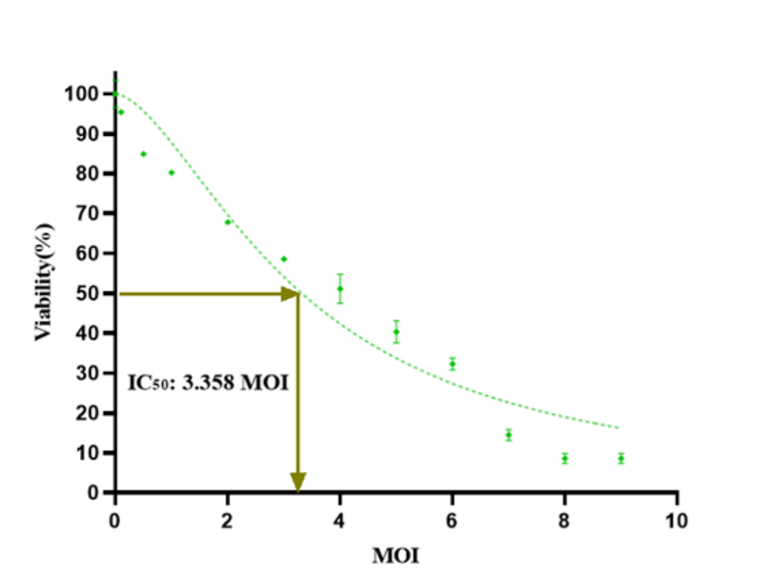
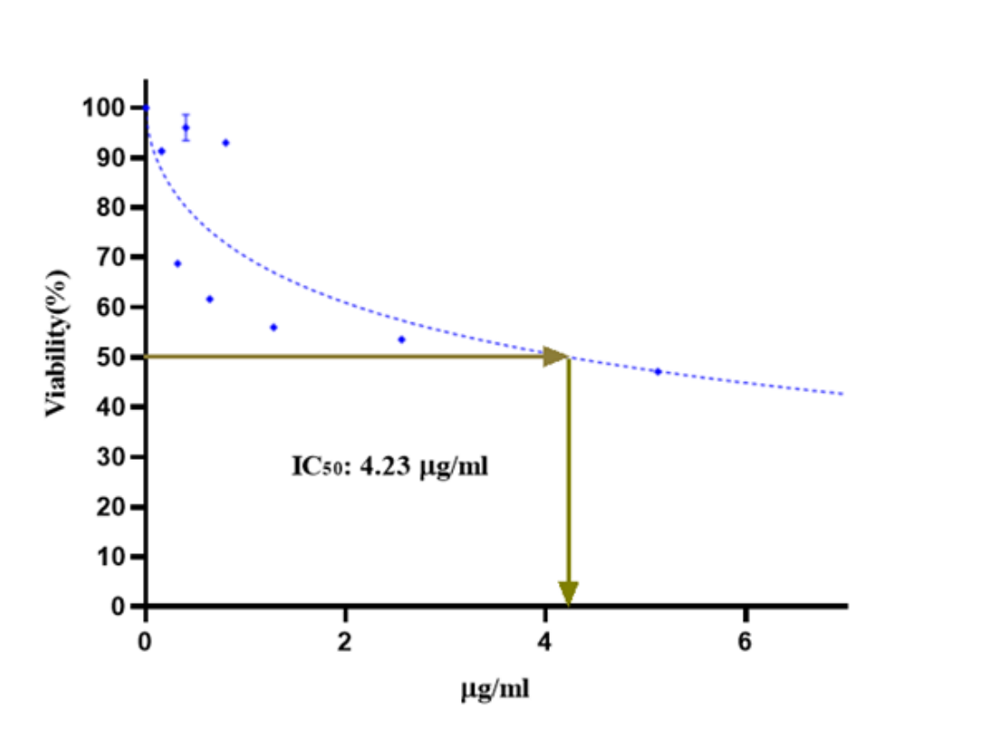
Figures 1 and 2 show the inhibitory effect on the growth of cancer cells after treatment with Newcastle disease virus and Lactobacillus casei extract using the MTT assay. According to the graphs, both agents reduce the growth of colorectal cancer cells in a concentrationdependent manner. The IC50 for the Newcastle oncolytic virus is 3.358 MOI, and for Lactobacillus casei, it is 4.23 µg/ml (Figures 1 and 2).
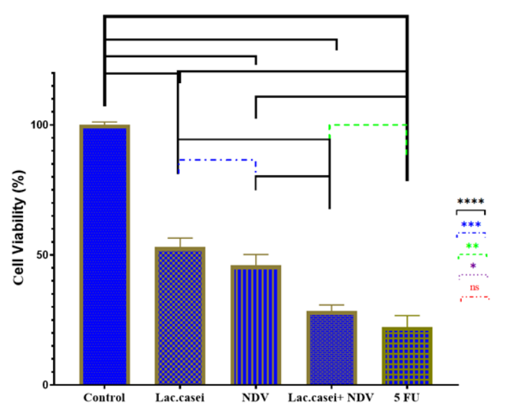
According to Figure 3, the synergistic anti-proliferative effects of the simultaneous treatment of Lactobacillus casei and Newcastle disease virus on HT-29 cells were evaluated using the MTT assay. A comparison was made between the results of 5-fluorouracil and the negative control group. The combined treatment group exhibited significantly higher cytotoxic effects compared to the control groups (Figure 3).
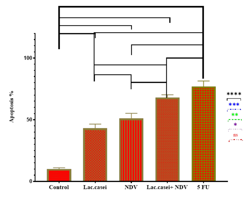
In Figure 4, the level of apoptosis resulting from the simultaneous treatment of Lactobacillus casei and Newcastle oncolytic virus was determined using fluorescent staining. The combined treatment group exhibited significantly higher apoptotic effects compared to the control groups (Figure 4).
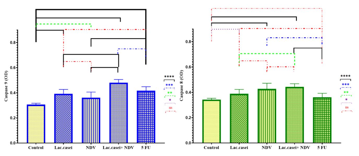
In Figure 5, the levels of caspase 8 and 9 production after treatment with Lactobacillus casei and Newcastle oncolytic virus, both alone and in combination, are shown. The results indicated that the Newcastle oncolytic virus and Lactobacillus casei extract increased the activity of both caspase 8 and 9. In other words, the oncolytic virus and the bacterium activate both apoptotic pathways (Figure 5).
Colorectal cancer is one of the most common malignancies of the digestive system, and diet plays a key role in its pathogenesis. There is abundant evidence that colorectal cancer is associated with an imbalance in the intestinal microflora [23]. Probiotics (such as Lactobacillus and Bifidobacterium) are live microorganisms that have numerous beneficial effects on health, and in addition to their positive gastrointestinal effects, they can help prevent colorectal cancer through various mechanisms [24]. There is no certainty in the treatment of cancer, and researchers are discovering new cancer treatment methods every day. For over a century, physicians have been interested in using viruses for cancer therapy, and in recent years, a small but growing number of patients have begun to benefit from this approach [25]. Some viruses tend to infect and destroy tumor cells. This group, known as oncolytic viruses, includes viruses that are found in nature as well as those that have been modified in the laboratory to effectively replicate in cancer cells without harming healthy cells [26]. So far, only one oncolytic virus, a genetically modified form of herpesvirus, has been approved by the Food and Drug Administration (FDA) for the treatment of melanoma, although a number of viruses are being evaluated as potential cancer therapies in clinical trials [27]. An increasing number of studies suggest that certain oncolytic viruses may work by stimulating an immune response in the body against cancer. When a virus infects a tumor cell, it replicates itself until the cell bursts. The dying cancer cell releases substances such as tumor antigens, which allow the cancer to be recognized by the immune system [28]. Based on the literature review conducted, the aim of the present study is to investigate the apoptosis induction pathway caused by Lactobacillus casei extract in combination with Newcastle disease virus in human colorectal cancer. The results of the current study indicated that the Lactobacillus casei extract and Newcastle disease virus led to an increase in the rate of apoptosis and a reduction in the proliferation of colorectal cancer cells. The results also showed that Newcastle disease virus and Lactobacillus casei extract induce apoptosis through both intrinsic and extrinsic pathways.
Kim et al. examined the effects of cellular components of ten different probiotics on eleven types of cancer cells. Their results indicated that probiotics had an inhibitory effect on cancer cells and attributed this effect to their peptidoglycans [29]. Lee et al. reported in their study that the cytoplasmic extracts of Lactobacillus casei and Bifidobacterium have a direct effect on inhibiting the growth of cancer cell lines. Since the uncontrolled proliferation of cancer cells is a major issue in cancer patients, any factor that can inhibit their proliferation could be beneficial for prevention and halting their progression. The best way to inhibit or suppress cell proliferation is to induce programmed cell death through apoptosis, as it will not cause inflammation in the body and cells adjacent to tumors, and as a result, it will be an immune factor for suppressing tumors [30]. The results of Baldwin et al.’s research also suggest that Lactobacillus acidophilus and Lactobacillus casei can enhance the induction of apoptosis in the LS513 carcinoma cell line and can be used as adjuvants in chemotherapy [31]. Other studies have shown that Bifidobacterium lactis induces apoptotic responses in genotoxic carcinogens shortly after the induction of carcinogens in the descending colon of rats [32]. Cell death by apoptosis is an important regulatory process for protection against oncogenes.
Additionally, numerous groups of scientists have reported that compounds derived from Lactobacillus acidophilus prevent the progression of colorectal cancer in animal models [33]. As a result, probiotics can be considered as a safe agent for combating cancer with no side effects. Probiotics enhance cytotoxic activity and are used as an adjunctive therapy alongside chemotherapy. Mehrabian et al. stated that Lactobacillus isolated from Tarkhina has a high anti-mutagenic and anti-cancer effect, which is indeed attributed to the strengthening of the immune system by probiotics [34]. Dolati et al. showed that the extract of Bacillus coagulans has inhibitory effects on the MCF-7 breast cancer cell line in a concentration-dependent and time-dependent manner. Meanwhile, lower cytotoxic effects were observed in normal human foreskin fibroblast cells. The increase in the expression of BAX, caspase 3, and caspase 9 genes, along with the reduction of the anti-apoptotic gene BCL-2, confirmed the induction of apoptosis in cancer cells, supported by flow cytometry results [35]. Sivamaruthi et al. (2020) proved that the use of probiotics is beneficial in the management of colorectal cancer treatment [36]. Recently, several alternative therapeutic approaches have been explored to overcome drug resistance issues, with virotherapy emerging as a promising strategy. The idea of using viruses in cancer treatment was proposed in the early 1960s. Since then, several groups of DNA and RNA viruses with oncolytic properties have been identified, including the Newcastle disease virus.
The Newcastle disease virus was first discovered in 1927 in Newcastle upon Tyne and causes a highly contagious disease in birds and poultry [37]. The use of the Newcastle disease virus in cancer treatment was first reported by Cassel and Garret in 1965, and since then, interest in using the Newcastle disease virus as an anti-neoplastic therapy has steadily grown. The Newcastle disease virus, or avian paramyxovirus serotype 1, belongs to the Paramyxoviridae family and causes severe Newcastle disease in poultry and wild birds worldwide [38]. However, the Newcastle disease virus is non-pathogenic for mammals, making it a promising viral therapeutic agent for human malignancies. The oncolytic activity of the Newcastle disease virus has been studied since 1952, and the National Cancer Institute has listed the Newcastle disease virus among complementary and alternative therapies [39]. The interferon response prevents the replication of the Newcastle disease virus in healthy cells. However, the Newcastle disease virus induces oncolytic properties due to the uncontrolled division and high mobility of cancer cells. Several strains of the Newcastle disease virus have shown efficacy in in vitro, in vivo conditions, and in phases I and II of clinical trials [40]. Keshavarz et al. (2020) showed that treatment with Newcastle oncolytic virus significantly reduced TC1 cell proliferation and increased their apoptosis rate. They also showed that the apoptosis of TC1 cells induced by the oncolytic Newcastle disease virus is mediated by the production of reactive oxygen species. In summary, their results indicated that Newcastle oncolytic virus is a useful therapeutic candidate as a selective anti-tumor agent for the treatment of cervical cancer [41]. Al-Ziaydi et al. (2020) showed that the combined treatment of the oncolytic Newcastle disease virus with D-mannohaptulose resulted in greater inhibition of tumor cell proliferation compared to monotherapy with D-mannohaptulose. This suggests a new strategy for breast cancer treatment through the inhibition of glycolysis by reducing hexokinase levels [42].
In a study conducted in 2018 by Lin et al. in China, it was observed that the use of the oncolytic coxsackievirus resulted in a reduction in the growth of endometrial cancer (EC) cells [43]. In a study conducted in 2003 by Bell et al. at the American Center for Respiratory Disease Research, it was shown that oncolytic viruses have a cytotoxic effect on Ehrlich tumor cells and, by binding to these cells and entering them, cause cytopathic effects (CPE) and kill tumor cells [44]. In another study conducted in 2018 at the University of Illinois, USA, by Abdullahi et al., it was shown that oncolytic viruses are capable of replicating in Hepatocellular Carcinoma cell lines. They induce CPE that leads to the destruction of these tumor cells, while not affecting normal, non-cancerous human fibroblast cell lines [45]. In a study conducted in 2008 at Hebrew University in Jerusalem by Yaacov et al, the effects of attenuated oncolytic viruses on tumor cells of mouse and human lung were investigated in vitro. The study revealed that attenuated oncolytic viruses selectively induce CPE in tumor cells of both mouse and human lung compared to non-tumor lung cells, leading to the selective destruction of tumor cells [46]. In a study by Ahmad et al. (2020), the cytotoxic and anti-cancer effects of oncolytic viruses on bladder cancer cell lines were investigated. The study demonstrated that oncolytic viruses can induce apoptosis in bladder cancer, effectively eliminating these tumor cells [47]. Kazimirsky et al. (2016) reported that the use of mesenchymal stem cells infected with Newcastle virus enhances the anti-cancer effects of the Newcastle virus on glioblastoma. Additionally, these researchers noted that this increased anti-cancer effect is associated with an elevated secretion of TRAIL, which acts as a cytotoxic agent [48].
Based on the obtained results, it appears that the use of both therapeutic agents (probiotics and oncolytic viruses) leads to a reduction in cell proliferation and an increase in the percentage of apoptosis in cancer cells. Furthermore, the effectiveness of these two agents in combination therapy groups enhances each other’s effects, suggesting that they have synergistic effects. Therefore, it can be concluded that the extract of Lactobacillus casei and the Newcastle disease virus have significant potential to be introduced as a new strategy for cancer treatment alongside chemotherapy and radiotherapy.
The authors declare no conflict of interest. This research received no external funding.
The researchers would like to express their gratitude to all those who contributed to the progress of this study.
Conceptualization, L.T., and H.E.; methodology, H.E.; validation, H.E., L.T., and M.D.; formal analysis, M.D.; investigation, M.E.; resources, S.K.A.; data curation, M.D.; writing—original draft preparation, M.E.; writing—review and editing, S.K.A.; supervision, H.E.; project administration, H.E. All authors have read and agreed to the published version of the manuscript.
This study involved in vitro experiments using cellular models and did not include human participants, human data, or animal subjects. The research was conducted in accordance with all applicable international, national, and institutional guidelines for the care and use of laboratory materials.