
The diagnosis of various liver tumors remains, nowadays, challenging even if new diagnostic and prognostic tools are continuously developed [1–5]. In this context, contrast-enhanced ultrasound (CEUS) has been a widely used imaging method in Europe and Asia for over 10 years. CEUS consists of an ultrasound that employs contrast agents for increasing the accuracy of the investigation, typically injected intravenously. It can be compared with other contrast-enhanced imaging methods, such as CT or MRI, and it possesses the advantage of a real-time assessment of different focal liver lesions, improving their detection and characterization [1,2]. CEUS has become more popular in recent years than CT or MRI due to the fact that it has almost no adverse reactions, does not make use of any type of ionizing radiation, and can be employed successfully in patients with impaired renal function [1].
CEUS uses a special type of contrast agent that consists of a microsphere with an albumin, phospholipid, or surfactant-based shell and a gas filling, which subsequently forms a microbubble [2]. These microbubbles do not usually surpass 7 μm in size and have a mean diameter of 2.5 μm. These small sizes allow the microbubbles to freely circulate in the capillaries and impede them from passing through the vascular endothelium into the interstitial space, thus rendering them exclusively intravascular [2,6,7].
The microbubbles are excreted in two different ways depending on their components – the shell is metabolized in the liver, while the gas filling is eliminated through the lungs in about 10 to 15 minutes, even in patients with chronic obstructive pulmonary disease. This characteristic allows the utilization of these contrast agents in patients with renal failure, thus posing no risk of nephrotoxicity, when other contrast-enhanced methods such as CT or MRI are not indicated [2,6,8,9].
There are many contrast agents available around the world, but the most conventional are the sulfur hexafluoride microbubbles (SonoVue, Bracco), the perflutren lipid microspheres (Definity, Luminity), and the perfluorobutane microbubbles (Sonazoid).
SonoVue is the contrast agent most often used in Western countries and comprises a phospholipid shell that surrounds the sulfur hexafluoride microbubbles, resulting in a diameter of 2.5 μm. The agent is usually injected as a bolus of 2.4 mL in a peripheral vein (the antecubital vein), together with a 5 mL to 10 mL saline solution, using a large enough needle (more than 20-gauge) to ensure that the bubbles do not rupture during the procedure [1,2,6].
Sonazoid is mainly employed in Japan, Norway, and South Korea, and uses perfluorobutane microbubbles. Although it is similar to the other contrast agents used in CEUS, unlike those, Sonazoid can be phagocytized by the Kupffer cells and allows an additional evaluation of the liver in the Kupffer phase or the post-vascular phase. The agent can persist up to several hours in the liver and spleen after its vascular phase [1,6].
Even though the most common clinical application of CEUS is the characterization of focal liver lesions and differentiating between malignant and benign masses, there are studies and guidelines that show there are numerous other uses for this technique. As an intravascular contrast-agent imaging method, CEUS can provide information about different vascular diseases that may affect the carotid arteries or the aorta and other abdominal blood vessels, or it can help assess the thrombosis of the portal vein [10,11].
CEUS plays an important role in the imaging of the extravascular structures, mainly cavities, either physiological, such as the peritoneal, pleural cavities, biliary and gastrointestinal tracts, or non-physiological, including cysts, abscesses, or diverticula [12,13]. Thus, CEUS is used in gynecology to evaluate the uterus and the fallopian tubes, in nephrology and urology to help diagnose ureteral reflux and to discern between bladder or renal tumors and cysts or testicular masses. Excluding the liver evaluation, CEUS also has other applications in the assessment of the gastrointestinal tract.
It is successfully used in patients with inflammatory bowel diseases to estimate disease activity or with fistulae and abscesses, and at the same time, it can be administered orally in order to examine the bowel lumen [12–14].
CEUS can be successfully used for assessing different focal liver lesions seen on standard ultrasound and is indicated for the following: hepatic nodules larger than 10 mm (for nodules smaller than 10 mm CEUS can be attempted by an expert examiner); LI-RADS-graded liver lesions as LR-3, LR-4 or LR-M on CT or MRI: monitoring enhancement pattern changes over time for LI_RADS-graded lesions as LR-3 or LR-4; detecting arterial phase hyperenhancement (APHE) when it is absent on CT or MRI, but still suspected; guiding liver biopsies or treatments for difficult to visualize lesions, aiding in choosing the right lesion to be biopsied or reevaluating prior biopsied lesions with inconclusive histopathology; assessing treatment response; making the differential diagnosis between a tumor inside a vein or a simple thrombus [15].
One of the most important indications for CEUS is liver mass characterization and not just detection, with some studies showing an equivalence or even a superiority compared to CT or MRI scans. This ultrasound can also help in differentiating benign from malignant nodules [16].
The most common benign lesions that can be diagnosed using CEUS are hemangiomata, focal nodular hyperplasias (FNH) and hepatic adenomata. Malignant masses which are evaluated by CEUS are hepatocellular carcinomata (HCC) and non-hepatocellular tumors, that include metastases, intrahepatic cholangiocarcinomata (ICC) and lymphomata [16–18].
Liver lesions in CEUS can be described using two features that appear in different phases of the ultrasound: arterial phase hyperenhancement (APHE) and washout [1,16].
If there is no washout present and the enhancement in the arterial phase is sustained, there is a high probability that the mass is benign. In comparison, malignant masses show a decline in the enhancement (washout) in the portal or late phase. Furthermore, the intensity and the timing of the washout can be employed to differentiate between types of liver malignancies.
HCC nodules are most likely to display a late (in over 1 minute) and weak washout, while non-hepatocellular malignancies exhibit early (in less than 1 minute) and intense washout. In order to detect liver metastasis, a sweep of the entire liver in the portal venous phase is essential [1,2,16,17,19]. The benign lesions are usually described by specific patterns of enhancement in the arterial phase. Some examples of these patterns are peripheral discontinuous nodular enhancement (hemangiomata), stellate vessels with centrifugal filling (FNH) and centripetal filling (some adenomata) [16,20].
To correctly perform an ultrasound using contrast agents, firstly, it is necessary to have a properly trained examiner who should be able to recognize focal liver lesions and vascular disorders, to assess the effect of a treatment over a lesion and to compare different findings with other imaging methods such as CT, MRI or PET-CT. Secondly, the ultrasound equipment needs to have appropriate hardware, for example, scanners that can cancel soft-tissue enhancement, and software that can display both the standard ultrasound image and the CEUS image simultaneously [15,16]. The patient should be in a supine position with the abduction of the right arm, and before injecting the agent, the examiner should be aware of the medical history of the patient, of the clinical aspects and also of any existing cysts or calcifications that may be wrongly interpreted as malignant [1,12,15,16,21].
For liver imaging, the recommended dose of SonoVue, the most frequently used contrast agent, is 2.4 mL. This value can be decreased by 20-25% if the patient has a low BMI in order to avoid the excessive enhancement of the liver or it can be increased if the lesion is very superficial [15]. The substance is then injected through a large IV line (20-gauge minimum) as a bolus in the span of 2-3 seconds, immediately followed by a saline flush of 5-10 mL [2,7,15].
Interpretation of CEUS is made by evaluating the three vascular phases that follow the injection of the contrast agent. These phases appear as a result of the double vascularization of the liver, namely the hepatic artery and the portal vein [21].
The arterial phase begins at 10 to 20 seconds after injection and ends at 30-45 seconds and appraises the arterial vascular supply of the liver lesion by degree and pattern [1,21]. The portal venous phase begins immediately after the arterial phase at 30-45 seconds and can last up to 120 seconds (2 minutes). This phase marks the entry of the contrast agent in the portal vein and is characterized by diffuse and maximal enhancement of the liver parenchyma [1,21]. The late phase is represented by the amount of time passed from the end of the previous phase until the clearance of the contrast agent from circulation and it can vary from 2 to 8 minutes (120–480 seconds), depending on the type and dose of the agent [1,21]. The post-vascular phase only appears if Sonazoid is injected and it is the result of the contrast loading of the Kupffer or other phagocytic cells. This particular phase usually starts after 8 minutes (480 seconds) following the initial injection and can be seen even after several minutes or hours [1,21] (Table 1).
| START | END | |
|---|---|---|
| Arterial | 10-20 s | 30-45 s |
| Portal venous | 30-45 s | 120 s |
| Late | >120 s | Approximately 4-8 min (bubble disappearance) |
| Post-vascular | >480 s | Approximately 30 min |
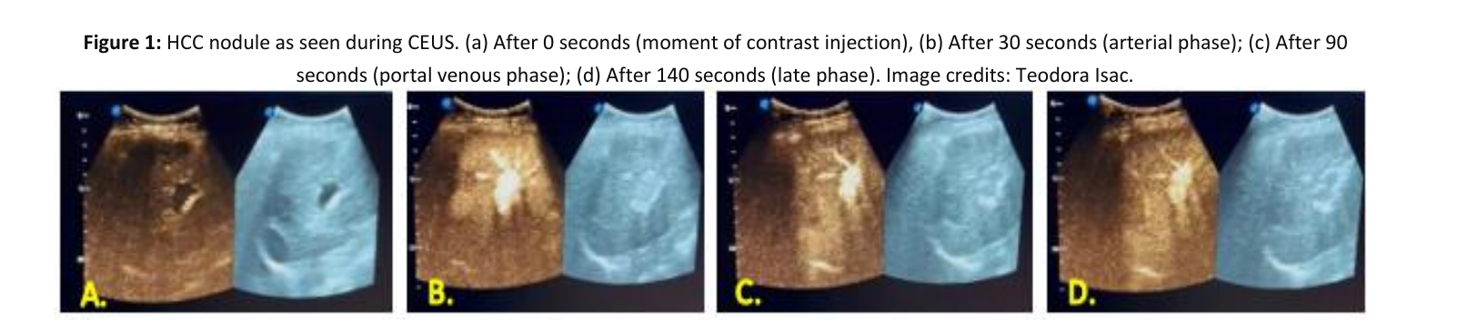
In the following images, an HCC nodule can be seen during a contrast-enhanced ultrasound. The hyperenhancement in the arterial phase and the late and mild washout in the later phases should be noted (Figure 1).
The contrast agents used for CEUS have a few major advantages in comparison to those used for CT or MRI. The most important characteristic is the increased safety of administration, with almost no side effects reported in the literature. There is no evidence of either cardiotoxic, nephrotoxic or hepatotoxic events, nor any interference with the thyroid function as they do not contain iodine and no need for any specific laboratory testing or fasting before the procedure [2,15,21]. Therefore, these contrast agents can be harmlessly administered to patients with renal failure and CEUS can be considered the first-line imaging method for these particular cases [2,21]. Some studies show that when adverse reactions do appear, they are: headaches, chest pain or chest discomfort and nausea, all other symptoms being extremely rare [21,23,24].
Compared to CT contrast agents, CEUS agents have a significantly lower incidence of hypersensitivity events and the probability of an anaphylactoid-type reaction is extremely low, with a rate of 0.001% and a rate of 0.0006% of fatalities after SonoVue administration [2,21]. Some studies show that CEUS is also safe for use in pediatric patients or pregnant women [21]. Following these observations in the population, the 30-minute period of monitoring previously recommended after injection of contrast agents was eliminated [15].
CEUS has the advantage of being a real-time investigation with a higher sensitivity for detecting APHE in focal liver lesions than CT or MRI and is one of the most important imaging methods for identifying HCC nodules [6,21]. CEUS can categorize a rapidly enhancing hemangioma as LR-1, often wrongly diagnosed as LR-3 or LR-4 by CT or MRI, and can also differentiate an arterioportal shunt from a true HCC nodule, which can confuse the previously mentioned types of scanning [6].
CEUS was successfully employed for guiding treatment procedures or for evaluating treatment response. When performing a US-guided thermal ablation of a liver nodule, often an HCC one, CEUS showed a rate of 95.2% of patients with complete tumor ablation after a single procedure compared to 32% of patients after using a standard US. The rate of tumor recurrence was reduced to 16% as compared to 48% and the patients that benefited from CEUS guidance showed better progression-free survival rates [16,25,26]. The sensitivity of CEUS for post-treatment assessment is more than 80% and the specificity nears 100%, in some studies exceeding other imaging modalities, and, if used immediately after the ablation, is able to evaluate hemorrhages or hepatic infarctions [26].
Another treatment procedure after which CEUS can be used to determine residual tumor is transarterial chemoembolization (TACE). When employed after 4 weeks, it can successfully differentiate between a real active tumor and post-procedural inflammation. Some studies show that CEUS can visualize residual HCC nodules after only 1 day following the procedure, with higher rates of detection than CE-CT after 1 month [25]. It also has the advantage of detecting smaller areas of viable tumor that cannot be correctly appreciated by CT or MRI [26].
Though limited, there are studies that attest to the use of CEUS for evaluating patients one week after transarterial radioembolization (TARE) based on its ability to detect changes in tumoral perfusion [26].
Some studies evaluate CEUS as a monitoring tool for systemic therapies and their effects on tumor angiogenesis [25].
Even if CEUS is one of the most efficient imaging methods for liver nodules, there are some limits that need to be taken into consideration.
First of all, being a real-time evaluation, it is not possible to examine more than one lesion at the same time or to visualize the entire liver in the arterial phase; for this instance, a second bolus of contrast agents is needed for the same patient [2,15]. For patients with multiple liver nodules, contrast-enhanced CT or MRI is recommended [22].
Secondly, areas that are difficult to examine on a standard ultrasound are most likely the same when using CEUS and it can be troublesome when the lesion is smaller than 10 mm or is located subdiaphragmatic or very deep [15,22]. Small, simple cysts can be misinterpreted as metastatic lesions because both of them appear hypoechoic on CEUS [2].
For nodules that require complete imaging staging or monitoring of changes in size over time, CT or MRI are more likely to be of use, because CEUS alone is either not sufficient or it is very difficult to capture exactly the same image as the first time on serial US exams [6,22,27].
Even though CEUS is better than standard US, it is still examiner-dependent and the quality of the images may differ with movement artifacts or other interferences [2]. Some limits may also appear in the technique itself, for example, artifacts related to the dosing of the contrast agent, destruction of the microbubbles by the US beam, or inadequate tissue suppression as seen in a fatty liver [16].
In order to create standardized reports for liver nodules, mainly HCC, seen on CEUS examinations, a scoring system has been designed called LI-RADS (Liver Reporting and Data System) [1,28,29]. It helps improve communication between different examiners and reduce confusion and diagnostic errors in patients with high risk for HCC [28]. The nodules observed with CEUS are then classified as: LR-1 (definitely benign), LR-2 (probably benign), LR-3 (intermediate malignancy probability), LR-4 (probably HCC) and LR-5 (definitely HCC) [1,28] (Table 2). Because CEUS is also able to evaluate other types of liver masses, additional categories have been added: LR-M (malignant, but not specific for HCC), LR-TIV (tumor present in the vein) and LR-NC (not categorized due to omission or image degradation) [1,15,30,31]. The sensitivity of CEUS for detecting HCC lesions is dependent on the mass size. For nodules of less than 2 cm, the sensitivity can vary in different studies from 53.6% to 83.3% and for nodules bigger than 2 cm, it ranges from 91.3% to 94.5%, with a mean of approximately 78.3% and a specificity of around 93.1% [32].
If a nodule shows rim-like APHE, which means the enhancement is more intense in the periphery, forming a halo around it, the category according to CEUS LI-RADS should be LR-M and they are usually liver metastasis or other malignant lesions, but not HCC. If the nodule has a peripheral discontinuous globular pattern, it should be included in the LR-1 category as a benign hemangioma [1,33].
| APHE (arterial phase hyperenhancement) | NO | YES (not rim and not peripheral discontinuous globular) | ||
|---|---|---|---|---|
| Rim APHE or early washout (<60 s) or marked washout – LR-M Peripheral discontinuous globular – LR-1 (hemangioma) |
||||
| Nodule size | Nodule size | ≥20 mm | <10 mm | ≥10 mm |
| No washout | LR-3 | LR-3 | LR-3 | LR-4 |
| Late and mild washout | LR-3 | LR-4 | LR-4 | LR-5 |
In a study that took place between January 2010 and December 2018, 382 patients (with a total of 464 liver lesions) with chronic liver disease at high-risk for hepatocellular carcinoma were included [28]. The exclusion criteria were: transarterial chemoembolization, radiofrequency ablation, and systemic treatment for HCC [28]. The 464 liver lesions were divided into: 68 non-HCC non-malignant lesions (hemangiomata, simple cysts, fatty infiltration, regenerative nodules), 359 HCCs and 37 non-HCC malignant lesions (liver metastases, other types of malignancies). By using CEUS LI-RADS, all of the 464 nodules were classified and the percentage of HCC was established for each category. From the total number of lesions examined, 22 of them were part of the LR-1 category and the other 8 lesions were classified as LR-2. Among those, the HCC rate was 0%. 26 lesions were included in the LR-3 class, but 11 (42.3%) of them were HCC. From the 106 nodules that were included in the LR-4 category, 85 (80.2%) were also confirmed to be HCC. 264 nodules from the total of 464 were categorized as LR-5 by means of CEUS and the HCC rate in this category was 258 (97.7%). For the remaining lesions, 11 were classified as LR-TIV and all of them (100%) were confirmed as HCC, and 38 were classified as LR-M; only 5 (13.2%) of those actually were HCC [28].
The sensitivity of the LR-5 category for the diagnosis of hepatocellular carcinoma was 71.9%, with a specificity of 94.3%, a positive predictive value of 97.7%, a negative predictive value of 49.5% and an overall diagnostic accuracy of 76.9% [28].
By combining the LR-4 and LR-5 categories, they managed to improve the diagnostic accuracy to 90.7%. In this case, the sensitivity was 95.5%, the specificity 74.3%, the positive predictive value 92.7% and the negative predictive value 82.9% [28].
This study has once more shown that CEUS is comparable to CE-MRI and CE-CT in the diagnosis of hepatocellular carcinoma and can be used as a first step to describe liver lesions in high-risk patients during the HCC surveillance. Moreover, using CEUS instead of CT/MRI limits the use of unnecessary imaging techniques in the case of non-malignant tumors [28].
Differentiating an intrahepatic cholangiocarcinoma from a hepatocellular carcinoma in patients with liver cirrhosis can be difficult. The CEUS pattern of the intrahepatic cholangiocarcinoma in this study was a heterogeneous and rim-like enhancement in the arterial phase and a portal venous and late vascular phase washout. The key feature of the HCC is represented by the arterial hyperenhancement of the nodule, followed by a late (after one minute) or absent washout [28]. CEUS has shown a good performance in the diagnosis of HCC, with good sensitivity and specificity using the LI-RADS algorithm. However, an HCC nodule lower than 2 cm has shown a lower sensitivity (56.3%) as compared to 78.9%, representing the sensitivity of HCC nodules higher than 2 cm.
In spite of all the limitations of CEUS presented in this study, the CEUS LI-RADS algorithm for HCC diagnosis remains a useful noninvasive clinical practice [28].
In conclusion, for a nodule to be diagnosed as HCC, it needs to show an APHE, not rim-like and not peripheral discontinuous globular patterns and late and mild washout, as compared to other malignant lesions of the liver categorized as LR-M, that reveal a rim APHE and early, marked washout [30,31,33].
CEUS could help differentiate AFP-negative hepatocellular carcinoma from other liver malignancies. A total of 99 patients with liver disease and AFP-negative primary liver malignant tumors were included in a study that took place between January 2018 and February 2021. While the inclusion criteria were the following: serum AFP negative for all patients, the CEUS LI-RADS criteria applied to all patients and CEUS performed within 2 weeks before the liver surgery or biopsy, the exclusion criteria were the following: tumor lesion below 1 cm, thrombosis of the vein, multiple tumors, other treatment modalities in advance of the CEUS examination [28]. They performed CEUS using Siemens ACUSON Sequoia and Philips IU22/EPIQ7 and SonoVue as the contrast agent [33].
According to the results of the biopsy, the 99 intrahepatic lesions were divided into 38 cases of other malignancies (3 cases of HCC combined with ICC, 31 cases of ICC, 3 cases of lymphoma, 1 case of spindle cell sarcoma and 1 case of angiosarcoma) and 61 cases of HCC [33]. The study showed that there were no patients in the LR-1, LR-2 or LR-3 categories in both the HCC group and the OM group. In the HCC group, there were 13 patients in the LR-4 category, 38 patients in the LR-5 category and 10 cases in the LR-M category, while in the OM group, there were 3 patients in the LR-5 category and 35 cases in the LR-M category [33].
The table below shows the sensitivity, specificity and both positive and negative predictive values for the diagnosis of hepatocellular carcinoma or other liver malignancies using the CEUS features [33] (Table 3).
| HCC characteristics | Sensitivity | Specificity | Positive predictive value | Negative predictive value | Diagnostic compliance rate |
|---|---|---|---|---|---|
| APHE (but non-rim) | 95.1% | 63.2% | 80.6% | 88.9% | 82.8% |
| Mild washout | 88.5% | 65.8% | 80.6% | 78.1% | 79.8% |
| Late washout | 75.4% | 84.2% | 88.5% | 68.1% | 78.8% |
| LR-4 | 21.3% | 97.4% | 100.0% | 44.2% | 51.5% |
| LR-5 | 62.3% | 92.1% | 92.7% | 60.4% | 73.7% |
| Other primary malignancies (OM) | Sensitivity | Specificity | Positive predictive value | Negative predictive value | Diagnostic compliance rate |
|---|---|---|---|---|---|
| Rim APHE | 57.9% | 96.7% | 91.7% | 78.7% | 81.8% |
| Marked washout | 65.8% | 95.1% | 89.3% | 81.7% | 83.8% |
| Early washout | 84.2% | 82.0% | 74.4% | 89.3% | 82.8% |
| LR-M | 92.1% | 83.6% | 77.8% | 94.4% | 86.9% |
Although AFP is a serological marker of great importance in the early screening of HCC, there is a important number of patients who are AFP-negative and present intrahepatic nodules. Therefore, CEUS is a safe, convenient, cost-effective and radiation-free tool that can reveal microvascular vascularization of the tumor. However, it may be difficult to differentiate HCC nodules from ICC nodules in cirrhosis due to the overlap between the enhancement patterns [33].
This study shows that the LR-5 category has a specificity of 92.1% for HCC nodules that are AFP-negative and a specificity of 62.3%. The LR-M category represents an optimal way to differentiate other liver malignant lesions from HCC nodules. The LR-M category in this study revealed a sensitivity of 92.1% and a specificity of 83.6%. [33] Moreover, CEUS LI-RADS has a great application value for the diagnosis of HCC versus OM in patients at risk of HCC. CEUS LI-RADS can be used to determine the presence of focal intrahepatic lesions, even at an early stage when AFP is negative. Nevertheless, CEUS LI-RADS needs continuous improvement in order to diagnose and differentiate other liver malignancies efficiently [33].
Furthermore, CEUS proved to be helpful in the evaluation of the post-treatment response in patients who underwent transarterial chemoembolization (TACE) for HCC nodules. TACE is a therapeutic option widely used for unresectable hepatocellular carcinomas, given the fact that the majority of patients with HCC are usually diagnosed when the disease is at an advanced stage [34]. Patients who underwent TACE as treatment for HCC need to be accurately evaluated to establish the subsequent plan of therapy. CEUS can depict tumor necrosis after TACE, as early as 1-2 months, while the shrinkage of the tumor can take longer to occur [34]. This study wanted to describe the patterns of residual HCC on CEUS and compare the accuracy of CEUS with that of CT in the assessment of HCC response to TACE [34].
This study took place between February 2010 and June 2015 and included patients with HCC who had undergone TACE. They were divided into two groups: those who were assessed for the very first time after the procedure for tumor response and those who were suspected of tumor recurrence due to raised serum AFP.
The inclusion criteria in the study were the following: single mass or multinodular HCC less than 50% of the liver, entire nodule seen on standard US, nodule smaller than 15cm, while the exclusion criteria were represented by the presence of comorbidities (renal and heart failure), severe cirrhosis, HCC involving more than 50% of the liver and breastfeeding women. After applying the exclusion criteria, 50 patients with 70 liver lesions were evaluated by CEUS, but only 57 masses by CEUS, CT and MRI [34].
MRI detected residual disease in 36 masses out of 57; meanwhile, CEUS successfully identified 34 out of 36, resulting in a sensitivity of 94%. The specificity of CEUS compared to MRI was 100% regarding the 21 masses with no recurrent disease. The positive and negative predictive values of CEUS were 100% and 91% [34].
Compared to the multiphase CT, the sensitivity of CEUS was 94% (vs. 50%), while the specificity was 100% for both imaging tools. These results may be explained by the Lipiodol substance used for TACE, which may appear similar to a residual viable tumor. Therefore, the CT scan may be confusing, while CEUS, by not being affected by Lipiodol, can detect even tiny nodules as early as one week after TACE. After 4 weeks post-TACE, CEUS achieved superior sensitivity rates than CT and MRI (100% vs. 50%) [34].
CEUS could be considered an effective alternative technique for identifying early tumor response of HCC after TACE (using Lipiodol substance). However, it is recommended that, in the long term, CEUS be used together with MRI/CT for a better evaluation of the tumor response [34].
CEUS is a real-time imaging method used in many areas of interest, including the study of liver masses. It is shown in many studies that the sensitivity and the specificity of CEUS compared to CT/MRI are similar, or even superior, in the diagnosis and surveillance of hepatocellular carcinoma and other malignant liver nodules. The contrast agents used by CEUS are eliminated throughout the lungs, showing no evidence of hepatotoxic, nephrotoxic, or cardiotoxic events. In order to systematize the assessment of the HCC nodules, the CEUS LI-RADS classification was introduced.
Ultimately, although CEUS has its limitations, there are great perspectives in the appraisal of liver nodules using this imaging technique. The accessibility and safety of the procedure and the absence of the use of ionizing radiation reinforce the idea of using CEUS in the near future as a stand-alone technique in the diagnosis and evaluation of the response to TACE for the hepatocellular carcinoma nodules.
The authors declare no conflict of interest. This research work received no external funding. No AI tools were used to draft or edit the manuscript.
All authors have read and agreed to the published version of the manuscript.
Not applicable.