
Around 700 species of oral bacteria form the oral microbiome [1], a term used by molecular biologist and Nobel Prize laureate Joshua Lederberg [2, 3] to define the mixture of symbiotic, commensal, and pathogenic microorganisms present in our organism [4]. Periodontal disease is a chronic inflammatory disease [5] with origins in the dental plaque [6], being a result of microbial communities that become dysbiotic, corroborated with immune response impairment in periodontal tissues, leading to an inflammatory process that promotes progressive tissue destruction [7, 8].
In the beginning, the inflammation induced by anaerobic bacteria [6] leads to gingival bleeding, pain, and swelling, which are clinical traits of gingivitis, which affects almost 90% of the population and is considered to be the mildest form of periodontal disease [9, 10]. The progression of the disease, in the lack of appropriate treatment, leads to irreversible attachment loss of the periodontium and alveolar bone destruction, thus causing periodontitis [9], which, if left untreated, results in tooth loss [11, 12].
Periodontal disease affects not only the quality of life of the patients because of the associated symptoms and dysfunctions, but also has a considerable effect on society, even from an economic point of view [13], becoming a global health burden [14]. Moreover, by favoring the transfer of periodontal pathogens and pro-inflammatory agents to other tissues, periodontal disease has been linked to systemic diseases [15], including diabetes and heart disease [9], respiratory infections [16], Alzheimer’s disease [17], neurodegenerative diseases[18], autoimmune diseases, and even malignancy [19].
Recent research has shown that the development of periodontal disease is not promoted only by bacterial, but also by viruses, environmental factors (e.g., smoking) and lifestyle of the host, and it involves genetic mechanisms and immune response [5, 7], new studies focusing on the development of alternative therapies such as vaccines or antioxidant agents to improve the prevention of the disease [20].
The activation of inflammasomes represents one of the core pathways linked to inflammatory diseases [21]. The nucleotide-binding oligomerization domain (NOD-), leucine-rich repeat (LRR-) and pyrin domain (PYD)-containing protein 3 (NLRP3) inflammasome [22], found in the cytosol, is a supramolecular complex that comprises receptor protein NLRP3, adapter protein ASC (apoptosis-associated speck-like protein containing a caspase recruitment domain) and caspase-1 [23]. These three components of the inflammasome have different functions. While receptor protein NLRP3 can detect endogenous danger signals and enlist downstream targets [23, 24], ASC links NLRP3 to caspase-1, which is involved in cytokine release and pyroptosis (inflammatory cell death) [22, 24, 25] via gasdermin D [24]. NLRP3 can be activated by cellular alterations [23], microbial infections, or environmental stimuli [26]. The activation of NLRP3 leads to protease caspase-1 activation, which in turn promotes the production of cytokines IL-1β and IL-18, thus upholding inflammation [26]. Furthermore, abnormal activation of NLRP3 promotes chronic inflammation, hence being involved in the development of numerous diseases [25], such as cardiovascular diseases [27], metabolic diseases [25], autoimmune diseases [28], cancer [25], periodontal disease [29], and, as it was recently discovered, COVID-19 [30].
So, the NLRP3 inflammasome is responsible for the transformation of IL-1β, one of the most important pro-inflammatory cytokines linked to periodontal disease, into its biologically active form [31]. This mediator of inflammation can induce osteoclast differentiation, thus playing a crucial role in alveolar bone resorption, specific to periodontitis. Moreover, NLRP3 activation mediates osteoblasts’ apoptosis via IL-1β and IL-18, affects the fibroblasts in the periodontal ligament via IL-1β and IL-6, and is involved in leukocytes’ regulation in inflamed periodontal tissue [32].
Research shows that leptin and reactive oxygen species (ROS) can activate inflammatory pathways, including the NLRP3 inflammasome [33]. Leptin, a peptide hormone produced primarily by adipocytes [34], is involved in the regulation of food intake, additionally having an important role in proinflammatory immune responses [35]. It is also produced by the salivary glands, which are responsible for leptin storage and secretion as well [36]. In periodontal disease, leptin has been shown to be able to regulate the ratio between osteoprotegerin and receptor activator of nuclear factor kappa beta (RANKL) in gingival fibroblasts [37], hence influencing bone remodeling [38]. In a recent study conducted on mice, Han et al revealed that leptin promotes macrophage polarization in periodontitis via NLRP3 inflammasome [39]. Furthermore, leptin was found to promote oxidative stress (OS) [40].
OS has already been proven to be one of the key players in periodontal disease development and progression, contributing to tissue destruction and tooth loss [41]. OS is characterized by the loss of balance between oxidants and antioxidants, promoting the formation of ROS and free radicals, which can play an important role in inflammation, despite their reduced lifespan [42]. In periodontal disease, the activation of the innate immune system produces a storm of proinflammatory cytokines and overproduction of ROS [41]. On the other hand, in chronic inflammation, such as the one present in periodontitis, overproduction of ROS, mainly by neutrophils [43], can not only induce OS, and activate proteolytic enzymes but can also activate different signaling pathways [44].
In order to investigate the complex mechanisms involved in the pathogenesis of this condition, the main objective of our study was to determine the levels of the NLRP3, leptin, and TAC (total antioxidant capacity) in the saliva of periodontitis patients.
The present cross-sectional study included 44 patients with periodontitis, diagnosed according to the 2018 AAP/EFP Classification of Periodontal & Peri-implant Diseases [45, 46].
• Presence of at least 14 teeth on both dental arches combined;
• Interdental clinical attachment loss (CAL) is detectable in ≥2 non-adjacent teeth or buccal/ oral CAL ≥3 mm with periodontal pocket depth >3 mm is detectable in ≥2 teeth;
Exclusion criteria for the study group:
Among the 46 patients initially recruited from the Periodontology Department of the Faculty of Dentistry, “Carol Davila” University of Medicine and Pharmacy, and two private practices, 2 were excluded because they had active autoimmune diseases, which could have altered the results, given the fact that inflammation is also at the center of these pathologies.
The control group was represented by 19 clinically and biologically healthy adults with no periodontal disease, other oral disorders, and any known systemic pathology.
All the participants in this study were informed about the aim of the study and their voluntary participation. It was also explained to them that by agreeing to take part in the research, they would have no additional risks to their periodontal treatment, nor any costs or benefits of any kind. Moreover, the subjects included in the study were assured of their anonymity in the processing and publication of the data obtained. Furthermore, they were informed of the right to freely withdraw from this study at any time without any type of repercussions or penalties. After they completely understood the nature and purpose of this research, they signed the informed consent. This study was approved by the Ethics Committee of the Carol Davila University of Medicine and Pharmacy (22976/17.08.2022).
All subjects included in our study received written information regarding the collection procedure before sampling. Saliva was collected during their next appointment, in the morning, between 8 and 10. They were recommended to avoid food and drink intake, as well as any oral hygiene method, for 12 hours before the collection of the sample. If this was not possible, they had to refrain from eating and drinking for at least 60 minutes before sampling.
Additionally, smoking and chewing gum were also restricted in the last hour before the sample collection, while physical effort was forbidden 30 minutes before the collection. Another requirement was to avoid using lipstick or similar products that morning. In order to remove food scraps and exfoliated cells, each participant in the study rinsed their mouth with distilled water before the collection of the samples.
During the procedure, they were advised to relax, not swallow, and not communicate with others. Three mL of unstimulated total saliva was collected from each participant in the study. Once retrieved in sterile tubes, the saliva was transported to the laboratory on ice in a special transport box for biological samples. Samples were then centrifuged at 3000 rpm for 15 minutes and aliquoted. Until further analysis, they were kept in the freezer, at -80°C.
The three biomarkers investigated in this study were determined using the Enzyme-linked immunosorbent assay (ELISA) and commercial kits (DRG Instruments GmbH, Germany – TAC and leptin; Abbexa LTD, Cambridge, UK for NLRP3). The determination of all parameters was performed according to the instructions of the manufacturer.
All the data from the study were analyzed using IBM SPSS Statistics 26 (IBM, USA). Data were expressed as means ± std. Deviation, as well as medians, depend on the distribution’s normality. The Shapiro-Wilk test was used to evaluate data distribution. The nonparametric Mann-Whitney U test was then applied to compare quantitative independent variables between the groups. Correlations were assessed using Spearman’s rho correlation coefficient. A p-value lower than 0.05 was considered statistically significant.
The patient group included 44 subjects, among whom 21 were males (47%). The mean age was 51.8 ± 9.9 years, with a median of 51.5 years. The average number of remaining teeth in the patient group was 21.8 ± 4.6, with a median of 23. Eleven patients (25%) suffered from hypertension, diabetes mellitus, hyperthyroidism, or osteoporosis. The control group was represented by 19 healthy subjects, among whom 5 were males (26%). Their mean age was 28.5 ± 13.8 years, with a median of 19 years.
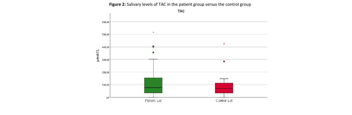
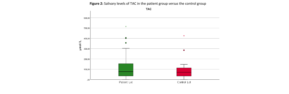
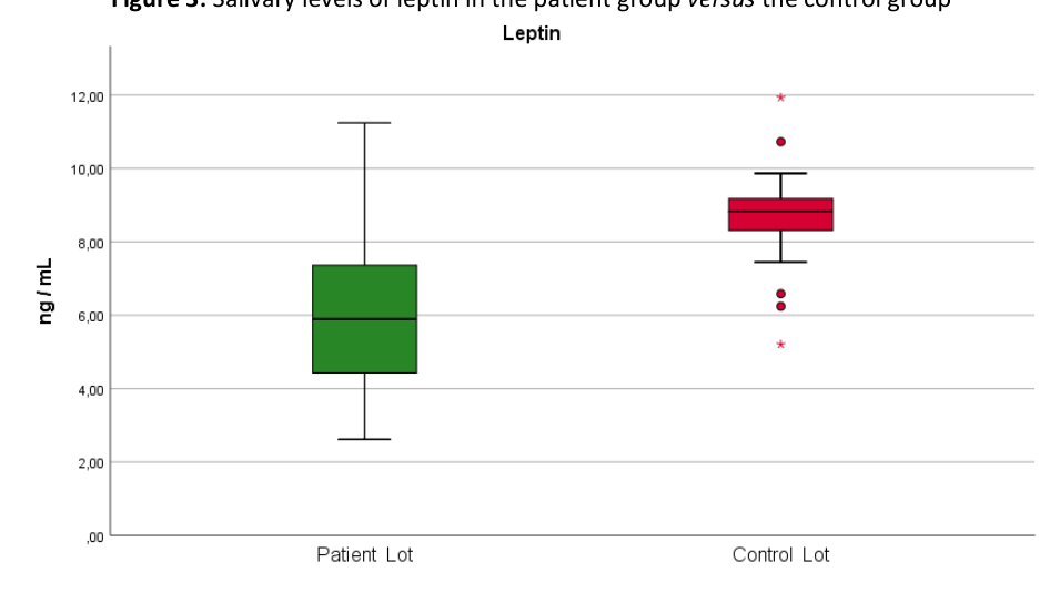
The results obtained show that when all patients with periodontal disease were compared with the control group, NLRP3 had statistically significantly increased levels in the study group (Table 1, Figure 1). Although TAC was also increased in the patients compared to the healthy subjects, no statistically significant differences were found (Figure 2). At the same time, leptin was significantly decreased in patients with periodontal disease compared to the control group (Figure 3).
| Biomarker | Group | Median | Mean ± Std. Deviation | p-value* |
|---|---|---|---|---|
| NLRP3 | Patient | 1.1372 ng/mL | 1.117 ± 0.332 ng/mL | < 0.00001 |
| Control | 0.7382 ng/mL | 0.776 ± 0.208 ng/mL | ||
| TAC | Patient | 76.741 µmol/L | 114.127 ± 119.398 µmol/L | 0.491 |
| Control | 70.838 µmol/L | 94.553 ± 101.928 µmol/L | ||
| Leptin | Patient | 5.894 ng/mL | 6.124 ± 2.064 ng/mL | < 0.00001 |
| Control | 8.827 ng/mL | 8.618 ± 1.478 ng/mL |
*Mann-Whitney U Test
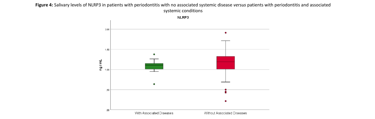
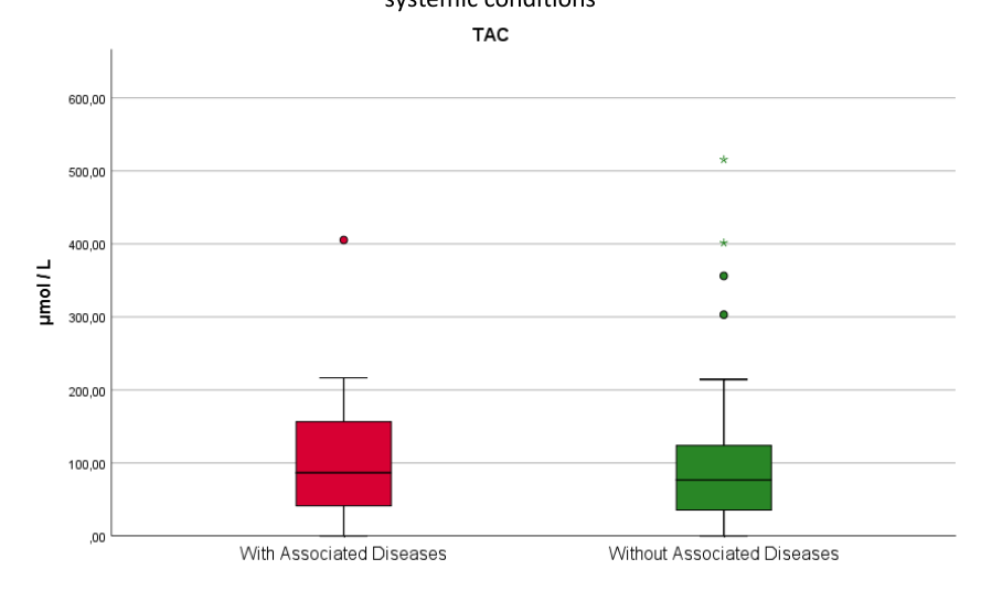
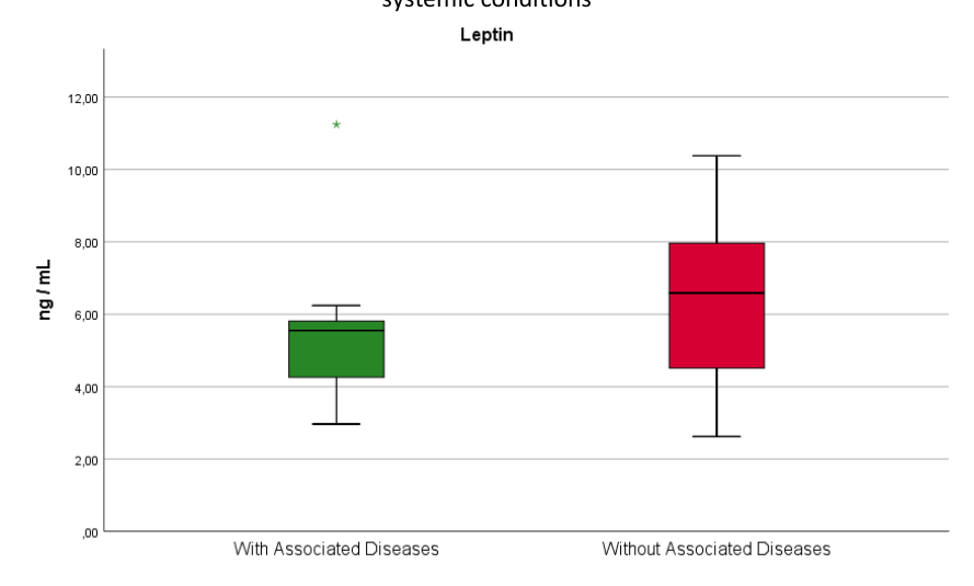
None of the three determined parameters displayed significant differences when comparing patients with periodontitis with no associated systemic disease versus patients with periodontitis and associated systemic conditions (Table 2, Figures 4, 5, 6).
Spearman’s rank correlation coefficient was used to test intragroup possible associations between salivary parameters. No significant correlation was found between salivary levels of leptin and NLRP3 when including all patients or when analyzing just patients with periodontal disease without associated systemic conditions. Also, no significant correlation was found between salivary levels of NLRP3 and TAC or TAC and leptin when including all patients or when analyzing just patients with periodontal disease without associated systemic diseases. No statistically significant correlations were found between the concentrations of the analyzed biomarkers and the number of remaining teeth when taking into consideration all periodontal disease patients, nor when including only the patients without associated systemic diseases (Table 3).
| Biomarker | Group | Mean ± Std. Deviation | p-value* |
|---|---|---|---|
| NLRP3 | No Associated Diseases | 1.13 ± 0.369 ng/mL | 0.356 |
| Associated Diseases | 1.077 ± 0.189 ng/mL | ||
| TAC | No Associated Diseases | 112.041 ± 112.434 µmol/L | 0.828 |
| Associated Diseases | 120.387 ± 115.215 µmol/L | ||
| Leptin | No Associated Diseases | 6.346 ± 2.011 ng/mL | 0.115 |
| Associated Diseases | 5.457 ± 2.174 ng/mL |
*Mann-Whitney U Test
| Correlation | Spearman Rho | p-value |
|---|---|---|
| Between the number of remaining teeth and the NLRP3 levels for all patients | 0.175 | 0.255 |
| Between the number of remaining teeth and Leptin levels for all patients | 0.095 | 0.541 |
| Between the number of remaining teeth and the TAC levels for all patients | -0.158 | 0.305 |
| Between the number of remaining teeth and the NLRP3 levels for patients without associated disease | 0.223 | 0.213 |
| Between the number of remaining teeth and leptin levels without associated disease | -0.063 | 0.729 |
| Between the number of remaining teeth and leptin levels for patients with associated diseases | 0.378 | 0.252 |
| Between the number of remaining teeth and the TAC levels for patients without associated disease | -0.088 | 0.626 |
| Between leptin and NLRP3 in all patients | 0.147 | 0.340 |
| Between leptin and NLRP3 in patients with no associated diseases | 0.234 | 0.189 |
| Between TAC and NLRP3 in all patients | -0.171 | 0.267 |
| Between TAC and NLRP3 in patients with no associated diseases | -0.131 | 0.467 |
| Between TAC and leptin in all patients | -0.168 | 0.276 |
| Between TAC and leptin in patients with no associated diseases | -0.059 | 0.746 |
Saliva has already proven to be a reliable diagnostic, prognostic, and monitoring fluid for both oral and systemic diseases, being very rich in molecules and macromolecules that can be used as biomarkers [48]. Furthermore, the collection procedure is easy, it is more efficient in terms of costs, has a lower risk of cross-infection compared to the blood, and most importantly, the sampling is noninvasive [48, 49]. However, when determining the salivary levels of these biomarkers, we have to keep in mind that their concentrations are frequently decreased compared to the blood. Additionally, there are several factors that can influence their levels, including the moment of the day or the level of stress [49].
In recent years, inflammasomes have represented a major research interest due to their potential to determine an inflammatory response by integrating signals from different pathogens and altered cells [50, 51]. The most studied of the inflammasomes in systemic diseases, NLRP3, has also gained attention regarding its connection with periodontal disease [52]. The dysbiosis and inflammation associated with periodontal disease lead to the activation of the components of the NLRP3 inflammasome. Apart from promoting pyroptosis and neutrophil recruitment, once activated, NLRP3 promotes the overproduction of cytokines and antibodies, as well as the release of prostaglandin E2. In the end, all these events result in the destruction of the periodontium [53].
The results of previous studies performed on animal models were not conclusive. Yamaguchi et al induced periodontal disease in mice by orally injecting them with Porphyromonas gingivalis, showing that this microbial species, involved in the inflammatory process characteristic of chronic periodontitis, activated the NLRP3 inflammasome, leading to a response from the cells of the innate immune system and involvement of the IL-1 family of cytokines [54]. Nevertheless, a recent study by Rocha et al. performed on mice with induced periodontal disease revealed different results, showing that the inflammation associated with periodontitis and the consequent bone resorption was not determined by NLRP3, which played a minor role in the process, but rather by caspase-1 [55]. The results of our study showed statistically significant increased salivary levels of NLRP3 in patients with periodontitis compared to the control group (p< 0.00001, Table 1, Figure 1), suggesting a possible association of NLRP3 with the pathogenesis of this oral condition. Furthermore, the data we obtained did not show any correlation between NLRP3 salivary levels and the number of remaining teeth on the dental arches (Table 3).
When comparing the salivary levels of NLRP3 in patients with periodontitis and without systemic diseases to those who have associated systemic diseases, we were unable to find any statistically significant difference (Table 2). These results indicate that NLRP3 levels are mainly due to the presence of periodontal inflammation and not to the coexistence of other pathologies. Isola et al reported that patients with periodontitis and patients with periodontitis and type 2 diabetes had increased NLRP3 levels, both in the serum and saliva, not only compared to healthy controls, but also versus patients who had type 2 diabetes, but did not have periodontal disease [53]. Using samples from the gingival crevicular fluid and gingival biopsies, Garcia-Hernandez et al. showed that patients with periodontitis and uncontrolled type 2 diabetes displayed upregulated genes and proteins associated with NLRP3 activation compared to patients who had only periodontitis [56].
A systematic analysis from 2020 concluded that the levels of leptin both in plasma and saliva reported in the analyzed studies were too different, and although a decrease in salivary levels of leptin was observed in periodontal patients compared to healthy subjects, that data was not sufficient to establish the relationship between the disease and levels of leptin [57].
Similar to other studies, our results showed that leptin had significantly decreased levels in the saliva of patients with periodontal disease compared to those of the control group (p< 0.00001, Table 1, Figure 2) [58]. Leptin has already been shown to activate NLRP3 inflammasome in other inflammatory diseases, including systemic lupus erythematosus, and autoimmune disease characterized by the presence of the inflammatory process [59]. Despite both NLRP3 and leptin showing increased levels in periodontitis, we could not find a correlation between the two biomarkers.
TAC was defined by Miller as the “measure of the antioxidant capacity of all antioxidants in a biological sample and not just the antioxidant capacity of a single compound” [60]. Since ROS are naturally counteracted by antioxidants, determining the total antioxidant capacity has become one of the common and important ways to monitor the effect of oxidative stress in periodontal disease. However, the results regarding TAC levels in periodontal disease are controversial, with research showing both significant increases in periodontitis patients compared to healthy controls, as well as no significant differences [61].
On the other hand, Toczewska et al reported lower levels of TAC in both the saliva (which was a mixture of non-stimulated and stimulated saliva) and the gingival crevicular fluid of periodontal patients [62]. A decrease in the antioxidant capacity of patients with periodontal disease was also reported by Diab-Ladki et al [63]. Moreover, similar results were obtained by Brock et al, although their data did not show a statistically significant difference for salivary levels [64]. In our study, the determination of TAC displayed higher salivary levels in periodontitis patients compared to the control group (p=0.491, Table 1, Figure 3), but no statistical significance was found. This difference could be explained by the fact that the control group consisted of healthy subjects who had a mean age lower than that of the periodontitis patients. Limberaki et al previously reported that, contrary to expectations, younger people have lower TAC levels in the serum than older people [65].
Previous research revealed that while mitochondrial dysfunction and ROS overproduction can promote NLRP3 activation, their existence is not imperative for this to happen. Additionally, DNA damage caused by ROS can activate the inflammasome, but its release can happen downstream of the activation of the inflammasome [66]. This could explain why our results showed no statistically significant correlation between the salivary levels of NLRP3 and those of TAC.
Our study has several limitations, including the study groups being relatively small. Additionally, there was a significant mean age difference between the study group and the control group. Moreover, although saliva was collected following the same recommendations for all participants in the study, the salivary flow of each participant could have played a role in the level of the biomarkers [67]. Nevertheless, they reveal that the data obtained by our research group suggests the involvement of NLRP, leptin, and oxidative stress in periodontitis.
The results obtained in the present study are promising, but they incite further research that should be more focused on the relationship between these biomarkers and other factors that could influence the outcome, for example, the different types of bacteria involved in periodontal disease. Moreover, analyzing not just the saliva of the patients, but their gingival crevicular fluid could provide better insights. Future perspectives could be offered by determining these biomarkers in dynamic, before and after periodontal treatment, as well as correlating them to clinical parameters. All these represent objectives for our future research, which would offer a more complete picture of the mechanisms involved in periodontal disease development and progression and thus, show how NLRP3 activation can promote periodontal tissue destruction and bone loss.
The authors declare no conflict of interest. This research was funded by Carol Davila University of Medicine and Pharmacy, Bucharest, Romania, through Contract no. 33PFE/30.12.2021 funded by the Ministry of Research and Innovation within PNCDI III, Program 1—Development of the National RD system, Subprogram 1.2—Institutional Performance—RDI excellence funding projects.
Conceptualization, I.-I.S.-S., A.C.D; methodology, A.-S.D, D.M., B.-F.M; software, R.I., S.C.B; validation, T.-C.S., A.M.C..; formal analysis, D.M., S.C.B; investigation, T.-C.S., B.F.M; resources, T.-C.S., A.M.C.; data curation, D.M., B.-F.M; writing-original draft preparation, I.-I.S.-S., T.-C.S., R.I.; writing-review and editing, I.-I.S.-S., D.M., A.-S.D, A.C.D; visualization, R.I., A.M.C.; supervision, A.-S.D, A.C.D; project administration, I.-I.S.-S., A.C.D.
The study was conducted in accordance with the Declaration of Helsinki and approved by the Ethics Committee of the Carol Davila University of Medicine and Pharmacy (22976/17.08.2022).
Informed consent was obtained from all subjects involved in the study.