
The Global reports estimates that ischemic heart disease is the leading cause of cardiovascular death in the world (9.44 million deaths in 2021 alone). Coronary artery disease (CAD) was the main cause of death in both the Romanian female (379 deaths per 100.000) and male (362 deaths per 100.000) in 2019 [1]. Acute Myocardial Infarction (AMI) is a heterogeneous group of diseases with similar clinical manifestations [2]. The complex predisposition for AMI includes medical history (e.g., hypertension, dyslipidemia), genetic (e.g., prothrombin FII or ADAMTS7 polymorphisms), epigenetic, anthropometrical (e.g., obesity), behavioral (e.g., sedentary lifestyle, smoking), and other factors (e.g., age, gender, ethnicity) [3].
The RO-STEMI study was the first Romanian registry for AMI with ST-segment elevation (STEMI) published in 2010 and had 3 phases: RO-STEMI-1 (1997-2001), RO-STEMI-2 (2002-2005), and RO-STEMI-3 (2006-2009) [4]. During the 13 years of follow-up, there was an increasing trend in the number of AMI patients with comorbidities (e.g., hypertension, type 2 diabetes, dyslipidemia) and a slight decrease in the percentage of smokers [4]. Based on the results obtained in this study, in August 2010, the Romanian National Program for primary angioplasty in AMI was implemented, which is still ongoing.
A subsequent study from RO-STEMI carried out between 2011-2015 identified new predictive factors for in-hospital mortality in AMI patients who underwent interventional treatment (e.g., Killip class, depressed left ventricular ejection fraction – LVEF, anterior location of infarction) [5].
During the global crisis caused by the COVID-19 pandemic, it became evident that the prevention and management of CADs need to be improved [6].
Percutaneous coronary intervention (PCI) is an invasive procedure that aims to revascularize ischemic myocardial tissue. “Dr. Carol Davila” Central Military Emergency University Hospital represents one of the 6 medical units from Bucharest that are involved by weekly rotation in PCI of patients with AMI, according to the management guide for AMI of the Ministry of Health.
This study aims to analyze the risk factors’ distribution for AMI in a group of subjects and to compare results with data published in the last 15 years for the Romanian population.
This study was based on medical records of the patients with AMI (the AMI group) or with suspicion of AMI (the control group). All subjects presented themself to the Emergency Department of “Dr. Carol Davila” Central Military Emergency University Hospital, Bucharest between 01.11.2021 and 30.04.2022. The AMI group refers to patients with suggestive acute clinical symptoms (e.g., chest discomfort, pain, pressure, tightness, heaviness, or burning), AMI-specific EKG morphology, and elevated cardiac enzymes [7]. The control group included subjects with symptoms suggestive of AMI but with normal EKG and cardiac enzymes. Echocardiography, Killip stage and echocardiographic LVEF were interpreted according to international criteria during their hospital admission [8]. The LVEF values were distributed in 3 groups according to international criteria: heart failure with reduced ejection fraction (HFrEF), heart failure with mildly reduced ejection fraction (HFmrEF) and heart failure with preserved ejection fraction (HFpEF) [8]. Levels of creatine kinase (CK) >145U/L, creatine kinase-MB fraction (CK-MB) >25 U/L, and high sensitive troponin T (hs-cTn) > 1.3 pg/ml were considered abnormal levels. Subjects who were under 18 years of age, not able to consent, or pregnant were not included.
We used odds ratios (O.R.) with 95% confidence intervals (CI) for comparing categorical variables, and the significant values were considered if the p-value was lower than 0.05. Statistical analysis was done using R Studio, version 2023.09.1 Build 494.
The characteristics of subjects included in the study are presented in Table 1. The gender distribution in patients and control groups presents no statistically significant difference. The average patient’s age was higher than in the control group (p<0.05). We identified three individuals with COVID-19 infection in the AMI group and 6 individuals in the control group (p>0.05). We found no statistical differences between the groups of patients who lived in the urban or rural areas regarding the risk for AMI. Also, the risk factors associated with CAD were investigated in the medical records of 138 patients (Table 1).
| Characteristics | AMI group | Control group |
|---|---|---|
| Male: Female | 50/19 | 39/30 |
| Average age | 61.66±14.42 (19-92) | 54.20±18.25 (18-87) |
| Male age | 59.28±14.65 (19-84) | 58.64±17.72 (18-87) |
| Female age | 67.94±11.96 (41-92) | 48.43±17.56 (19-78) |
| Rural / Urban | 23/46 | 24/45 |
| Covid Yes/No/Unknown | 3/66/0 | 6/11/52 |
| Hypertension | 58 | 27 |
| Dyslipidemia | 45 | 14 |
| Smoking | 34 | 18 |
| Diabetes | 17 | 10 |
| Heart failure | 15 | 12 |
| Cardiac pathological antecedents | 16 | 11 |
| Neurological pathological antecedents | 8 | 6 |
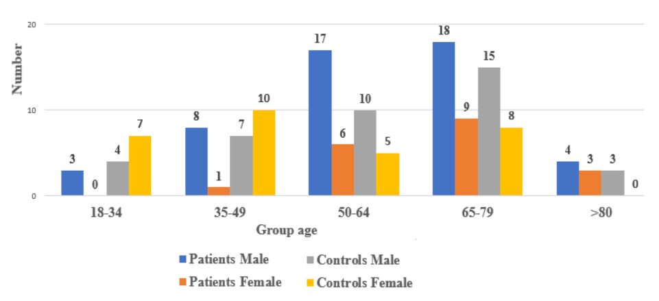
The majority of AMI patients have had at least one traditional risk factor for this disease. We observe statistically significant differences between the two groups (Figure 1), for females in the 35-49 age group (p<0.05), and for females in the 18-34 age group (p<0.001). Anterior chest pain was the most frequent clinical symptom reported by AMI patients (91% vs 14% in subjects from the control group).
Almost half of patients presented to the Emergency Department in the first six hours after the onset of acute symptoms (49% presented in the first 6 hours; 33% within 6-12 hours; 17% more than 12 hours after the onset of symptoms). We observed that in the first 12 hours from the onset of symptoms, 94% of male patients and 52.6% of female patients presented in the ED (p<0.001). The EKG morphology analysis showed that the most frequent localization of AMI was anterior (39.1%) and inferior (23.1%). The values for CK [663 U/L (range 11-4024) vs 90 U/L (range 17-3443)], CK-MB [88.5 U/L (range 6-586) vs 13 U/L (range 6-107)] and hs-cTn [4658 pg/ml (range 1.3-50000) vs 1.3 pg/ml (range 1.3-66.5)] levels were higher in AMI than in control group (Table 2).
| Cardiac markers | Subgroups | AMI male | AMI female | Control male | Control female |
|---|---|---|---|---|---|
| CK | CK1 (10-145 U/l) | 4 | 6 | 29 | 28 |
| CK2 (146-2000 U/l) | 32 | 12 | 9 | 2 | |
| CK3 (> 2000 U/l) | 14 | 1 | 1 | 0 | |
| CK-MB | CK-MB1 (≤ 25 U/l) | 10 | 4 | 34 | 30 |
| CK-MB2 (25-200 U/l) | 27 | 14 | 5 | 0 | |
| CK-MB3 (> 200 U/l) | 13 | 1 | 0 | 0 | |
| hs-cTn | hs-cTn (≤1.3 pg/ml) | 0 | 0 | 17 | 24 |
| hs-cTn (1.4-50000 pg/ml) | 39 | 15 | 22 | 6 | |
| hs-cTn (> 50000 pg/ml) | 11 | 4 | 0 | 0 |
The CK values showed statistically significant differences between the CK1 and CK3 subgroups (p < 0.001). The values of hs-cTn show significant differences between AMI patients with CK MB 1 and CK-MB 2 values (p<0.001). We observed that in the group of patients, the median CK values are related to gender, with men having 870.5 U/L (range 54-3781) and women having 456 U/L (range 11-4024) (p<0.05). The values for CK-MB and hs-cTn were not related to the subject’s gender. The majority of patients investigated were treated with PCI (81.16% that went primarily to PCI and 18.84% of patients received thrombolysis before PCI) and 4.36% died before receiving interventional treatment. Regarding the result of PCI, it is observed that 4 out of 66 AMI patients who presented with clinical signs and symptoms of infarction had no obstructive lesions of the coronary arteries; these patients were included in the category of AMI with non-obstructive coronary artery (MINOCA).
From the AMI group, 61 patients were discharged with improved status (the median hospitalization period was 7 days; range 1-70 days), and 5 patients died during hospitalization (median 7 hospitalization days; range 1-19 days). Sixteen subjects from the control group were hospitalized (23.2%) and had a median of 6 hospitalization days (range 2-24 days). No deaths were reported in this subgroup of patients.
LVEF was preserved only in 17.4% of AMI patients and Killip 1 was established for 73.9% of patients from them (Table 3). The bivariate analysis of the number of deaths shows that it is statistically associated with the Killip and LVEF class at admission (p<0.01) and also with the number of hospitalization days (p<0.05) (Table 3).
| Cardiac markers | Subgroups | AMI male | AMI female | Control male | Control female |
|---|---|---|---|---|---|
| CK | CK1 (10-145 U/l) | 4 | 6 | 29 | 28 |
| CK2 (146-2000 U/l) | 32 | 12 | 9 | 2 | |
| CK3 (> 2000 U/l) | 14 | 1 | 1 | 0 | |
| CK-MB | CK-MB1 (≤ 25 U/l) | 10 | 4 | 34 | 30 |
| CK-MB2 (25-200 U/l) | 27 | 14 | 5 | 0 | |
| CK-MB3 (> 200 U/l) | 13 | 1 | 0 | 0 | |
| hs-cTn | hs-cTn (≤1.3 pg/ml) | 0 | 0 | 17 | 24 |
| hs-cTn (1.4-50000 pg/ml) | 39 | 15 | 22 | 6 | |
| hs-cTn (> 50000 pg/ml) | 11 | 4 | 0 | 0 |
1 Fisher exact, p 2-tails: * Killip I+II vs. III+ IV: p < 0.01; ** HFpEF + HFmrEF vs. HFrEF: p < 0.01; *** Number of hospitalisation days: I + II vs. III: p < 0.05.
The period of the study covers the winter period and the fourth wave of the pandemic in Romania, which had the highest mortality reported for COVID-19 in our country [9].
In our study, the percentage of men is comparable to that reported in the RO-STEMI study (72.46% vs. 68.56%) [4]. The median age of patients was approximately 1.5 years lower than reported in the RO-STEMI study (61.66±14.42 vs. 63.39±12) [4]. Also, we observe a decrease in age, especially in men; in our study, the age of men compared to that of women is 8.66 years lower, compared to the RO-STEMI study, where it was only 6.51 years lower [4]. This trend is concordant with those reported for other populations [10].
Treatment of dyslipidemia and hypertension is a key step for reducing the risk of atherosclerotic cardiovascular disease; populations with hypertension and dyslipidemia are associated with an increased risk of AMI. For example, Anderson et al. reported that from AMI patients, 73.7% have hypertension and 73.4% have dyslipidemia [11]. Our data shows that compared to RO-STEMI, the frequency of hypertension (84.05% vs. 52.20%), smoking (49.27% vs. 47.00%), and dyslipidemia (65.21% vs. 38.4%) increased in patients with AMI from Romania in the last 10 years [4]. In our study, hypertension (O.R.= 8.20, p<0.0001) and dyslipidemia (O.R.= 7.36, p<0.0001) were strongly associated with AMI, although all AMI patients had specific treatment for hypertension or dyslipidemia. More subtle differences were observed for diabetes (24% vs 21.80%) and personal history of AMI (11.6% vs. 9.80%) [4].
Overall, the percentage of active or former smokers in the AMI group (49.27%) is concordant with figures reported by RO-STEMI (47.06%) [4]. However, we observed an increasing frequency of smoking at young ages (78.3% in the 18-34 age group and 76.4% in the 35-49 age group) in contrast with RO-STEMI (75% vs 73.25%). The frequency of smokers in the control group (26.1%) was higher than the value reported for the Romanian population (19.8%) or in other European populations [12]. In our control group, we don’t observe any significant difference between smoking and gender, although such differences were reported in Romania (30.6% of men and 7.5 % of women) [4] and in other European countries [12].
The frequency of anterior AMI is much higher compared to data previously reported for the Romanian population (31%), but the frequency of inferior AMI is similar (22%) [13]. There is a significant increase in patients who were treated by direct PCI (81.16% vs. 12.94%) and a halving of the percentage of patients treated with thrombolytic medication before the intervention (18.84% vs. 35.07%) compared to the RO-STEMI study [4]. In the year 2009, when a percentage of 30.82% of patients who benefited primarily from PCI within the RO-STEMI program was reported, the number of patients was continuously increasing, a fact that was confirmed by other reports [5,13]. The frequency of patients diagnosed with MINOCA identified in our study group is within the limits reported in the specialized literature of 5-15% [14].
Compared to the RO-STEMI 3 study, the percentage of patients with Killip 1 (73.9% vs. 68.34%), Killip 2 (13.04% vs. 17.65%), Killip 3 (2.9% vs. 7.93%), or Killip 4 (10.16% vs. 6.06) was similar [4]. The values of LVEF are different from those reported by others [15] and may represent a characteristic of the subjects included in this study. For patients with HFmrEF after STEMI, a need for specific attention and appropriate management was reported [15]. The percentage of patients who presented more than 12 hours after the onset of symptoms decreased by 5.18%. Compared to the pre-COVID-19 period, the percentage of patients who presented in less than 6 hours decreased by approximately 15%, and the percentage of patients who presented between 6 and 12 hours increased by 19.02%.
The values of patients from our study who were discharged with improved status were comparable to those reported by another study (9.20 ± 5.47 days) [13]. The percentage of 7.24% of deaths during hospitalization is below that reported by the RO-STEMI study (11.84%) and similar to that reported by other studies [5,13]. We did not observe statistical differences regarding the evolution of this AMI related to patients’ gender, although in other studies such associations were reported [16].
Also, the relation between the number of deaths and the Killip, LVEF class at admission, and the number of hospitalization days is in concordance with another report [5].
During the COVID-19 pandemic, a considerable reduction in hospitalization for AMI, more complex catheterization laboratory protocols, an increase in time to the catheterization laboratory associated with an increase in door-to-balloon times, which finally led to increased rates of in-hospital mortality [17]. These remarks can be explained by the fact that patients did not activate emergency medical systems because hospitals were perceived as dangerous places regarding the infection risk, and by the fact that patients with AMI had a significantly higher mortality in-hospital compared to those admitted before COVID-19, potentially due to late arrival to the hospital [17]. The complications determined by the COVID-19 infection and the difficulties in accessing the ambulance services during the lockdown period could increase the mortality due to AMI. All these observations can explain, at least partially, the reduced incidence of COVID-19 in patients with AMI from our Emergency Department. The decrease in the age of AMI onset observed during the COVID-19 pandemic can be explained by the increase in the incidence of cardiovascular risk factors and the fact that young people probably had more courage to call on medical services [18].
The COVID-19 pandemic created a subsequent impact on mortality in patients with AMI. In Romania, the difference in time delay and clinical outcome in patients with AMI during the pre-pandemic and COVID-19 pandemic period was not investigated in detail [19]. Several studies performed in different countries revealed significant differences in accessing medical care related to gender, population density, socioeconomic status, and infrastructure [20].
The main limitation of our study was that the number of patients and controls was relatively low in that period of time, so there should be further studies that include a larger number of subjects and investigate the connection between risk factors, COVID-19 infection, and myocardial infarction in the Romanian population.
The analysis of the data included in this study highlighted that the age at which AMI symptoms begin has decreased (men diagnosed with AMI were 8.66 years younger than women), and this can be explained by the increase in the incidence of some cardiovascular risk factors (e.g., hypertension, dyslipidemia, smoking). Compared to the RO-STEMI study, the number of patients presenting within 6-12 hours from the onset of symptoms has increased and most of the patients diagnosed with AMI had reduced and slightly reduced LVEF. The number of patients that received interventional treatment has increased compared to the RO-STEMI study and Killip class and LVEF are associated with mortality and hospitalization days. All these results of this study are useful for prospective studies regarding risk factors for myocardial infarction in the Romanian population.
The authors declare no conflict of interest. The study was supported by the Ministry of Research and Education, grant number PN 23 01 03 03.
No generative AI was used during the production of this article.
Conceptualization, D.A.-V., A.-T.O.-A and C.O.E.; methodology, D.A.-V.; software, T.M.; validation, D.A.-V., B.L.M., and T.M.; formal analysis, D.A.-V.; investigation, D.A.-V. and T.M.; resources, B.L.M. and T.M.; data curation, T.M.; writing-original draft preparation, D.A.-V.; writing-review and editing, A.-T.O.-A., T.M., and C.O.E.; visualization, T.M.; supervision, C.O.E.; project administration, B.L.M.; funding acquisition, B.L. All authors have read and agreed to the published version of the manuscript.
As a University Hospital, patients and controls sign informed consent upon admission, confirming their explicit acceptance of the use of their personal data and blood samples for scientific research purposes and clinical studies.
The study was conducted in accordance with the Declaration of Helsinki and approved by the Local Ethics Committee of the Central Military Emergency University Hospital “Dr. Carol Davila” (protocol number 641/07.11.2023).
Not applicable.