
This narrative review aims to provide readers with a brief, yet pinpointed overview as to the therapeutic potential of nutritional antioxidants. Given that oxidative stress has a key involvement in the pathogenesis of many neurological diseases (neurodegenerative, cerebrovascular, or inflammatory), targeting oxidative stress through such components (known to medicine for hundreds, if not thousands of years) may prove efficient. Moreover, this paper should be viewed as an incentive to inspire future research, preclinical and particularly clinical, in this field, so that the usage of such components may one day become a validated treatment with a probable wide spectrum. A step-by-step approach to nutrient intervention in neurological diseases is provided in Figure 1.
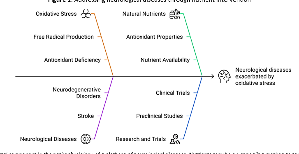
Comprehensive research has been conducted by consulting the PubMed database for relevant reports published between the inception of the archive and January 2025.
The following keywords have been used by themselves or in various combinations to achieve optimal results: “Parkinson’s disease”, “amyotrophic lateral sclerosis”, ”Alzheimer’s disease”, ”multiple sclerosis”, ”small vessel disease”, ”ischemic stroke”, “hemorrhagic stroke”, “inflammation”, “oxidative stress”, “reactive oxygen species”, “mitochondrial dysfunction”, “apoptosis”, “necrosis”, “excitotoxicity”, “lipid peroxidation”, “vitamin C”, “vitamin E”, “coenzyme Q10”, “black tea”, “green tea”, “taurodeoxycholic acid”, “flavonoids”, “curcumin”, “polyphenol”, “antioxidant”, “Ginkgo biloba”, “mecasin”, “citicoline”, “Artemisia”, “Silybum”, “curcumin”, “melatonin”, “saffron”, “vitamin D3”, “Gastrodia elata”, “lavender oil”.
Case reports, preprints, and standalone abstracts have been excluded. Only papers published in English were included in the selection.
Parkinson’s disease (PD) is a chronic neurodegenerative disease with a characteristic loss of the dopaminergic neurons in the pars compacta of the substantia nigra [1]. Literature has shown that environmental factors (herbicides, pesticides (such as paraquat), industrial pollutants, neurotoxins (1-methyl-4-phenyl-1,2,3,6-tetrahydropyridine or MPTP) and its metabolite (1-methyl-4-phenylpyridinium), as well as genetic factors (mutations of DJ-1, Parkin, PINK1) [2] involved in PD pathogenesis induce oxidative stress (OxSt) in the mitochondria [3], reduce the production of adenosine triphosphate (ATP), facilitate the generation of reactive oxygen species (ROS), and finally lead to the apoptosis of dopaminergic cells [2].
Pathogenesis
OxSt contributes to the degeneration of dopaminergic cells in PD [4]. Also, OxSt is tightly linked to other pathogenic mechanisms involved in PD (mitochondrial dysfunction, excitotoxicity, and inflammation); therefore, it is difficult to ascertain whether it is a primary factor in PD or a consequence of the aforementioned mechanisms [3].
In PD, lipid peroxidation is upregulated, and antioxidant pathways (superoxide dismutase — SOD, catalase, glutathione peroxidase — GPx, ceruloplasmin, vitamin E, vitamin C, copper, zinc, selenium) may be insufficient to protect the cells from pathological oxidation [4]. Increased OxSt in association with diminished antioxidant protectors leads to the loss of neurons in the pars compacta, hence the development of PD [4]. Studies have proven lower levels of vitamin C and E in PD patients versus control groups, launching the hypothesis of increased consumption of these vitamins in PD due to OxSt [4]. Excessive ROS production, mitochondrial transport changes, and increased iron deposition in the pars compacta of the substantia nigra are key pathways in PD pathogenesis [1]. There is a close link between ROS formation and mitochondrial dysfunction [1]. Cellular death in PD is thought to be connected to excessive lipid peroxidation, occurring as a consequence of increased oxidation and expenditure of compensatory mechanisms [1]. ROS have a direct cytotoxic impact, as well as an indirect one by impacting mitochondrial complex I, with higher lipid peroxidation [1]. The reduction of this complex leads to the production of more ROS [1].
In PD, α-synuclein deposition induces an activation of the microglia in the pars compacta, thus activating nicotinamide adenine dinucleotide phosphate oxidase (NADPH-oxidase) [1]. The latter’s consequences are, on the one hand, ROS production in the extracellular compartment afflicting nearby neurons, and on the other hand, redox signaling in the microglia, amplifying the pro-inflammatory response with the induction of chronic inflammation [6].
The presence of excessive OxSt in PD is supported by studies that have proven an increase in carbonyl derivatives [7], in indicators of lipid peroxidation [8], and 8-hydroxy-2′-deoxyguanosine (a marker of nucleic acid damage) [9].
Nutrients
Given the involvement of OxSt in the occurrence of PD and the fact that PD treatment (especially L-dopa) may determine, after a long period of usage, the appearance of free radicals, the administration of multiple antioxidant agents in high dosage along with L-dopa may increase the latter’s efficiency [10,11].
In experimental studies, vitamin E was proven to protect cells from OxSt-related damage in PD and the chronic administration of high doses of vitamin E may be a therapeutic strategy in PD treatment or prevention [3,12]. In other experimental studies, vitamin E diminished the OxSt induced by midbrain iron deposits in PD patients, suggesting it may have neuroprotective effects in these patients [13].
Other observational studies on human subjects suggested that a combined administration of high doses of vitamins E and C may determine a slowed PD progression through their antioxidant effect (14), although their efficacy was not proven in double-blind randomized clinical trials (RCTs) [15].
Taurodeoxycholic acid is an antioxidant agent, decreasing ROS reduction, an effect that has been proven when in vivo PD lab models were used [16].
Coenzyme Q10 has an important antioxidant role in the mitochondria and the lipid membrane, some of its antioxidant effects being mediated by its interaction with α-tocopherol. A phase 2 clinical trial on the efficiency of the antioxidant effect of the coenzyme on early PD patients (i.e., patients who did not require specific therapy) versus placebo showed a decrease in the treated patients’ disability [17].
Black and green tea, due to high polyphenol concentration, exert an antioxidant effect and may have a neuroprotective and neuroregenerative effect in this disease, although clear evidence to support this assertion, derived from clinical research, is scarce [18].
Flavonoids enhance the endogenous activation of antioxidant enzymes, inhibit the peroxidation of lipids, and reduce the production of inflammatory mediators [19]. Due to all these actions, flavonoids exert a favorable effect on dopaminergic neurons and mitigate PD manifestations in laboratory studies [19].
An association between PD and a lack of adherence to the Mediterranean diet suggests a potential neuroprotective effect of this diet, through an undetermined mechanism that may attenuate the OxSt and inflammation, which are both recognized pathogenetic factors in PD [20].
Alzheimer’s disease (AD) is a neurodegenerative affliction characterized by the dysfunction of memory, attention, and other cognitive processes; it is the most common form of dementia [21]. Small vessel disease (SVD) may affect microvasculature systemically or mainly in the brain, the latter constituting a devastating cause of stroke and dementia worldwide [22,23].
Pathogenesis
AD mainly determines a synapse affliction, with the development of senile plaques mainly containing β-amyloid (Aβ). Aβ derives from the proteolysis of the amyloid precursor protein performed mainly by the enzymatic actions of β- and γ-secretase, with an imbalance between production and clearance causing the Aβ buildup [21]. The lesions produced by OxSt in AD are associated with an accumulation of Aβ [11]. Aβ buildup is also a central feature of the cerebral amyloid angiopathy form of SVD, to which AD is intimately connected; some might argue that the two constitute an inseparable spectrum [22]. The other form of SVD, deep perforator arteriolosclerosis, centrally features a blood-brain barrier (BBB) dysfunction, subsequent morphological and functional changes to the vascular wall, as well as myelin impairment [22,23].
Aβ determines the release of ROS by attacking mitochondrial enzymes (particularly cytochrome c oxidase), leading to mitochondrial dysfunction, increasing superoxide radicals, conversion to hydrogen peroxide (H2O2), the release of cytochrome, affecting the production of ATP and cellular apoptosis [21].
OxSt increases with aging and it is an important causal factor in the appearance of AD and other neurodegenerative age-related diseases, such as SVD. Moreover, OxSt is thought to bridge the connection between SVD and AD [24]. It is not yet fully understood how neurochemical factors influence the appearance of age-related cerebral lesions, but more and more data has shown the involvement of biometals (Cu, Fe, Zn) in Aβ accumulation. Copper is a potent mediator for the hydroxyl radical, and the high-affinity link between copper and Aβ (via histidine and tyrosine), as well as between zinc and the amyloid precursor protein and Aβ may contribute to the increased OxSt in AD [11].
Apolipoprotein E is a lipid transport molecule believed to be related to ROS and directly to lipid peroxidation in AD [25]. Carriers of apolipoprotein E4 more often have cerebral amyloid angiopathy [24].
To conclude, the involvement of OxSt in AD pathogenesis is supported by the high concentrations of Cu, Fe, Al, and Hg in the brain parenchyma, a high level of lipid peroxidation and DNA oxidation, as well as a decrease in polyunsaturated fatty acids, in cytochrome c oxidase activity, and energy metabolism [11]. OxSt could become a promising link between AD and SVD; therefore, research focusing on OxSt may decrease the neurological manifestations of both illnesses [24].
Nutrients
In preclinical research, caffeine has been associated with a tendency to lower the Aβ levels in early (familial) AD, because of its antioxidant properties [26]. Studies on cerebral amyloid angiopathy models are lacking.
Curcumin is an antioxidant and anti-inflammatory agent (inhibiting cyclooxygenase and lipoxygenase) that has been shown to reduce carbonyl derivatives, facilitating Aβ disaggregation, with possible beneficial effects in AD in animal studies [26]. Curcumin may also aid endothelial function, which is frequently impaired in SVD, as it has been shown to diminish the risk of stroke in animal models [27].
Vitamin E is a potent antioxidant that prevents oxidizing lesions induced by Aβ in cell cultures; it also delays memory impairment in animal studies on AD [28]. Some RCTs suggested vitamin E, through its antioxidant effects, may delay functional disability in patients with mild to moderate AD [29]. Vitamin E tocotrienols have demonstrated a positive impact on white matter lesions (associated with SVD) in an RCT [30].
Polyphenols (such as anthocyanins from berries, catechins and flavonoids from tea, curcumin from turmeric, and resveratrol from grapes) have an antioxidant effect with possibly associated neuroprotection, observed in preclinical models of AD [26]. Dizziness improved in SVD patients taking polyphenols [31].
The association between AD and a lack of adherence to the Mediterranean diet indicates that the latter may have a neuroprotective effect through an as-of-yet incompletely understood mechanism, potentially including an influence on OxSt and inflammation, both involved in AD pathogenesis [32]. Compliance with a Mediterranean diet lowered the burden of white matter lesions in SVD patients [33].
Due to their antioxidant properties, alkaloids such as groenlandicine, berberine, and palmatine from Coptis chinensis rhizomes may find their use in AD treatment [26]. Berberine promotes endothelial health and has been proven to support proper arterial elasticity in human subjects [34].
Milk thistle (Silybum marianum) diminishes OxSt and memory decline in laboratory studies of AD [35]. Milk thistle extracts also favored proper endothelial function in mice [36].
Ginkgo biloba, through its capacity to purge ROS, may reduce Aβ aggregation in AD, which is suggested by certain in vitro studies [26], but on the other hand, this theory has been contested in the literature [37]. Currently, there is little proof to support the routine use of Ginkgo biloba in AD patients [26]. Ginkgo extract had a beneficial yet limited effect on cognitively impaired mice models of SVD [38].
Melatonin overcomes apoptosis, prevents Aβ from interfering with mitochondrial DNA, diminishes lipid peroxidation, and, in high concentrations, protects from the OxSt induced by Aβ [39]. However, more studies are needed to prove its efficiency in delaying progression in early AD stages or in AD prevention [39]. Melatonin was shown to be a potential BBB integrity protector in cell cultures [40].
Coenzyme Q10 is an antioxidant factor that maintains mitochondrial membrane potential during OxSt, while also protecting neural cells from excessive Aβ deposition in laboratory studies of AD [26]. It also eliminates peroxyl radicals and may prove efficacious in AD [17].
Amyotrophic lateral sclerosis (ALS) is a progressive degenerative disease defined by a loss of central and peripheral motor neurons [41].
Pathogenesis
The pathophysiology of ALS onset is quite complex, probably heterogeneous, and not fully understood [42]. OxSt is a common link between various pathogenetic mechanisms of ALS, contributing to: (a) mitochondrial dysfunction: high ROS levels may augment mutations in mitochondrial DNA, and mutations of the Cu-Zn-SOD1 mitochondrial gene are synchronous with increased OxSt; (b) protein aggregation: OxSt is a contributor to the mechanisms of abnormal aggregation of SOD1; (c) axonal transport: certain fragments of axonal neurofilaments are targeted by OxSt [43].
Environmental or modifiable risk factors may be the source of OxSt, incriminated in ALS pathogenesis: (a) smoking induces lipid peroxidation, DNA damage, and cellular death, smokers having a higher risk of ALS (sporadic form); (b) chemicals used in agriculture (pesticides, organophosphates) are linked to ALS and induce mitochondrial dysfunction and ROS production [44].
It is not clear whether OxSt is the main cause of ALS or if it is a consequence of all the other pathogenetic mechanisms involved, but the existence of SOD1 gene mutation among familial cases suggests OxSt is central to the pathophysiology of genetic forms of ALS, at the very least [43].
OxSt could also interfere with ALS pathophysiology by deteriorating the mitochondria that support presynaptic transportation and, hence, affecting presynaptic mechanisms involved in neurotransmission [45]. ROS and reactive nitrogen species (RNS) alter RNA processing, generating mitochondrial dysfunction that eventually leads to neuronal death [46].
In ALS, indicators of OxSt are increased, such as carbonyl derivatives (in sporadic forms), 3-nitrotyrosine (a marker of oxidative lesions, with a higher expression in sporadic forms as well as familial forms associated with SOD1 mutation), 8-hydroxy-2′-deoxyguanosine (a sign of damaged DNA), 4-hydroxynonenal (showing lipid peroxidation) [43].
Nutrients
Laboratory studies have proven that mecasin (also known as KCHO-1, a herbal extract) diminishes OxSt via the gp91phox and MAPK pathways in ALS patients. Mecasin improved motor function and delayed ALS onset in study models [47].
EGb761, a standardized Gingko biloba extract with antioxidant and neuroprotective effects, could prove useful in treating ALS patients [48].
To prevent OxSt from interfering in ALS pathogenesis, various antioxidant agents have been tested, such as vitamin E and vitamin B complex; however, there were no significant proven benefits [43].
Multiple sclerosis (MS) is a chronic, inflammatory disease of the central nervous system [49,50], defined by perivenous lymphocytic infiltration and macrophage presence in the cerebral parenchyma [51].
Pathogenesis
MS neuropathology is characterized by focal demyelinating lesions and axonal lesions. Neurodegeneration is induced by inflammation and mitochondrial dysfunction, with ROS production as a common element. ROS promotes transendothelial migration of leukocytes, contributing to axon and oligodendrocyte degeneration [52].
ROS occurs mainly by the activation of macrophages, microglia, and astrocytes and are produced through NADPH-oxidase, myeloperoxidase, and inducible NO-synthase. ROS leads to neuron, axon, myelin, and oligodendrocyte destruction and may contribute to mitochondrial dysfunction, which in turn exacerbates ROS production, augmenting MS lesion appearance [52]. Through lipid, protein, and DNA oxidation, OxSt leads to cellular dysfunction and, finally, to necrosis or apoptosis [53]. In the early stages of MS, OxSt is associated with microglial activation and the inflammatory process, whereas in advanced stages, it is linked to mitochondrial dysfunction, pathological iron accumulation in cerebral structures and its release in demyelinating lesions [54]. Myelin and oligodendrocytes are the most damaged by OxSt in this disease, a phenomenon that has been supported by the cytoplasmic accumulation of oxidated lipids and nuclear DNA alterations [54].
Nutrients
Melatonin presents neuroprotective effects and may decrease the OxSt in individuals with secondary progressive MS [55] by ROS neutralization and induction of antioxidant enzymes [49]. Coenzyme Q10 potentially diminishes OxSt and stimulates the activity of antioxidant enzymes in relapsing-remitting MS [56].
Saffron extract attenuated OxSt in experimental studies on MS [57].
Vitamin D3 has antioxidant properties and has been shown to halt lipid peroxidation in MS literature [58].
Ischemic stroke (IS) is the most widespread type of cerebrovascular affliction and a relevant cause of death and disability worldwide. Intracerebral hemorrhage (ICH) is about 30% of all non-traumatic strokes worldwide, with a higher disability rate compared to IS [59]. The major cause of ICH is small vessel disease, a complex microvascular affliction of the brain [23].
Pathogenesis
Normally, the BBB protects against neurotoxins. After ischemia occurs, various mediators of inflammation, as well as other potentially neurotoxic molecules, cross the altered BBB to the brain parenchyma, contributing to neuronal lesions [60]. A breakdown of the BBB also occurs in ICH, as well as an enhanced proinflammatory response with a release of ROS [61,62].
OxSt is an important factor involved in the pathophysiology of IS [63]. Immediately after a cerebral injury is produced in IS as a result of vascular obstruction, the rapid increase of ROS determines the extension of the ischemic area [64]. OxSt also occurs in ICH and may surprisingly have a disseminated impact, such as the induction of glomerular dysfunction [65].
The brain is particularly sensitive to oxidative lesions. The main sources of ROS in the brain are the mitochondrial respiratory chain, NADPH-oxidase (especially isoforms 2,3,4 that are notably expressed in the central nervous system) and xanthine oxidase (XO). Superoxide (O2−), physiologically produced by the mitochondria, is converted via SOD to H2O2 and acts as a cellular messenger, with a role in neuronal signaling in the central and peripheral nervous system. Oxygen depletion in ischemic neurons favors anaerobic glycolysis, with higher lactate levels and acidosis. The latter contributes to OxSt, providing H+ and hence facilitating O2− conversion to H2O2 [60].
After cerebrovascular flow is reestablished, the mitochondrial respiratory chain is reestablished with the generation of a new peak of ROS [63], contributing to cerebral reperfusion lesions [60]. O2− is involved in reperfusion lesions and NADPH-oxidase contributes to its production [60]. Mitochondrial malfunction has also been detected in ICH around the hematoma [66].
During cerebral ischemia, ATP is broken down to hypoxanthine, which accumulates in the ischemic cerebral parenchyma, and during the reperfusion phase, XO facilitates hypoxanthine oxidation to xanthine and subsequently to uric acid, a process during which O2− and H2O2 are generated [60].
An increase in excitotoxic amino acids such as glutamate in cerebral lesions as well as in the perihematoma region in ICH also favors the production of ROS [63,67].
Excessive ROS leads to lipid peroxidation and a deterioration of proteins and nucleic acids, hence the lesion area is extended and finally, necrosis and apoptosis occur [63]. Lipid peroxidation is the main pathway through which ROS impacts neurons: phospholipase A2 is activated by ROS and subsequently releases arachidonic acid, further enhancing the production of ROS. By impacting lipids, ROS produces aldehydes, dienals, and alkanes, thus provoking neuronal apoptosis. The process of DNA oxidation has an early onset in ischemia, and, although it is initially reversible, then an irreversible deterioration of DNA and cellular death appear due to all the ROS production pathways and a blockage of antioxidant mechanisms [60].
Nutrients
Vitamin C, playing the role of an oxygen donor, can revert oxidative processes [60]. Vitamin E is a lipid antioxidant capable of inhibiting lipid peroxidation and acting as a ROS scavenger [68]. In an experimental study, vitamin C and E administration diminished lipid peroxidation and IS volume [69], but their therapeutic effects were not proven in human observational studies [68,70,71]. Vitamin C plasma levels are reduced in ICH patients [72] and administering vitamin C and E in combination with ICH rat models had positive effects [73].
Coenzyme Q10 is a component of the mitochondrial transport chain, the latter being involved in ROS production [74]. Coenzyme Q10 builds up in the mitochondria, having a powerful antioxidant effect, proving its efficacy in neuronal protection in IS experimental models [75]. Coenzyme Q10 therapeutic strategies in ICH are also under investigation [76].
Citicoline is a natural component that may reduce the release of fatty acids during lipid peroxidation [74]. Citicoline has demonstrated protective effects in experimental IS studies [77], but these results were not obtained in IS patients [78]. Small double-blind trials on citicoline in ICH have shown an increase in muscular strength, however, this needs to be proven in larger studies [79].
Wormwood (Artemisia absinthium), a plant of the Asteraceae family, increases ROS purging and diminishes lipid peroxidation, managing to reduce IS volume in experimental literature [80]. Eupatilin, an Artemisia-derived flavone with antioxidant properties, had neuroprotective effects by reducing inflammation in the microglia in ICH cellular models [81]. Ginkgo biloba extract reduces lipid peroxidation and protects neurons against OxSt in experimental studies [82]. There is no convincing data to support its routine use in IS patients [83]. A systematic review on ginkgo biloba extract in ICH patients has recently shown promise for this therapy, but further validation is needed [84].
Gastrodia elata is a plant belonging to the Orchidaceae family with antioxidant activity, capable of inhibiting glutamate neurotoxicity [85]. In animal studies, it confers neuroprotection against cerebral lesions caused by ischemia and reperfusion [86]. In animal models, lavender oil proved to have antioxidant properties, reducing ROS levels in ischemic or reperfusion lesions [87]. Cannabinoids, mainly cannabidiol, have been associated with neuroprotective effects and the studies with rat cortical neuron cultures exposed to toxic levels of glutamate support a further investigation of these compounds in different pathologies, including ischemic stroke [88,89]. Also, cannabidiol is increasingly investigated in other disorders, including psychiatric illnesses, due to its anxiolytic, antidepressant, and antipsychotic properties, and the benefit of being administered as an adjuvant to current treatment, thus potentially decreasing the rate of treatment resistance [90,91].
To summarize, whereas it may play a variety of roles in the pathogenesis of various neurological diseases, oxidative stress is a worthy target to consider in future therapeutic strategies. Natural nutrients may have beneficial effects that can be impactful, pending more validation in the literature. Vitamin E appears as a versatile option, with potential neuroprotective effects in PD, a possible functional role in AD, and an antioxidant capacity in both ischemic and hemorrhagic stroke, although its clinical role requires further, solid proof. Coenzyme Q10 could also be useful to PD, AD, MS, and stroke patients, with a vast potential awaiting exploration. Ginkgo biloba might also hold promise in AD and stroke, but there is limited evidence to support its routine use, despite clinicians nowadays often adding it to therapeutic regimens. Pending improved technologies to refine and standardize natural extracts, as well as solid and validated methodology usage in RCTs, novel therapies may include nutrients in a plethora of neurological diseases.
Aβ = β-amyloid; AD = Alzheimer’s disease; ALS = amyotrophic lateral sclerosis; ATP = adenosine triphosphate; BBB = blood-brain barrier; GPx = glutathione peroxidase; ICH = intracerebral hemorrhage; IS = ischemic stroke; MPTP = 1-methyl-4-phenyl-1,2,3,6-tetrahydropyridine; NADPH = nicotinamide adenine dinucleotide phosphate; OxSt = oxidative stress; PD = Parkinson’s disease; RNS = reactive nitrogen species; ROS = reactive oxygen species; SOD = superoxide dismutase; SVD = small vessel disease; XO = xanthine oxidase.
The authors declare no conflict of interest. No artificial intelligence automatically generated text was inserted in this manuscript, and no image was previously published in another journal or is under consideration of being published elsewhere. This research received no external funding.
Conceptualization I.I.E., M.M.M., S.M.G., D.D.; methodology M.M.M, D.D, R.N., C.I.S., I.I.E; software M.M.M, I.I.E.; validation M.M.M, S.M.G., D.D,; formal analysis M.M, D.D., I.I.E, S.M.G; investigation M.M.M., I.I.E, resources I.I.E., M.M.M.; data curation M.M.M.; writing—original draft preparation, I.I.E., M.M.M., R.N., C.I.S., D.D.; writing—review and editing I.I.E., M.M.M., R.N., C.I.S, D.D.; visualization M.M.M.; supervision M.M.M, D.D., I.I.E; project administration M.M.M, D.D., S.M.G. All authors have read and agreed to the published version of the manuscript.
Not applicable.
Not applicable.