
A severely diseased tricuspid valve (TV) is classically treated by tricuspid valve repair (TVr) or, when the valve is too damaged, by tricuspid valve replacement (TVR) in classical open-heart surgery. In the case of TVR usually, a biological valve is used considering the thrombo-embolic risk, however, the life expectancy of this valve is limited due to degeneration [1]. Tricuspid stenosis is not an infrequent late complication of TVR. The 8-year follow-up of 31 patients showed that 1 out of 3 patients developed degeneration of the prosthesis with an average gradient above 5 mmHg, and 21% of patients needed surgical redo [2]. Redo surgery is associated with very high risk, with reported early mortality after TVR being 37% in patients undergoing TVR after prior TV repair [3,4]. The off-label use of transcatheter aortic valve prostheses for tricuspid valve–in–valve implantation within dysfunctional surgical tricuspid valve bioprosthetics has been described in small reports and has become of interest nowadays [5,6] especially in patients with high surgical risk or contraindications for surgery [7,8].
We describe the case of a 64-year-old female patient who presented in our department for fatigability and dyspnea with mild exertion. She had a past medical history of surgical excision of a right ventricle myxoma in 2008, followed by TVR with a biological prosthesis for residual severe tricuspid regurgitation in 2011, VVI pacemaker for iatrogenic grade III atrioventricular (AV) block in 2011 followed by lead fracture and DDD pacemaker upgrade with one lead in the right atrium and second lead in the coronary sinus in 2016. Between 2016 and 2021 the patient had several admissions for heart failure symptoms and degeneration of the tricuspid valve bioprothesis was diagnosed with moderate-severe regurgitation and moderate stenosis. The patient developed progressive signs of cardiac failure despite the administration of high doses of diuretics.
At the time of admission, the patient had signs of peripheral congestion and hepatomegaly. Lab tests showed hepatic failure, low albumin, thrombocytopenia, prolonged prothrombin time, and a BNP of 144 pg/mL. Repeated echocardiography showed severe stenosis of the bioprosthesis (max/medium gradient of 20/10 mmHg) and moderate-severe regurgitation. After a careful evaluation of the case by our Heart Team (which includes cardiac surgeons, a cardiologist, an interventional cardiologist, and an anesthesiologist), the patient was contraindicated for open heart cardiac surgery due to an evaluated very high surgical risk. An off-label transcatheter valve–in–valve procedure was considered and an Angio-CT was performed to confirm the feasibility. After careful clinical, biological, and anatomical evaluation, the procedure was scheduled, with the patient’s consent.
The procedure was performed by transfemoral right vein approach with a percutaneous Proglide suture implanted before sheath insertion. The 16 F system delivery sheath was inserted without events. A long 0.035 regular wire was advanced with the aid of a JR catheter, passing the wire through the stenotic TV, and positioning a Pigtail catheter in the right ventricle (RV). An exchange was performed with the tip of a Safari wire in the apex of the RV. A 26 mm balloon valvuloplasty catheter was then advanced and pre-dilatation was performed with good expansion of the balloon. Then the valve system was introduced into the sheath with a 180-degree counterclockwise turn by inserting the valve system with the Edwards E logo facing downward. The passage through the valve proved to be very difficult with prolapse of the guidewire in the superior vena cava (SVC) even with the use of the flex features of the system.
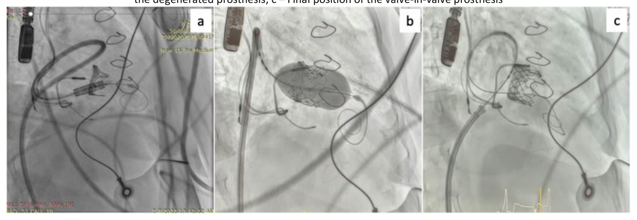
After several maneuvers, a 90-degree clockwise turn allowed us to obtain a loop into the RV as seen in Figure 1 (a), that stabilized the system and proved to be useful to cross with adequate alignment the degenerated valve, 3 mm below the ventricular edge of the surgical valve frame. A very slow inflation during implantation was performed. This gave us the possibility in the last part of the implant, when the valve tended to protrusion into the RV, to control the position and obtain a good final result as observed in Figure 1 (b, c). The procedure was finalized with the closure of the implanted Proglide suture. There was a good postprocedural result with no para-valvular leak and a mean gradient of 2 mmHg as observed in Figure 2. No vascular complications were encountered during and after the procedure.
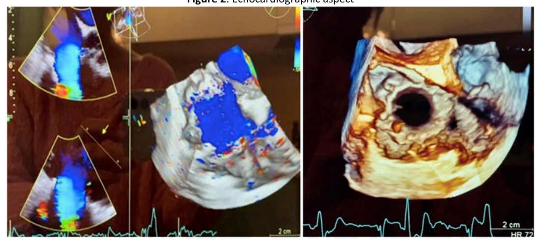
The patient had a favorable postprocedural evolution with symptom improvement. She was discharged on the 4th postprocedural day. At 1 month follow-up the patient had good clinical status (NYHA functional class II). Biologically, there was a significant decrease in natriuretic peptides and normalization of the hepatic markers. On echocardiography, there was no para-valvular leak, with a stationary mean gradient of 2 mmHg.
With the technical evolution of percutaneous valvular systems and clinically proven real benefits of transcatheter aortic valve implantation, a new treatment option has become available for patients with inoperable tricuspid valve stenosis [9]. The calcified degenerated native valves have a rough inner surface that acts as an effective anchor for the prosthesis [10]. The irregular inner surface and metal frame of a degenerated bio-prosthetic have similar characteristics and form an ideal docking station for a transcatheter valve. This has allowed valve-in-valve implants, mostly in the aortic position [11-13]. The first case of transcatheter valve-in-valve tricuspid valve replacement was described by Hon et al [14]. Their approach was through a thoracotomy and direct puncture of the right atrium [14]. This has definite advantages regarding the delivery of the device but remains more invasive. The first transcatheter tricuspid valve-in-valve procedure was performed by Leen van Garsse, aided by an extracorporeal circulation machine [15]. Today, with the aid Edwards Sapien valve system, the procedure result is similar and offers real benefits in severe clinical cases.
A successful procedure is highly dependent on the preprocedural evaluation of the patient. A good selection is mandatory in out-of-label cases. Confirmation of the tricuspid stenosis can be made by echocardiography with additional data from the trans-esophageal evaluation. A transvalvular gradient across the tricuspid valve of more than 5-10 mmHg at a heart rate of 70 beats per minute suggests severe stenosis. Gradients are affected by the patient’s current hemodynamic status and evaluation needs to be performed with the patient under maximal medical therapy. Additional parameters may be evaluated: a pressure half-time of more than 190 msec, right atrial enlargement, and plethoric inferior vena cava suggest severe TS. Right ventricular performance is exceedingly difficult to assess after surgical tricuspid valvular replacement. Published data suggests that TAPSE and S’ wave may be inaccurate parameters when we need to evaluate right ventricular function, at least in the first year after surgery, being highly dependent on the load volume [16]. However free wall longitudinal strain is a valid parameter even after immediate surgery. The mechanism of the prosthesis failure can be analyzed by echo and additional data on the stability of the surgical implanted prosthesis and paravalvular leak are mandatory to evaluate the procedural success. Also, clinical and echocardiographic assessment needs to eliminate the presence of acute endocarditis which is often caused by bacteria entering the bloodstream, adhering to damaged or abnormal heart tissue, and forming colonies [17-19].
The sizing of the valve is made considering three aspects: the dimensions and type of the surgically implanted valve with the aid of the ViV software developed by the Minneapolis Heart Institute Foundation, the Angio-CT sizing, and the echocardiographic findings. As we demonstrated in this case the presence of hepatic failure appears to regress after the treatment, and this preprocedural condition should not be a contraindication.
The presence of cardiac stimulation leads may need judgment before the procedure, but several case reports suggest that the presence of a transvalvular lead in a valve-in-valve tricuspid procedure is safe and does not affect the functionality of the pacemaker. In our case, however, the ventricular lead was positioned in the coronary sinus, a position that reduced the risk of electric disturbances and paravalvular leak after the implant.
The procedure is done by general anesthesia considering the high dependency of the atrioventricular plane during positioning and implant of the valve with the respiratory activity, general anesthesia allows the control of respiratory movements with apnea if needed during the implant. Conscious sedation and local anesthesia may be considered in selected cases when good anatomy indicates a straightforward procedure.
There are three ways for valve delivery: the transfemoral, transjugular, and direct surgical trans-atrial approach. The decision is made considering the anatomy of the tri-cuspid plane: the valve is vertical, and the transfemoral approach is indicated. Nowadays, with the complex features of the systems that allow us flex movements, the trans-atrial and transjugular approaches are almost abandoned, transfemoral approach allowing good crossing and alignment in the valve [20]. The transjugular approach may remain a good option in case of inferior vena cava thrombosis. The trans-atrial approach is nowadays considered too invasive and is almost abandoned.
The transfemoral approach that we perform is a gradual one, with a first 7F approach, one Proglide suture implanted, and an 8F sheath insertion. As demonstrated in MitraClip studies [21], pre-closure of the femoral vein with Proglide suture is feasible and safe for the 14-16 F access needed for a tricuspid valve-in-valve procedure, considering the pressure and volume vein overload status before the valve treatment.
The insertion of the 16 F large system sheath is performed by gentle movements on a stiff wire, with the positioning of the sheath 5 to 10 cm below the entrance into the right atrium to allow free movement without tension of the system.
The crossing of the valve may be done with the aid of an angulated catheter (JR or vertebral catheter) with a regular J wire, a strait wire, or a hydrophilic wire depending on the degree and anatomy of the valvular complex. After the passage into the right ventricle, two final positions of the wire can be chosen: one in the apex of the RV and another one in the pulmonary artery. There are significant differences between the two positions: the RV position has a higher risk of complications by ventricular rupture and pericardial effusion but allows a better coaxially of the system in the valve with better control during the implant; the pulmonary position may give better support with the minus of poor coaxially and a higher risk of distal pulmonary perforation with pulmonary hemorrhage.
The more vertical the tricuspid plane is, the more the RV position is indicated. The positioning of the stiff wire should be made on a pigtail catheter to diminish the possible trauma on the RV muscle.
If many studies proved the benefit of a direct implant in the aortic position without pre-dilatation, in a tricuspid position during a valve-in-valve procedure we consider that pre-dilatation with a similar-sized balloon is mandatory to anticipate the behavior of the system in the valve, to facilitate the passage through the valve with the system and to anticipate the movement of the system during the gentle inflation.
As we already anticipated for the Edwards systems the insertion should be made opposite to an aortic implant so that the flex movement is possible on the left and not on the right side. This allows typical anatomies to align and cross into the valve without major difficulties. As we presented in our case prolapse of the wire into the right atrium (RA), especially in enlarged anatomies, is possible and in such case, there are two strategies to be tried: one is using a stiffer wire such as Lunderquist Extra-stiff wire by Cook Medical, eventually positioned into the PA- this strategy needs to be prepared in advance because once the system is inserted repositioning of another wire is virtually impossible. If this doesn’t allow the positioning of the system in the TV the technique that we used was a gentle two-people synchronized maneuver with the return of the system in an aortic position, developing a loop with the wire in the RA and crossing the valve from the lateral wall of the RA using the loop as a stabilizer to increase pushability (a maneuver similar for the right heart catheterization by femoral approach in severe tricuspid regurgitation (TR) with the loop-technique with the Swan-Ganz catheter).
Once the system is positioned there is no need for rapid pacing during inflation, but several aspects may be considered: the inflation needs to be very slow to allow small adjustments, especially in the last part of the inflation when the rigidity of the full inflated balloon may change the position of the valve in the AV plane. Small movements of the wire and the system in slow inflations can achieve perpendicular lines on the valvular plane with a perfect result. Also, during implantation, apnea may help in maintaining the position.
We consider that transesophageal echocardiography (TEE) during the implant is mandatory to evaluate the result of the procedure, and the presence of the paravalvular leak, and evaluate complications such as pericardial effusion. There is no consensus about an antithrombotic treatment after a valve-in-valve procedure in the tricuspid position. We believe that the low regimen pressure of the right heart indicates us to consider oral anticoagulant treatment (OAC) treatment (without consideration of the presence or absence of sinus rhythm) associated with at least 6 months of clopidogrel. The regimen should be adapted to the patient, evaluating the bleeding risk. This strategy is highly debatable and further studies are needed to define the correct antithrombotic treatment.
In conclusion, tricuspid valve-in-valve implantation using the Edwards SAPIEN valve system is a feasible and effective procedure for selected patients with failing bio-prosthesis where redo open heart surgery presents considerable risk. The procedure proved a good clinical improvement for the patients in NYHA III or IV functional class at baseline, with consistent results at 1-year follow-up. Further studies are needed to analyze patient selection, implant technique, results, and antithrombotic treatment following a valve-in-valve procedure in the tricuspid position.
The authors declare no conflict of interest. No artificial intelligence automatically generated text was inserted in this manuscript, and no image was previously published in another journal or is under consideration of being published elsewhere. This research received no external funding.
Conceptualization, H.M. and M.R.; methodology, H.M., V.A.I., A.G., E.N., G.G. and M.R.; software, A.G., E.N., A.B., A.Z., M.S.S. and A.B.; validation H.M., V.A.I., A.G., E.N., A.B., A.Z., M.S.S., G.G., M.R. and A.B.; formal analysis, H.M., V.A.I., A.G., E.N., A.B., A.Z., M.S.S., G.G., M.R. and A.B.; investigation, H.M., V.A.I., A.G., E.N., A.B., A.Z., M.S.S., G.G., M.R. and A.B.; resources, H.M., A.B., M.R. and A.B.; data curation, H.M., V.A.I., A.G., E.N., G.G. and M.R.; writing—original draft preparation, H.M., A.G., A.B., A.Z., M.S.S., M.R. and A.B.; writing—review and editing, H.M., V.A.I., A.G., E.N., A.B., A.Z., M.S.S., G.G., M.R. and A.B.; visualization, H.M., V.A.I., A.G., E.N., A.B., A.Z., M.S.S., G.G., M.R. and A.B.; supervision, H.M., V.A.I. and G.G.; project administration, H.M., G.G. and M.R; All authors have read and agreed to the published version of the manuscript.
Not applicable.
Informed consent was obtained from all subjects involved in the study.