
In this era, bacterial resistance to antibiotics is considered the most important health problem in developed and developing countries. Antibiotic resistance is a phenomenon in which bacteria become resistant to the effects of antibiotics and make the treatment of infections caused by these bacteria difficult or impossible [1]. Antibiotic resistance can arise from a variety of mechanisms, including preventing drug access to the bacterial site of action, changes in the structure and protection of antibiotic receptors, and direct modification or inactivation of antibiotics. Excessive and incorrect use of antibiotics has led to antibiotic resistance, which is a growing global health crisis [2]. According to the currently available studies, antibiotic resistance in Staphylococcus aureus (and Escherichia coli strains has increased significantly in patients but also in preclinical populations. Today, the resistance of different strains of these bacteria against penicillin, gentamicin, clindamycin, tetracycline, and vancomycin has been reported in various studies [3,4]. Therefore, due to the increase in antibiotic resistance, as well as the high cost of producing new antibiotics, the lack of new antibiotics entering the market, and environmental problems, there is an urgent need to produce, discover, design, and increase the availability of alternative drugs for antibiotics around the world [5,6].
Probiotics and their derivatives are among the alternative medicines that have been studied recently. Until today, the antibacterial effects of these alternative agents have been studied on many antibiotic-resistant bacteria, including Escherichia coli, and the results of these studies have been very promising. It has also been found that these factors are very effective in treating various diseases, such as necrotizing enteritis, periodontitis, and diarrhea resulting from bacterial infection [7,8]. Also, the favorable effects of probiotics on the body, including improving immune system function, reducing inflammation, improving digestion, reducing the risk of bacterial diarrhea, and improving cardiovascular system function, and their significant positive effects in livestock and poultry have attracted scientists’ attention to
probiotics and their derivatives [9,10]. Lactobacillus is a genus of the Lactobacillaceae family. Lactobacilli are the most important food fermenters and are used in most cases as part of the natural flora or as starter cultures. This genus of bacteria produces lactic acid along with Leuconostoc, Pediococcus, Lactococcus, and Streptococcus, and for this reason, they belong to lactic acid bacteria or LAB. The size of lactobacilli varies in different species. Lactobacilli have different nutritional requirements for vitamins, amino acids, etc. Gram-positive bacilli are catalase-negative, lack cytochrome oxidase, their metabolism is fermentative, and they can survive in the presence of oxygen; because they have peroxidase activity to inactivate hydrogen peroxidase, they are therefore microaerophilic or facultative anaerobes. Most lactobacillus species are non-motile and have peripheral flagella when motile. They are present in the digestive system of healthy humans and reduce the acidity of the environment by converting sugar into lactic acid and are a small part of the intestinal flora [11]. Lactobacilli produce various substrates with antimicrobial properties, such as hydrogen peroxide, organic acids, inhibitory enzymes, antimicrobial compounds, bacteriocin, and biosurfactants [12]. Lactobacilli are also used in the pharmaceutical industry to produce pharmabiotics, which are probiotics used for therapeutic purposes. In addition, lactobacilli are used in the dairy industry to produce fermented foods such as cheese and yogurt [13,14]. They are also used as a biological preservative due to their strong antimicrobial activity against many microorganisms [12]. Lactobacillus rhamnosus is a probiotic bacterium of the Lactobacillus genus that has been studied for its potential health benefits. For example, Lactobacillus rhamnosus GG is a strain of Lactobacillus rhamnosus that has been shown to have good therapeutic properties and can be used as a spray in spray devices [13]. Lactobacillus rhamnosus is one of the four probiotics that have been studied to limit their growth in different environmental conditions. This study showed that Lactobacillus rhamnosus is able to tolerate a wide range of stressful conditions. This feature of heat tolerance is one of the prominent features of this bacterium [15]. Also, a controlled clinical trial showed that probiotic supplementation in early pregnancy with Lactobacillus rhamnosus HN001 may reduce the incidence of gestational diabetes [16]. The therapeutic properties of this bacterium do not end here, as Lactobacillus rhamnosus Lcr35 is a strain of Lactobacillus rhamnosus that is used to treat diarrhea and prevent vaginitis [17].
Bacteriocins are peptides or bacterial proteins that are synthesized in the ribosome and have antimicrobial properties [18]. Bacteriocins perform their antimicrobial action by binding to the surface receptors of the target bacteria, and their function depends on the strain. Destruction of target bacteria occurs as a result of metabolic, morphological, and biological changes caused by bacteriocins [19].
Biosurfactants are amphiphilic biological compounds produced extracellularly or as part of cell membranes by various microorganisms. Commercially, biosurfactants are of special importance due to their use in various industries such as petroleum, petrochemical, food, pharmaceutical, cosmetic, agriculture, textile, papermaking, leathermaking, etc. For this reason, the microorganisms that produce these compounds are suitable candidates for the expansion of biosurfactant production due to their high surface-to-volume ratio and diverse biosynthetic capacity [1].
In medicine, swarming movement and biofilm formation are important actions in surface colonization by bacteria and increase the possibility of infections. Biosurfactants are produced to prevent pathogenic organisms from sticking to solid surfaces or to infection sites. Surfactin reduces the amount of biofilm formation. Accordingly, this study is conducted to isolate and purify bacteriocin and biosurfactant from Lactobacillus rhamnosus and investigating its antibacterial and anti-biofilm activity [2].
The standard strains of Escherichia coli (ATCC25922), Staphylococcus aureus(ATCC25923), and Lactobacillus rhamnosus PTCC1607 (ATCC7469) were lyophilized from Iran’s Fungi and Bacteria Collection Center, Iran Scientific and Industrial Research Organization.
Bacterial biosurfactant was prepared according to the method of Emmanuel et al. [19]. First, the bacteria were cultured on MRS agar medium and incubated for 48h at 37°C. Then, the single colonies created were transferred to MRS Broth medium and cultured. After 24h of incubation at 37°C, the tube containing bacteria was centrifuged at 8000 rpm at 4°C for 30 min. The supernatant was collected and analyzed for biosurfactant separation [20].
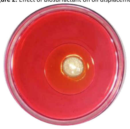
In order to perform the oil spreading test, one milliliter of oil was added to the surface of 20 milliliters of distilled water. Then, 0.5 ml of bacterial supernatant was added to it. The formation of a clear area is a sign of the presence of biosurfactants in the supernatant [19].
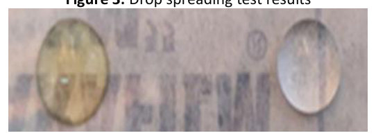
To test the droplet spreading, the first 25µl of biosurfactant was added as a drop on the parafilm, and the droplet of the desired substance was observed to spread on the parafilm. Similarly, 25µl of water was added to the parafilm, and changes were observed. Due to the low surface tension, the biosurfactant spreads and flattens on the parafilm while the water remains unchanged [19].
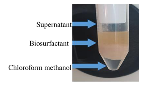
The supernatant obtained after centrifugation was collected, and its pH was adjusted to 2 using normal HCL 6. The supernatant was
kept overnight at 4°C. An equal amount of mixture of chloroform: methanol (1:2) was added to the supernatant and mixed. Then, the resulting mixture was kept overnight for phase separation. Three layers were formed in the desired phase mixture – the lower layer is chloroform: methanol, the middle layer is biosurfactant, and the upper layer is the supernatant. The formed biosurfactant (middle layer) was carefully removed with the help of a micropipette and transferred to appropriate microtubes. Then 1 ml of distilled water was added to the microtubes containing biosurfactant and vortexed completely. This mixture was centrifuged at 7000 rpm for 30 minat 4°C. The supernatant was discarded and the obtained pellet was dried for 24h and separated from the supernatant as a precipitate [20].
Bacteriocin extraction was performed according to the protocol of Anjana et al. [21]. First, the desired strain was inoculated in an MRS Broth culture medium (with a pH of 4) and incubated at 37°C for 48h at 150 rpm in order to reduce the pH. The inhibitory activity of organic acids produced in the fermentation medium was neutralized by 4N sodium. To remove bacterial cells, it was centrifuged at 5000rpm for 15 min. The supernatant now contains bacteriocin, the activity of which was examined. [22].
In this study, the bacteriocin was separated using two different methods:
In this method, first, the resulting supernatant was mixed with cold 2-propanol (isopropyl alcohol) 50% by volume in the vicinity of ice. Then, the resulting mixture was centrifuged at 4 degrees with 15000rpm for 40 min. Finally, the sediment obtained was dissolved in citrate phosphate buffer (final concentration 0.1 M and pH 4), and its bactericidal activity was investigated [22].
In this method, first, the obtained supernatant was precipitated with the help of ammonium sulfate at 4 degrees Celsius. The resulting sediment was centrifuged with the previous conditions. Then, the precipitate was dissolved in potassium phosphate buffer (final concentration 0.06 M and pH 7), and finally, its bacteriocin activity was checked [22].
The sample obtained from the previous step was dialyzed for 48h with potassium phosphate buffer (0.06 M and pH=7, at 4 degrees Celsius), and then using Nessler’s reagent, the removal of ammonium ions from the solution was proven. Bacteriocin activity was evaluated against the biofilm of sensitive strains.
For this purpose, first, 250, 500, and 1000mg of the obtained biosurfactant were dissolved by dimethyl sulfoxide with a concentration of 0.1% (1000 μl/liter), and the bacteriocin obtained was dissolved by potassium phosphate buffer (final concentration of 0.06 M and pH 7). Three different concentrations of 250, 500, and 1000 mg/ml were prepared from each of them. Then, uniform paper discs with a diameter of about 5 mm were prepared and then sterilized. Then 0.5 McFarland concentration was prepared from Staphylococcus aureus and Escherichia coli. With the help of the sterile swap and grass cultivation method, the desired pathogens were cultured on Müeller Hinton agar culture medium. After cultivating the desired bacteria on the Müeller Hinton agar culture medium, sterile paper discs impregnated with 50 µl of biosurfactant or bacteriocin were placed in certain places on the Müeller Hinton agar with the help of forceps. The desired plates were examined after incubation at 37 degrees Celsius for 24 hours in order to form a halo of non-growth around the paper discs [23].
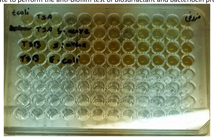
The 96-well plate was used to perform this test. At first, the overnight culture of standard Staphylococcus aureus and Escherichia coli was prepared in TSA culture medium enriched with 0.2% glucose, and then the resulting single colonies were prepared in TSB culture medium enriched with 0.2% glucose to prepare a suspension with light absorption. 0.1 was used at 625 nm wavelength.
In the 96-well plate, one row was assigned to Escherichia coli and one row to Staphylococcus aureus. For each row, one column was assigned to the positive control (containing 200 µl of bacterial suspension) and one column to the negative control of 200 µl of TSA culture medium enriched with 0.2% glucose. Also, 100 µl of bacterial suspension was added to the column of each row and 100 µl of different concentrations of bacteriocin and biosurfactant (250, 500, and 1000 mg/ml) were added to each column. Then, the 96-well plate was transferred to the incubator and incubated for 24h at 37°C.
After the incubation period, the contents of the wells were aspirated with the help of a sampler, then re-aspiration of the wells was done first with the help of PBS buffer and then with sterile physiological serum. In the next step, 150 µl of pure methanol was added to the wells and aspirated after 10 min.
In the next step, 200 µl of 1% crystal violet was added to each well, and after 20 minutes, was aspirated, and the plates were washed with tap water. After drying the plate in the vicinity of free air, 150 µl of 33% glacial acetic acid was added to each well, and with the help of an ELISA reader, the optical absorption of the plate was measured at a wavelength of 625 nm.
In order to calculate the biofilm inhibition percentage by bacteriocin or biosurfactant, the formula ({100 × (optical absorbance of positive control well/optical absorbance of wells containing bacterial suspension and bacteriocin or biosurfactant)}-100 = inhibition percentage) was used (Figure 1) [24].
Statistical Program for Social Sciences (SPSS) software, version 17 (SPSS Inc., Chicago, IL, USA), was utilized for data analysis. All the data of this study were expressed as mean ± standard deviation. Also, one-way analysis of variance and Tukey’s test were used for data analysis.
Based on the results of the oil spreading test, adding biosurfactant to the oil caused the oil to move and form a transparent area of about 2 cm in the oil (Figure 2).
The results of this test showed that the biosurfactant was spread and smoothed, which indicated the low surface tension of the biosurfactant, while the water drop did not move (Figure 3). The results of this test together with the results of the previous test show the correct screening of biosurfactant.
The liquid containing 3 phases, according to Figure 4, was used to purify the biosurfactant. Biosurfactant pellets were also obtained based on the above method.
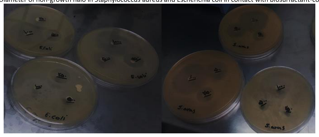
In the present study, the antibacterial properties of biosurfactants extracted by the disk diffusion method were investigated. Based on the creation of a non-growth halo around the discs containing biosurfactant, it was concluded that this substance has antibacterial properties. The diameter of the halo of non-growth around the disks by the separation of Staphylococcus aureus and Escherichia coli (Figure 5) studied, and the series carried out is included in Tables 1, 2, and 3.
| The halo diameter in the third series | The halo diameter in the second series | The halo diameter in the first series | Biosurfactant concentration (mg/ml) |
|---|---|---|---|
| 15 mm | 20 mm | 20 mm | 250 |
| 20 mm | 25 mm | 30 mm | 500 |
| 40 mm | 40 mm | 50 mm | 1000 |
| The halo diameter in the third series | The halo diameter in the second series | The halo diameter in the first series | Biosurfactant concentration (mg/ml) |
|---|---|---|---|
| 21 mm | 20 mm | 20 mm | 250 |
| 20 mm | 25 mm | 20 mm | 500 |
| 40 mm | 35 mm | 30 mm | 1000 |
| The halo diameter in the third series | The halo diameter in the second series | The halo diameter in the first series | Bacteriocin concentration (mg/ml) |
|---|---|---|---|
| 14 mm | 21 mm | 20 mm | 250 |
| 19 mm | 26 mm | 28 mm | 500 |
| 38 mm | 42 mm | 49 mm | 1000 |
The results of reading the 96-well plate and evaluating the inhibition percentage of biofilm formation showed that this substance has good anti-biofilm properties. The results of the reading of the ELISA device and the inhibition percentage of biofilm formation are included in Table 4.
| Escherichia coli | Staphylococcus aureus | Biosurfactant concentration (mg/ml) |
|---|---|---|
| 0.36 | 1.07 | 250 |
| 2.15 | 5.96 | 500 |
| 71.75 | 53.3 | 1000 |
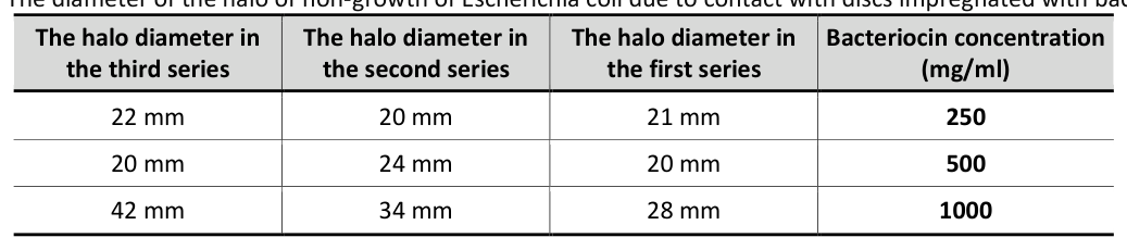
The diameter of the non-growth halo around the discs impregnated with bacteriocin was measured as an indication of bacteriocin’s antimicrobial activity. The diameter size of each halo of non-growth of Staphylococcus aureus and Escherichia coli is included in Table 3 and Table 5, respectively. Also, the images of the diffusion test results from the growth disk of Staphylococcus aureus and Escherichia coli can be seen in Figure 6, respectively.
| The halo diameter in the third series | The halo diameter in the second series | The halo diameter in the first series | Bacteriocin concentration (mg/ml) |
|---|---|---|---|
| 22 mm | 20 mm | 21 mm | 250 |
| 20 mm | 24 mm | 20 mm | 500 |
| 42 mm | 34 mm | 28 mm | 1000 |
By examining the results obtained from the reading of the ELISA device, it was found that Lactobacillus rhamnosus bacteriocin has anti-biofilm properties; these results on biofilm inhibition by bacteriocin are included in Table 6.
| Escherichia coli | Staphylococcus aureus | Biosurfactant concentration (mg/ml) |
|---|---|---|
| 5.06 | 7.74 | 250 |
| 46.32 | 16 | 500 |
| 76.65 | 58.9 | 1000 |
In this study, the biosurfactant and bacteriocin of Lactobacillus rhamnosus extract and their antibacterial and anti-biofilm properties were investigated in dilutions of 250 and 1000 mg/ml. The diameter of the halo of non-growth of Escherichia coli in contact with the disc impregnated with bacteriocin in concentrations of 250, 500, and 1000 mg/ml was 21 ± 1, 21.33 ± 2.3 and 34.66 ± 7.02 mm, respectively, and in Staphylococcus aureus was evaluated as 18.33 ± 3.78, 24.33 ± 4.72 and 43.66 ± 4.72 mm, respectively. The biofilm inhibition percentage of this material was for Escherichia coli in the three mentioned concentrations, respectively, 5.06, 46.32, and 76.65, and for Staphylococcus aureus in the three mentioned concentrations, respectively, 7.74, 16, and 58.9. Also, in this study, the diameter of the non-growth halo resulting from the diffusion test for biosurfactant around Escherichia coli in three different concentrations of 250, 500, and 1000 mg/ml was 20.33 ± 0.57, 66 ± 2.88, 21.21 and 35 ± 5 mm, respectively and in Staphylococcus aureus, it was evaluated as 18.33 ± 2.88, 25 ± 5, and 43.33 ± 5.77, respectively. Also, the percentage of biofilm inhibition was measured for Escherichia coli as 0.36, 2.15, and 71.75% in the three mentioned concentrations, and for Staphylococcus aureus, 1.07%, 5.96%,
and 53.3%, respectively. Based on the obtained results, it was found that the biofilm inhibition effect of bacteriocin and biosurfactant at a concentration of 1000 mg/ml on Escherichia coli was higher than on Staphylococcus aureus. Therefore, bacteriocin and biosurfactant of the desired bacteria have antibacterial and anti-biofilm properties against standard bacteria Escherichia coli and Staphylococcus aureus. The anti-biofilm properties of biosurfactant and bacteriocin extracted in the present study are quite promising and remarkable based on the obtained results. These results are consistent with the results of different studies that have reported anti-biofilm properties.
The results of the study conducted by Leslie et al. in 2021 show that bacteriocin isolated from Lactobacillus plantarum has antibacterial properties against Staphylococcus aureus [25], which is consistent with the present study. Also, the antibacterial activity of class III bacteriocin from Lactobacillus acidophilus against Staphylococcus aureus and Escherichia coli was also reported by Meng et al. in 2021 [26]. Also, in the study of Mohapatra et al. in 2021, the anti-biofilm property of bacteriocin obtained from Lactobacillus plantarum bacteria was investigated on Staphylococcus aureus and Pseudomonas aeruginosa. The results of this study indicated that this bacteriocin has anti-biofilm properties against these bacteria [27]. Inhibition of Escherichia coli biofilm formation by bacteriocin BM1157 was evaluated by Luo et al. in 2021. Based on the findings of the mentioned study, this bacteriocin has the ability to inhibit the biofilm formation of this bacterium [28]. Also, according to recent studies, bacteriocin obtained from Lactobacillus bovis can also inhibit the formation of Escherichia coli biofilm [29]. Escherichia coli biofilm inhibition is not limited to bacteriocins derived from lactobacilli. According to the study conducted by Shanks et al. in 2012, the bacteriocin isolated from Citrobacter freundii also has the ability to inhibit the formation of Escherichia coli biofilm [30]. In addition to Staphylococcus aureus and Escherichia coli, the antibiofilm properties of different bacteriocins have also been performed on other bacteria. In a study conducted by Menif et al. in 2013, the results proved that the biosurfactant produced by Bacillus subtilis SPB1 has antimicrobial activity and can be produced and optimized at an industrial level [31]. Also, the antibacterial activity of the biosurfactant produced by Streptomyces cellulase was recently reviewed by Panatola et al. In the mentioned study, the antibacterial property of Streptomyces cellulase probiotic biosurfactant against Escherichia coli and Staphylococcus aureus was reported [32].
The important point in the results of these reports is their agreement with the results of the present study, which shows that biosurfactants can be considered as a potential antibacterial substance. Also, it seems that in addition to Lactobacillus rhamnosus, the biosurfactant of other existing probiotics can also be studied and used.
The anti-biofilm property of this substance is one of the other important things mentioned in the studies. Considering the fact that the formation of biofilm is very effective in the rate of survival, pathogenicity and resistance to antibiotics, obtaining anti-biofilm substances is of double importance [33]. The results of similar studies show that biosurfactants have good anti-biofilm properties. A clear example of this is the study conducted by Ednan et al. in 2023, which shows that the biosurfactant extracted from Lactobacillus acidophilus has a high anti-biofilm effect against Gram-negative pathogenic bacteria such as Pseudomonas aeruginosa, Serratia marcescens, and Chromobacterium violaceum [34].
Meanwhile, Lactobacillus rhamnosus has been studied more than other Lactobacillus in terms of biosurfactant production and its various antibacterial properties. The high ability of biosurfactants obtained from this bacterium to destroy bacteria and its easy engineering for large-scale production has attracted the attention of scientists to this substance extracted from Lactobacillus rhamnosus [35]
The high anti-biofilm biosurfactant property of this bacterium has been investigated in various studies. For example, Lactobacillus rhamnosus glycolipid biosurfactant was extracted, and its anti-biofilm properties were investigated by Patel et al. In the mentioned study, this material had high anti-biofilm properties against Staphylococcus aureus, which is consistent with the present study [35]. In a similar case, the anti-biofilm properties of this substance extracted from Lactobacillus rhamnosus against Enterococcus faecalis were reported by Walried et al. [36]. In the study conducted by Samabantamoorthy et al. in 2014, the biofilm inhibition percentage of Lactobacillus rhamnosus biosurfactant against Escherichia coli and Staphylococcus aureus was reported to be 80 and 93% with a concentration of 25 mg/ml of this substance at a wavelength of 595 nm [37]. Although there is a general agreement between the present study and the study conducted by Samabanthamurthy in terms of understanding the anti-biofilm properties of biosurfactants, the inhibition percentage of these two studies is not the same. This mismatch can be due to the difference in light absorption, the difference in biosurfactant concentration, the type of origin strain, and the amount of biofilm formation by bacteria. The study conducted by Nataraj et al. in 2021 is another example of the effect of Lactobacillus rhamnosus biosurfactant on eliminating biofilm and bacteria.
In this study, the effect of different concentrations of this substance on the biofilm and bacteria of Staphylococcus aureus ATCC25923 (the standard bacterium examined in this study) and Staphylococcus aureus resistant and sensitive to methicillin was investigated. Although the anti-biofilm property of this study is consistent with the current study, the important point is the difference in the diameter of the non-growth state of the present study from the previously approached study, where the diameter of these states was found to be larger.
This difference can be due to reasons such as the difference in Lactobacillus rhamnosus strains (in the mentioned study, the GG strain was exclusively used). The different methods of evaluating the lack of growth halo (in the mentioned study, the diffusion method from the well was used instead of the diffusion from the disk) and the difference in biosurfactant concentration may explain the discrepancies .
The results of this study showed that bacteriocin and biosurfactant isolated from Lactobacillus rhamnosus have high antibacterial and anti-biofilm properties that can be used as an alternative drug to antibiotics. Due to the increasing antibiotic resistance, it is necessary to use alternative drugs to deal with this phenomenon, such as the one suggested by the results of this study. Also, in the future, these solutions can have potential applications in approaching environmental problems, solving challenges within the food industry, and enhancing the tehrapeutic armamentarium to fight diseases.
The authors declare no conflict of interest. No artificial intelligence automatically generated text was inserted in this manuscript, and no image was previously published in another journal or is under consideration of being published elsewhere.
This article was extracted from the master’s thesis of Mrs. Roya Hamidi from the Islamic Azad University of Tabriz branch with the thesis number 102290793474175162620141 and the design with tracking code 162620141. The authors are grateful to the Islamic Azad University of Tabriz branch for their assistance in approving and implementing the project.
All authors participated in the presentation of the original idea and design, search for sources and review of articles, initial writing or revision of the article, and all agreed with the final version of the present article and took responsibility for the accuracy of the content.
Not applicable.