
The specialized literature lacks scientific studies on the functional evaluation and neuromusculoskeletal recovery in Lyme Disease (LD). Approximately 80 articles from the past decade are available in databases, primarily focusing on early detection and antibiotic treatment. No studies provide information on potential methods for functional evaluation or physical therapy-based recovery for musculoskeletal dysfunctions caused by inflammation secondary to Borrelia infection.
LD is caused by the bite of a tick infected with Borrelia. The disease progresses through three stages and can result in multisystem complications. When the nervous system is affected, it is referred to as neuroborreliosis, which may manifest as meningitis, mononeuritis multiplex, or cranial neuritis, including inflammation of the vestibulocochlear nerve [1]. Exclusive involvement of the vestibular system can lead to dizziness and balance instability [2]. Musculoskeletal symptoms can appear at any stage, including early localized infection, often resulting in patients being misclassified into the incorrect stage [3].
We describe motor control and the potential effects of systemic inflammation caused by borreliosis, followed by the stages of assessment and recovery, and the conclusions drawn from these steps.
Although the exact mechanism by which LD causes small fiber neuropathy is unknown, several potential mechanisms of neuronal damage are considered. These include the direct neurotoxic effect of the spirochete, neuroinflammation, and increased autoimmunity affecting the peripheral nervous system (PNS) with subsequent impacts on muscles, joints, and specific post-infection pains. However, a clear causal link between neurological symptoms and Borrelia infection cannot be
Among the genospecies of Borrelia burgdorferi sensu lato: Borrelia burgdorferi sensu stricto (s.s.), Borrelia garinii and Borrelia afzelii, Borrelia burgdorferi s.s. has the strongest potential to damage joints [5]. It is the most common cause of LD in the United States [3, 6], while infections with this pathogen are much less common in Europe [7-9]. The pathogenesis of LD is primarily driven by a strong inflammatory reaction [5], resulting in a perivascular collection of lymphocytes, macrophages, and scattered mast cells in the interstitial tissue, as typically observed in histopathological examinations of severe LD cases [9].
Previous studies have shown that late manifestations affecting the nervous system or joints can appear months or years after the initial infection, presenting symptoms such as fatigue, difficulty walking, posture disorders, and muscle pain. These symptoms are also present in the subject of our study, secondary to the diagnosis of borreliosis [10].
The examination and treatment of patients with motor control disorders are based on theories of motor control that guide the understanding of movement mechanisms [11-14]. In our study, we utilize systems theory as a foundation for clinical applications. Systems theory posits that movement arises from the interaction of various processes, including perceptual, cognitive, and motor processes, as well as the interactions between the individual, the activity, and the environment.
The severity of symptoms observed in all the patients with LD that we have evaluated has led us to pay more attention to this condition. As a result, our study focuses on evaluating and recovering a patient with LD using methods and applications informed by previous scientific studies. We analyzed a patient with sequelae following LD, including postural, walking, and static and dynamic balance disorders, muscle fatigue, changes in balancing synergies, walking base, and the projection of the General Center of Gravity (GCG). We conducted initial and final tests after a series of 10 and 20 sessions of specific physical therapy exercises.
Pioneers in the study of motor control and movement, such as Bernstein and Latash [15-17], along with Charles Sherrington (1857 – 1952), Thomas Graham Brown (1882-1965), and Walter Hess (1881-1973), have significantly contributed to our understanding of joint freedom, redundancy, and muscle-articular synergies. Charles Sherrington contributed to the understanding of inhibition, spinalmuscle connectivity, and the sensory-motor cortex. Thomas Graham Brown researched central movement programming and pattern generation mechanisms. Walter Hess studied the central coordination of the autonomic system and anticipatory motor behavior. Bernstein, recognized posthumously by Western colleagues, demonstrated a central hierarchy of postural control mechanisms and distributed, parallel processing of movement.
Bernstein also participated in developing theories of motor programs, recognizing that understanding neural control of movement requires knowledge of the system’s characteristics in response to external and internal stimuli [16]. His distributed model of motor control showed that central commands could produce different motions due to external forces and initial conditions. His student, Mark Latash, furthered this work by demonstrating that synergies evolve to ensure flexible and stable performance in motor activities [17].
Postural control for stability and body orientation in space requires a complex interaction between the musculoskeletal and neural systems. This includes joint amplitudes, intersegmental biomechanical relationships, muscle physiology, spinal flexibility, neuromotor processing, synergies, perceptual and sensory processes, and their central integration.
Research has concluded that postural control is not managed by a single system but depends on various intrinsic and extrinsic factors [18]. Initial studies highlighted the importance of maintaining the center of mass (COM) within certain limits for static posture. However, subsequent studies demonstrated that stability limits are not fixed and vary with individual characteristics or actions undertaken. When recovering balance, individuals tend to restore COM disturbances rather than segmental alignment disturbances, suggesting that COM is a key variable controlled by the central nervous system during postural control.
Further studies have shown that analyzing the relationship between the center of gravity (COG) and the center of pressure (COP) provides a better picture of stability [19-24]. Their interaction has been considered and used to measure and estimate posture control effectiveness in our study.
As actions have both stability and orientation components, and certain actions prioritize orientation over stability, research by Shumway-Cook & McCollum [25] and Horak & Macpherson [26] has demonstrated that stability and orientation vary with each action. The body can behave like an inverted pendulum, employing various strategies (ankle, hip) for both static and dynamic balance [21].
Bernstein’s assertion that “Movement coordination is the process of mastering the redundant degrees of freedom of a mechanism in motion” [16] encapsulates the essence of motor control theories used in our study.
While bipedal standing is characterized by minor postural imbalances, dynamic balance involves the integration of sensory information from the visual, vestibular, and proprioceptive systems in conditions of postural instability.
There are several theories about how sensory organization adapts to a changing environment, with two primary ones being: the intermodal theory of sensory organization by Stoffregen and Riccio [27], which posits that all sensory systems equally and continuously contribute to postural orientation; and the sensory weighting model, which suggests that the central nervous system adjusts the importance of each sensory system based on their accuracy for orientation and postural stabilization.
Torres and Oviedo [28] examined the structure and organization of muscle synergies for balance control, highlighting that a muscle can contribute to multiple synergies, each with a unique role. They demonstrated that muscle synergies can be continuously modulated by feedback signals to create postural adjustments, indicating that the central nervous system uses both pre-programmed synergies and continuous feedback control to regain stability.
Nashner et al. [29] investigated the selection of sensory information by the CNS for postural control by designing a dynamic platform with a moving visual wall to study sensory interactions in maintaining posture and balance. This approach allowed for the measurement and recording of posture and balance under various conditions.
Several studies on normative values using static platforms have examined the performance of normal subjects under varying sensory information conditions [29-33]. These studies have shown that adults and children over seven years old (the age of vestibular maturation) maintain balance well under all conditions of sensory disturbance.
The inability to maintain a stable static posture has significant functional consequences in neurological pathologies. It necessitates using the arms and hands for balance, detracting from daily activities [34-38]. Moreover, postural instability and the inability to stand on one leg for more than six seconds increase the risk of falls threefold.
Springer’s 2007 study [39] showed that balance disorders are prevalent in patients with specific diseases or traumas, particularly head injuries, peripheral neuropathies, or acquired neurological conditions. Neurological pathologies can disrupt one of the postural control strategies (e.g., loss of pre-programmed synergies) while leaving the other (continuous feedback control) intact, necessitating complementary studies to distinguish between these aspects and propose specific solutions.
For the main types of static/dynamic balance (postural control/anticipatory and reactive postural control), numerous clinical tests measure performance, including the unipedal stance and computerized posturography. Our study used such tests to assess the patient’s condition at the beginning and end of the recovery program.
Unipedal standing is a highly challenging situation for humans and allows researchers and clinicians to assess the effects of aging, disease, trauma, and the influence of multiple sensory systems. The single-leg stand test, which requires minimal equipment, measures balance aspects and shows abnormal values in conditions such as peripheral neuropathies and intermittent claudication. [35,36]
Various studies demonstrated the importance of maintaining unipedal posture and balance by examining corticomuscular coherence between the ankle joint and the sensorimotor cortex. This study showed that the sensorimotor cortex is directly involved in postural control during unipedal standing, compensating for high postural demands due to the low support base. Normative values in the literature indicate a significant decrease in unipedal stance time with age and pathology [40, 41].
Alain Berthoz et al. [20] demonstrated that during complex movements, the head is intermittently stabilized under gaze and fixation control, allowing it to act as an inertial guidance platform for controlling multisegmental movements. The otolithic organs, the saccule, and utricle, detect the linear accelerations of the head and provide a gravitational reference used not only to correct posture but also for perceptual actions requiring spatial orientation, including mental map rotation based on sensory information from all senses.
The interaction between motor and sensory processes is bidirectional, and the importance of sensory information for motor control was established many years ago. The traditional feedforward and feedback control mechanisms could not fully explain the coactivation of the alpha-gamma muscle loops, necessitating alternative theories such as the equilibrium point hypothesis and the hierarchy of synergies to explain how correct posture is maintained during external destabilizations [42].
Merton proposed a model that adjusts the equilibrium point, where the CNS controls both speed and position during rapid segmental movements. Degenerative, infectious, and metabolic diseases have disrupted the transmission of sensory information from the periphery to the CNS, affecting the parameters and components of posture and movement control. Disorders of nerve conduction from proprioceptors to the brain, as seen in neurological or diabetic patients, result in significant balance issues, making it difficult for patients to stand upright with their eyes closed on a static plane [42].
Posture, a term introduced by André Bregegère in 1955 [43], is defined as a momentary attitude or body position [44]. It involves the synergistic and coordinated action of the musculoskeletal system and the central and peripheral nervous systems to maintain body stability, balance, and constant relations between body segments and the environment [45-47]. Correct posture involves the alignment of all joints, and any misalignment can lead to muscle-joint imbalances [48].
Maintaining postural stability requires the co-participation and complementarity of actions from all support components, followed by motor actions (balancing, distribution of motor control tasks), which can be synchronous or asynchronous, simultaneous, or alternating depending on the demands [44,49]. Optimal body alignment in an upright position involves:
Head: Maintained by neck muscles. Insufficiency or habitual forward tilt increases muscle demand.
Trunk: Tends to fall forward due to its weight and the head’s weight, counteracted by vertebral muscles.
Shoulders: The scapular belt and upper limbs’ weight cause the shoulders to fall and scapulae to separate from the spine, counteracted by shoulder girdle muscles.
Pelvis: Key to maintaining correct posture. Anteversion is balanced by gluteal and posterior thigh muscles, whose hypotonia causes anterior trunk tilt. Retroversion is balanced by rectus abdominis, obliques, and iliopsoas muscles, whose hypotonia causes posterior tilt.
Lower limbs: Stability is ensured by plantar flexor muscles and triceps surae, especially the soleus [45].
Postural defects can lead to fatigue, muscle strain, and pain in later stages, potentially affecting vital organs, particularly in the abdominal region. Our study integrates current scientific conclusions and hypotheses to address parameters affecting motor control and static and dynamic postural control in our subject, considering the literature review.
Motor control theories are inherently unfinished, always allowing room for new information from recent studies on normal and pathological brain and PNS functioning. Neuroscientists identify the scientific basis for movement disorders, while clinicians develop applications of this research, as theories are considered more or less useful based on their problem-solving efficacy for patients with movement disorders.
In this context, we present a study where a patient with Lyme disease sequelae is compared to literature findings regarding posture, balance, and neuromuscular control of movement. Previous works by Schor D, Schor V et al.[50-52] introduced the BACKFIX Method, used for spine functional impairments in preventive care. This study evaluates its application to a complex disease like LD, characterized by neurological symptoms affecting balance, walking patterns, general movement control, and muscle strength imbalance.
We demonstrate the patient’s condition evolution using physical therapy with visual feedback on the rectangular force-amplitude model. Improvements in agonist-antagonist cooperation lead to enhanced muscle strength, functional muscle-joint balance of the spine, posture, and walking pattern. The patient exhibited muscle cramps in the calf and foot, pronounced thigh flexor fatigue during walking, insecurity in unipedal support, jerky walking with a wide support base, and reduced standing time in unipedal support.
We performed the following measurements:
We utilized the GPS System (Global Postural System) for computerized somatoscopy and computerized static bipodal balance tests, provided by UNEFS Bucharest (University of Physical Education and Sport from Bucharest), and the Ergocontrol System for musculoarticular functionality tests of the spine using the Backfix Method, technology provided by Medisport Company.
An initial evaluation and two intermediate evaluations were conducted. For the static one-legged balance test, we measured the hold time while standing on one leg. An initial evaluation and two intermediate evaluations were conducted. For the static one-legged balance test, we measured the hold time while standing on one leg
Computerized Somatoscopy Test
Evaluation of the Muscle-Articular Functionality of the Spine
The results of the computerized musculo-articular measurements regarding the muscular support of the spine are presented in Table 1. The movements were performed with difficulty and were jerky, which is characteristic of the neurological sequelae of the condition.
| Test segment | F(daN) | L(cm) | Lm(J) |
|---|---|---|---|
| Direct – abdominal Indirect – lumbar | 5.5 | 26 | 14.47 |
| Direct- Scapular girdle ½ anterior, thorax anterior Indirect – anterior and posterior muscle groups of the trunk | 9.31 | 49 | 45.45 |
| Direct- Scapular girdle posterior ½ of the trunk Indirect – anterior and posterior muscle groups of the trunk | 8.47 | 39 | 32.85 |
The recovery protocol involved 20 sessions of exercises utilizing visual feedback on force-amplitude curves, performed on the Ergocontrol System. This protocol aimed to restore agonist-antagonist cooperation within muscle groups and chains responsible for global body posture while also focusing on strength enhancement through movement control exercises targeting deficient muscle groups and chains.
Each session included walking exercises designed to integrate the improvements from the neuro-muscular movement control exercises, to enhance the walking pattern.
There was a noticeable improvement in overall body posture and a tendency towards optimizing the indices of musculo-articular functionality of the spine. The results from the intermediate evaluations demonstrate progressive improvements in all measured parameters, as detailed in Tables 2 and 3.
| Test segment | F(daN) | L(cm) | Lm(J) |
|---|---|---|---|
| Direct – abdominal Indirect – lumbar | 15.6 | 41 | 64.93 |
| Direct- Scapular girdle ½ anterior, thorax anterior Indirect – anterior and posterior muscle groups of the trunk | 181 | 61 | 110.29 |
| Direct- Scapular girdle posterior ½ of the trunk Indirect – anterior and posterior muscle groups of the trunk | 179 | 58 | 104.37 |
| Test segment | F(daN) | L(cm) | Lm(J) |
|---|---|---|---|
| Direct – abdominal Indirect – lumbar | 20.8 | 49 | 103.5 |
| Direct- Scapular girdle ½ anterior, thorax anterior Indirect – anterior and posterior muscle groups of the trunk | 21.5 | 66 | 140.7 |
| Direct- Scapular girdle posterior ½ of the trunk Indirect – anterior andposterior musclegroups of the trunk | 213 | 56 | 118.2 |
Spine Pathology and Accident Risk Scale:
• 0.50-0.75 = Medium Risk
Initial Personal Levels:
After Two Series of 10 Physical Therapy Sessions:
Chart number 1 illustrates the progress in muscle-articular functionality indices of the spine, specifically Thoracic Antero-posterior and Thoraco-lumbar, as assessed by Test 2 and Test 3. These improvements are also evident in the overall body posture, with notable
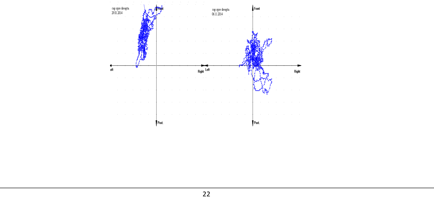
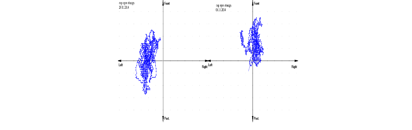
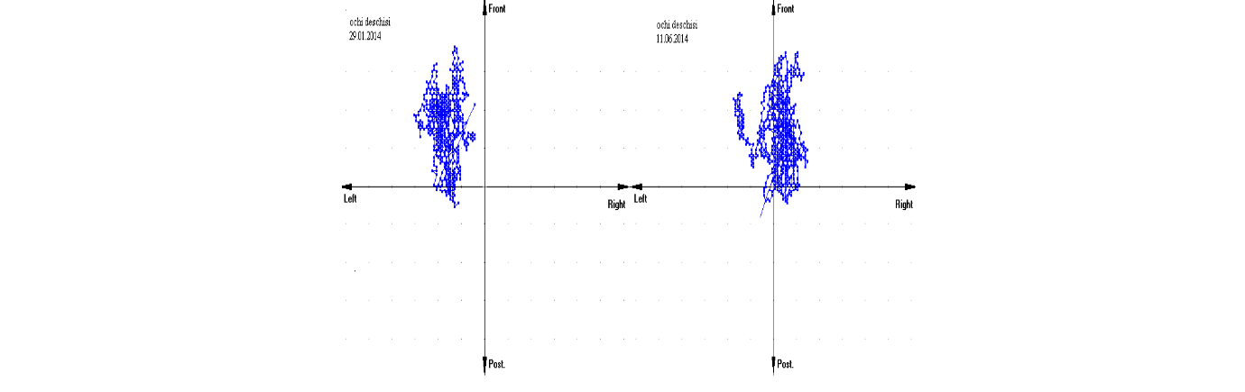
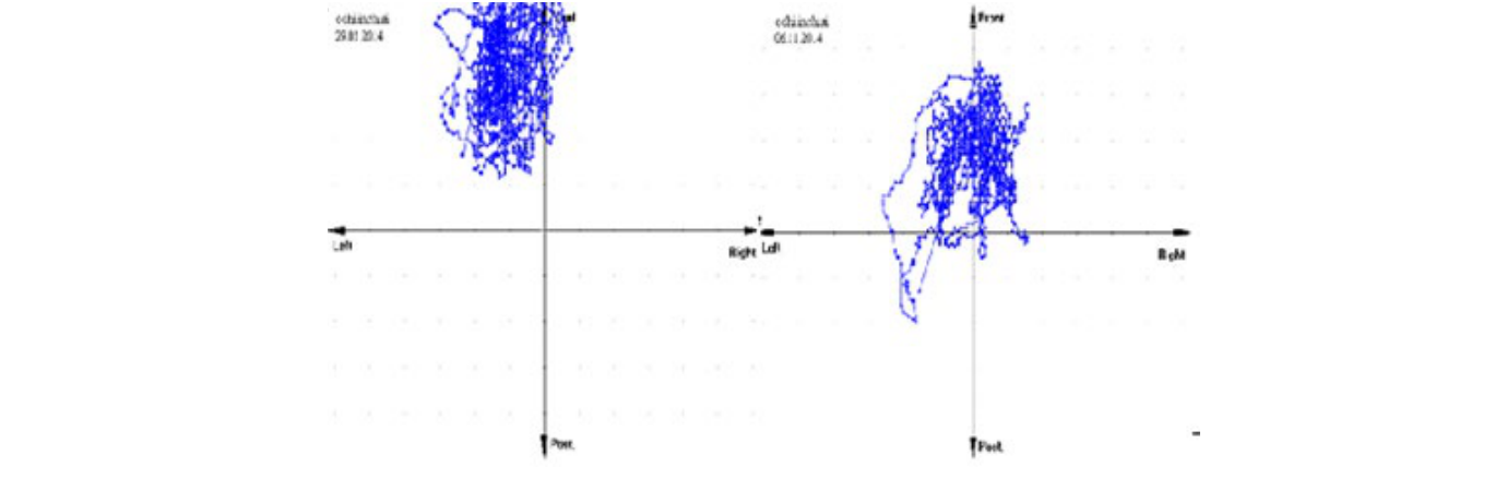
enhancements in the static balance and the projection of the GCG (see Figure 1).
| Segmental and inter-segmental ratio | Optimal values | Personal values 03.02.2023 | Personal values 27.02.2023 | Personal values 26.03.2023 |
|---|---|---|---|---|
| Thoracic anterior – posterior | 0.85 – 0.95 | 0.73 | 0.94 | 0.84 |
| Thoraco-lumbar | 0.85 – 0.95 | 0.31 | 0.59 | 0.73 |
Chart number 1: Indices of muscle-articular functionality of the spine – Thoracic antero-posterior and Thoraco-lumbar
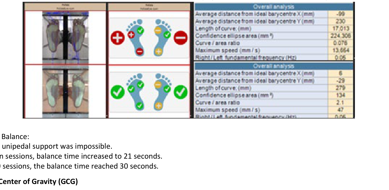
Projection: Figures 2, 3, 4, and 5 depict the improvement in the migration of the General Center of Gravity in four positions: standing with eyes open, standing with eyes closed, facing left, and facing right.
The key outcomes and benefits observed after completing two series of 10 physical therapy sessions in the study are presented in the following.
After Two Series of 10 Physical Therapy Sessions:
The BACKFIX Method:
• The risk of spinal pathology and accidents improved, moving from medium to low risk, with enhanced muscle chain synergies supporting proprioceptive and sensory system integration in unipedal stance.
The observed benefits from targeted physical exercises confirm previous studies on the uniqueness of bipodal and unipedal stance control, inter-segmental coordination, and the antigravity function of the leg extensors. Most cases of LD resolve after one course of therapy, although some patients experience gradual symptom relief even after treatment ends; therefore, follow-up assessments are recommended 3 months post-therapy. In some cases, symptoms persist despite antibiotic treatment, leading to the term “antibioticrefractory LD”, defined as persistent synovitis lasting more than 1 month after two four-week oral antibiotic courses or more than 2 months after intravenous ceftriaxone treatment [53].
Untreated arthritis may eventually resolve on its own, but it can persist for years, with some cases of LD lasting 7-8 years [54,55]. Potential treatments for patients with post-arthritis LD include non-steroidal anti-inflammatory drugs (NSAIDs), disease-modifying anti-rheumatic drugs (DMARDs), biologic agents, intra-articular steroids, and arthroscopic synovectomy [55,56].
The strengths of this study lie in its focus on the functional evaluation and rehabilitation of patients with Lyme disease-related neuromuscular issues, an area with limited prior research. The use of computer-assisted physical therapy, employing technologies like the GPS Posture-test System and BACKFIX Technology, allowed for a precise and objective assessment of balance and posture, leading to significant improvements in the patients. These technologies facilitated a more thorough understanding of musculoskeletal impairments and offered targeted interventions that improved both static and dynamic balance, posture, and muscle coordination.
However, some limitations must be noted. The rehabilitation was conducted on one patient, and this limits the generalizability of the results. Additionally, the long-term sustainability of the therapy outcomes remains uncertain, and further follow-up studies are needed to verify the persistence of improvements beyond the immediate post-treatment period. Another limitation is the lack of control, which would have strengthened the validation of the study’s findings.
Future research in this domain should focus on expanding the sample size and incorporating control groups for comparative analysis. Long-term studies are also crucial for understanding the chronic impact of rehabilitation techniques. Finally, integrating more advanced technologies, such as virtual reality or machine learning algorithms for personalized therapy adjustments, could further enhance patient outcomes in neuro-muscular recovery for Lyme disease.
This study demonstrates the effectiveness of computer-assisted physical therapy using technologies such as the GPS Posture-test System and BACKFIX Method in addressing the neuro-muscular and postural impairments associated with Lyme disease sequelae. Patients exhibited significant improvements in static and dynamic balance, posture, and musculo-articular functionality after completing two series of physical therapy sessions. The visual feedback provided during therapy sessions played a crucial role in restoring agonist-antagonist cooperation and enhancing motor control.
In conclusion, targeted physical therapy combined with advanced postural assessment technologies offers an effective means of improving functional recovery in Lyme disease sequelae patients, supporting their overall motor control and quality of life.
The authors declare no conflict of interest. The funders had no role in the design of the study; in the collection, analyses, or interpretation of data; in the writing of the manuscript; or in the decision to publish the results.
The current manuscript does not contain previously published materials or self-generated AI text.
Conceptualization D.S. and V.S.; methodology D.S.; software V.S.; validation D. S., L.G.E. and A.G.; formal analysis R.P.; investigation P.S.F.; resources S.S. and M.B; data curation R.R.G.; writing—original draft preparation D.S.; writing—review and editing L.G.E and A.G.; visualization R.R.G.; supervision L.G.E.; project administration D.S.; funding acquisition V.S. All authors have read and agreed to the published version of the manuscript elaboration.
The study was conducted in accordance with the Declaration of Helsinki, and approved by the Institutional Review Board of MEDISPORT Physical Therapy Clinic (11/30.03.2023).
Informed consent was obtained from all subjects involved in the study.