
Lymphedema treatment has significantly advanced in recent years, driven by technological innovations such as supermicrosurgery [1] and a deeper understanding of the disease’s pathophysiology [2,3]. Existing treatment algorithms are continually being re-evaluated [4], as is the optimal sequence of treatment modalities. Lymphedema treatment requires a multimodal approach that must primarily consider patient-related factors, including etiology, disease stage, individual anatomy, and medical history, as well as the experience of the treating physician and the technology available [2].
Patients with lymphedema strive for normalcy, and even compliant patients seek to minimize the time and effort required for compression therapy [5,6]. This is especially pertinent for highly functional areas, such as fingers, toes, hands, feet, and the head and neck, where treating the solid component of the disease poses considerable challenges [7].
PB Serum Medium is a synthetic bioengineered enzymatic mixture comprising three enzymes: lipase PB500, collagenase PB220, and lyase PB72K, produced by Proteos Biotech in Madrid, Spain. It has been utilized for treating lipodystrophic, scarred tissues, and localized, resistant adipose deposits.
Lymphedema is a progressive condition characterized not only by lymphatic fluid accumulation but also by structural tissue remodeling, including chronic inflammation, adipose deposition, and fibrosis. As the disease evolves, the relative contribution of this “solid component” increases, rendering the condition less responsive to therapies aimed solely at improving lymphatic drainage. This phenomenon is particularly evident in distal anatomical regions such as the hands, feet, and digits, where dense fibrotic changes and limited lymphatic reserve contribute to persistent deformity despite otherwise successful treatment.
Current management strategies follow a multimodal paradigm, combining complete decongestive therapy, physiologic microsurgical procedures such as lymphaticovenular anastomosis and vascularized lymph node transfer [1,3], and excisional approaches including liposuction. While these interventions are effective in reducing global limb volume, their ability to address localized, distal manifestations remains limited. Residual fibrosis and lipodystrophy in anatomically constrained regions continue to represent a significant therapeutic challenge and are frequently associated with persistent functional impairment and reduced patient satisfaction [4,8].
Pharmacologic and injectable therapies for lymphedema remain largely investigational, with no standardized or widely accepted agents available for clinical use [8, 9]. Among enzymatic approaches, hyaluronidase has demonstrated the capacity to degrade extracellular matrix glycosaminoglycans, thereby reducing interstitial resistance and facilitating fluid mobilization. In preclinical models, repeated hyaluronidase administration has also been associated with enhanced lymphangiogenesis in the context of vascularized lymph node transfer, suggesting a potential adjunctive role in lymphatic regeneration [10]. However, clinical translation remains limited, and its effects appear primarily directed toward the fluid component of lymphedema rather than established fibrosis or adipose tissue deposition.
Earlier investigations into systemic enzyme therapy provide indirect support for enzymatic modulation as a therapeutic strategy. Oral proteolytic combinations, including trypsin, chymotrypsin, bromelain, and papain, have demonstrated statistically significant reductions in limb volume in post-mastectomy lymphedema when compared to diuretic therapy [11]. Nevertheless, these studies predate contemporary multimodal treatment algorithms, lack anatomical specificity, and do not address localized, treatment-resistant disease. Furthermore, systemic administration limits the ability to achieve targeted effects within fibrotic or adipose-rich tissues.
Recent advances in lymphatic biology and regenerative medicine further support the rationale for localized therapeutic interventions. Experimental approaches such as nanofibrillar collagen scaffolds have shown the capacity to guide lymphatic regeneration [12], while molecular therapies involving VEGF-C and apelin signaling pathways have demonstrated reversal of established lymphedema in preclinical models [13]. Despite these promising developments, such strategies remain largely experimental and are not yet applicable in routine clinical practice. As a result, a therapeutic gap persists between established surgical techniques and emerging regenerative therapies, particularly in small, anatomically complex regions that are not amenable to further surgical intervention.
Within this context, locally administered, bioengineered enzymatic combinations represent a novel and mechanistically plausible approach. A multi-enzyme formulation combining lipase, collagenase, and lyase offers the potential to simultaneously target adipose hypertrophy and fibrotic extracellular matrix, the principal components of the solid phase of lymphedema. By promoting localized tissue remodeling, such an approach may improve compliance, reduce residual volume, and enhance functional outcomes in areas where conventional therapies have limited efficacy.
To our knowledge, no prior clinical reports have described the use of multi-enzyme injectable cocktails specifically for the treatment of localized, distal lymphedema. The present case series aims to address this gap by providing initial clinical observations on the feasibility and short-term outcomes of this targeted approach in patients with persistent, post-surgical residual disease.
This retrospective case series included three patients with extremity lymphedema who demonstrated persistent, localized distal swelling following standard surgical management. All treatments described were delivered as part of routine clinical care. No modifications to technique, dosing, or timing were implemented for research purposes.
Patients were eligible for inclusion if they met all of the following criteria:
Exclusion criteria were acute cellulitis, uncontrolled systemic comorbidities, pregnancy, anticoagulation therapy, active dermatologic pathology at the planned injection site, prior enzymatic therapy in the affected region, and inability to comply with follow-up.
PB Serum Medium (Proteos Biotech, Madrid, Spain) contains lipase PB500, collagenase PB220, and lyase PB72K. For each session, a full 20-mL kit was reconstituted according to manufacturer instructions. To reduce injection discomfort, 2 mL of 1% lidocaine was added; no further dilution was performed, yielding a final volume of 22 mL per session.
All procedures were performed in a minor procedure room under sterile conditions.
Each patient received three treatment sessions, administered at 2-week intervals. No deviations from standard clinical practice occurred in the cases included in this review.
All patients continued their existing complete decongestive therapy, including the use of flat-knit 30–40 mmHg compression garments, without interruption. Daily wear was maintained as per the pre-treatment routine. Patients were instructed in local hygiene at the injection sites, and adherence to compression was reviewed at each visit.
At each follow-up, patients were assessed for pain (numeric rating scale, 0–10), erythema, ecchymosis, warmth, paresthesia, skin breakdown, ulceration, and signs of infection or cellulitis. No laboratory testing was performed, as the treatment was local, conducted within routine clinical practice, and not expected to generate systemic effects.
Objective volumetric measurements (water displacement, perometry, bioimpedance) were not feasible owing to the small magnitude and highly localized nature of distal hand and foot deformities. This limitation is acknowledged.
Functional and quality-of-life outcomes were assessed through non-validated patient-reported impressions obtained during structured interviews and routine communication. Validated PROM instruments were not used.
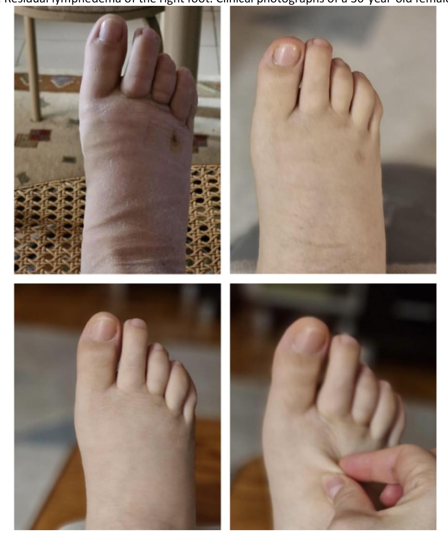
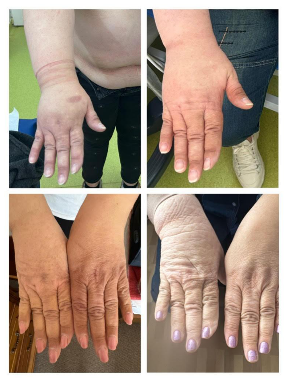
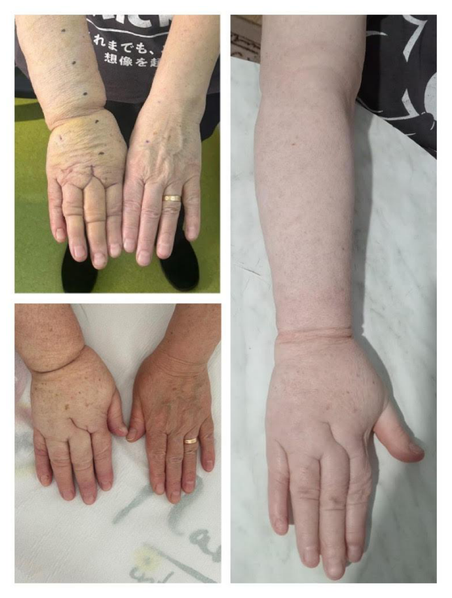
As this is a retrospective case series, clinical photographs and patient-reported outcomes were collected as part of routine clinical care. Some photographs were taken by patients themselves and vary in distance, angle, and lighting; all images were time-stamped and correspond to the documented treatment timepoints (pre-treatment, 1 month, and 6 months post-final session). Clinical photographs were obtained retrospectively as part of routine care. Because some images were patient-provided, standardized positioning, lighting, and camera distance could not be ensured. As a result, image quality and comparability vary.
Patient-reported outcomes were recorded via direct communication and messaging applications, reflecting perceived functional improvement and swelling reduction. In a prospective study, a standardized photography protocol and validated outcome instruments would have been employed; however, the current approach provides a descriptive record consistent with routine practice.
All enzymatic procedures represented standard-of-care management, and the retrospective analysis met institutional criteria for exemption from formal ethics committee review. All patients provided written informed consent for off-label use of PB Serum Medium, retrospective use of their clinical data, and publication of de-identified clinical images.
A 36-year-old female patient, BMI 20.07kg/m2, otherwise healthy, without chronic treatment, presented with secondary lymphedema, stage III (lymphography) / Stage 1 ISL (pitting oedema that improves with elevation) of the right lower limb, with a more pronounced volume discrepancy in the leg compared to the thigh, following trauma in early childhood. The patient’s history included a right femoral fracture, followed by a right ankle sprain. Initially, the patient underwent decongestive therapy [14], until LVA [15, 16] was performed in April 2023. Although good results were achieved initially, a relapse occurred, necessitating debulking liposuction [17] in May 2024. The volume discrepancy recorded prior to surgery was 900 cc, and the volume of aspirate removed was 700 cc. Although the debulking led to temporary relief, persistent swelling in the foot area remained problematic for the patient.
The patient subsequently received three sessions of PB Serum injections administered to the foot area. At one month post-treatment, the patient continued to experience episodic leg swelling but reported no residual swelling in the distal part of the foot (Figure 1). The results were maintained at six months, based on patient report, with supple skin, negative Stemmer sign, and requested further treatment of a small swelling around the ankle, an area outside of the initial treatment.
A 57-year-old female, BMI-29.72kg/m2, otherwise healthy, without chronic treatment, who underwent radical right mastectomy and right axillary lymphadenectomy in 2020, followed by radio and chemotherapy, presented with secondary lymphedema of the right upper limb, stage IV (lymphography)/ stage 1 ISL (pitting edema that improves with elevation). The patient had previously undergone LVA [15, 18] in 2023, at another clinic, with no significant improvement in limb volume discrepancy. We offered debulking surgery [17], in March 2024, which resulted in the removal of approximately 1500 cc of aspirate (the initial volume difference was 1700 cc). The patient was satisfied with the results; however, compliance with compression in the hand and finger areas was low due to difficulties in maintaining hand hygiene [7].
The patient subsequently received the same PB Serum injection regimen, involving three sessions administered to the dorsum of the right hand, spaced at two-week intervals. One month after treatment, follow-up indicated a significant reduction in swelling, localized to the treated area (Figure 2). Results are maintained at six months after treatment, based on patient report, with supple skin and a negative Stemmer sign.
A 62-year-old female, BMI-30.12kg/m2, also known with osteoporosis and vertigo, who underwent right radical mastectomy and right axillary lymphadenectomy in 1993, followed by radio and chemotherapy, developed upper right limb lymphedema 20 years after surgery. At presentation, she is staged with stage IV upper right limb lymphedema (lymphography)/ stage 2 ISL (pitting edema that does not resolve with elevation). She underwent MITESE (minimal invasive tissue excision and skin excision) of the right upper arm [3], with significant volume reduction in 2025. A volume of 2300 ml of fat was aspirated in one session, followed by excision of redundant skin. The patient proved extremely compliant and followed thoroughly the postoperative compression regimen [14]. Still, volume excess was present in the dorsum of the right hand and fingers.
Three sessions of PB Serum Medium were administered at two-week intervals, and results at one month after treatment ended showed marked volume reduction at the injection site, especially around interphalangeal joints, and improvement of hand function (Figure 3). At six months follow-up, the improvement was maintained, based on patient report, with supple skin and a negative Stemmer sign.
This manuscript reports preliminary clinical observations of an off-label enzymatic adjunct used for residual, localized lymphedema after standard surgical management, and situates these observations within the limited available literature on pharmacologic and injectable approaches to lymphedema. While debulking procedures achieved >80% excess volume reduction across cases, functionally limiting distal residual fibrosis persisted despite compression therapy compliance. These observations suggest that multi-enzyme cocktails may address a specific clinical niche currently underserved by existing modalities [3,5,17]. These three cases illustrate that patient satisfaction is not solely dependent on volume reduction [15].
One major limitation of this report is the selection of the enzymatic cocktail, which was based on prior experience with localized adipose tissue rather than on comparative testing of the three available enzymatic cocktails for lymphedema treatment—specifically, those targeting adipose tissue. Quality of life improvements were assessed through anecdotal and spontaneous patient reports, and the assessment of changes in the treated area was based on photographs and patient self-reporting [6], rather than objective measurements. Validated patient-reported outcome measures were not administered, which limits quantitative assessment of perceived functional benefit.
A key limitation of this case series is the lack of standardized photography. Several follow-up images were provided by patients, preventing strict control over lighting, angle, and distance. Future prospective studies should incorporate a standardized imaging protocol.
Our observations suggest that adding a new tool for addressing small but sensitive areas may significantly improve perceived quality of life. The dorsum of the foot or hand, toes, fingers, face, head and neck, and children’s extremities might benefit from injectable treatments rather than liposuction. Given that a compound targeting both fibrotic (collagenase) and adipose (lipase) tissue is already available on the market, investigating the role of enzymatic treatment within the lymphedema treatment algorithm [4] is warranted. Identifying the optimal enzymatic combination may also prove valuable if pathology examinations of the aspirate can determine whether fibrosis or adipose tissue is predominant. The potential benefit of this adjunct treatment, alongside physiological microsurgical approaches [19,20,21], merits further investigation.
An additional patient with sleep apnea and a heavy neck (lymphedema not formally diagnosed), treated in a similar manner, except no surgical procedure was performed initially, reported subjective improvement in breathing and sleep quality and returned for 2 more sessions for further improvement. This case signals a possible research field for head and neck lymphedema, where liposuction in fibrotic/ irradiated tissue might become problematic [3].
The initial clinical observations presented in the current article demonstrate the feasibility and consistent short-term improvement using an off-label multi-enzyme cocktail for distal lymphedema residuals refractory to standard care. While hypothesis-generating only, these findings support formal evaluation of injectable enzymatic therapy within contemporary lymphedema algorithms, particularly for anatomically challenging regions poorly addressed by surgery or compression. Future prospective studies with standardized imaging and validated patient-reported outcomes are warranted.
The author declares no conflicts of interest related to this study. No financial grants or funding were received for this research. The author has no industrial links, affiliations, or financial interests in any of the products, devices, or therapeutic substances mentioned in this manuscript, including PB Serum Medium (Proteos Biotech, Madrid, Spain). This study did not involve clinical trials or commercial devices requiring additional conflict declarations.
The author acknowledges the patients who provided informed consent for the publication of their clinical information and photographs for scientific and educational purposes. The author also acknowledges the clinical staff at Alexandru Gafencu Military Emergency Hospital for their support in patient care and data collection.
All patients signed consent forms for the release of their clinical information, including photographs, for publication in a scientific journal or presentation at a meeting.
Informed consent was obtained from all subjects involved in the study. Written informed consent has been obtained from the patient(s) to publish this paper.