
H. pylori is recognized as a human gastric pathogen that is a flagellated, microaerophilic, spiral-shaped, and gram-negative bacterium isolated from human gastric mucosa and was first recognized in 1982. It colonizes the stomachs of at least half of the world’s population. It is proposed that H. pylori is the main cause of gastritis and peptic ulcer and a risk factor for gastric malignancy in humans. Most cases colonized by these bacteria are asymptomatic. In symptomatic subjects, the antibiotic treatment is not completely effective due to antibiotic resistance.
According to the Food and Agriculture Organization of the United Nations and the World Health Organization, probiotics are defined as ‘living microorganisms, which when administered in adequate amounts confer health benefits on the host’ [1]. Mechanisms of probiotics include controlling intestinal microbial communities, inhibiting pathogens, stimulating epithelial cell proliferation, and immunomodulation [2]. Numerous studies have examined the precise role of probiotics for the eradication of H. pylori. Some research has shown that probiotics can’t eradicate H. pylori but can reduce its levels in the stomach [3]. It has been shown that probiotics can be used as a complementary therapy for the management of H. pylori infection [4]. Lactobacillus spp. are well-characterized probiotics that produce lactic acid and have demonstrated beneficial effects against gastrointestinal pathogens such as H. pylori [5]. It has been indicated that L. rhamnosus JB3 can decrease gastric inflammation by weakening H. pylori virulence in mice [6]. Some probiotic strains, such as Lactobacillus and Bifidobacterium, can inhibit H. pylori growth via bacteriocins or organic acid production [3]. Michetti et al. showed that gastric inflammation can decrease when animals were colonized by L. johnsonii La1 and L. acidophilus LB [7,8]. Probiotics can modulate cytokine secretion through signaling pathways that also affect immune cells, such as T lymphocytes, promoting their proliferation and differentiation [2]. In this study, H. pylori and two probiotic strains were administered to infected mice, and immune responses were monitored to investigate the immunomodulatory effects and to prospect possible mechanisms of Lactobacillus strains in suppressing H. pylori infection.
A clinical strain of H. pylori obtained from the Alzahra University collection was grown on Brucella agar (Merck, Germany) supplemented with 5% defibrinated sheep blood and antibiotics under microaerophilic conditions at 37 °C for 72 h, and then identified by biochemical tests and Gram staining. L. acidophilus ATCC 4356 and L. rhamnosus PTCC 1607 were obtained from the Organization of Industrial Research of Iran. Lactobacillus spp. were routinely grown on de Man, Rogosa, and Sharpe (MRS) agar (Merck, Germany) in an anaerobic chamber at 37 °C [9].
Pathogen-free male C57BL/6 mice weighing between 20 and 22 g were obtained from the Razi Institute, Karaj, Iran. All experiments were performed on mice in accordance with the Animal Ethics Committees of AL Zahra University. Mice groups and the number of mice in each group were determined by the “Resource Equation” method [9]. The mice ranged in age from 6 to 8 weeks. All mice, in 6 groups, were maintained in a pathogen-free environment, with standard brightness (12 h of darkness/light), standard aeration, and free of contamination at a temperature of 21±2 ° C and 55±5% humidity (with free access to chow and sterile water).
It should be noted that the H. pylori stomach colonization test was performed first. For the determination of colonized bacteria, gastric tissue was removed and homogenized in Brucella broth (with 5% FCS), then cultured on the Brucella blood agar medium and incubated for 5 days (microaerobic; 37°C conditions). After incubation, colony recognition, Urease action, oxidase, and catalase tests were performed.
Mice were divided into 6 groups of five, and the treatments were performed as follows:
Group 1- 100µl (microliters) of a concentration of 1 ×10⁸ CFU / ml L. acidophilus was given to mice by gavage (orally) for three consecutive days under the same conditions.
Group 2- 100µl of concentration of 1 ×10⁸ CFU / ml L. acidophilus was given to mice by gavage (orally) for three consecutive days under the same conditions. Then, for three days, 100µl of a concentration of 1× 10⁹ CFU / ml H. pylori was given to mice by gavage.
Group 3- 100µl of concentration of 1 ×10⁸ CFU / ml L. rhamnosus was given to mice by gavage (orally) for three consecutive days under the same conditions.
Group 4- 100µl of concentration of 1 ×10⁸ CFU / ml L. rhamnosus was given to mice by gavage (orally) for three consecutive days under the same conditions. Then, for three days, 100µl of a concentration of 1× 10⁹ CFU / ml H. pylori was given to mice by gavage.
Group 5- 100µl of a concentration of 1× 10⁹ CFU / ml H. pylori was given to mice by gavage for three consecutive days under the same conditions.
Group 6- Five mice were considered the control group under the same conditions as the other treated mice, except that they received no bacteria and were given only sterile phosphate-buffered saline (PBS).
It should be noted that the booster doses for the above treatments were administered two weeks after the initial treatments, under the same conditions. 14 days after the last treatment, the animals were killed ethically (anesthesia with peritoneal injection of diazepam and 10% ketamine), and their spleen was removed.
Spleen samples were thoroughly washed with normal saline and stored in numbered microtubes containing 1 ml of sterile normal saline in a -80 ° C freezer.
Levels of IFN-γ and IL-4 expression were measured by real-time PCR. Total RNA was extracted from 25-50 mg of spleen tissue according to the manufacturer’s protocol (Pars-tous Biotechnology, Iran). To determine the quantity and purity of the extracted RNA, the optical density of the samples at 260/280 was measured.
The extracted RNA was used to synthesize cDNA using the First-Aid Reverse Transcription Kit (Fermentas). In brief, 5-10µg of the extracted RNA, 1µl of oligo dT, 1µl of the random hexamer, and 15-20 pmoles of sequence-specific primers were adjusted to 14µl by DEPC water. The resulting composition was heated (65° C for 5 min), then cooled on ice and mixed rapidly. Then, 4µl of RT 5 x Buffer, containing 10 mM dNTPs, 1µl DTT, and Revert UP™ II Reverse Transcriptase were added to each microtube. It was then heated at 50 °C for 60 minutes and incubated at 95 °C for 5 minutes to deactivate the reverse transcriptase (terminal steps were performed in the thermocycler). The cDNA was stored in a freezer and used for quantitative PCR reactions.
The real-time PCR reactions were performed to analyze the expression levels of IFN-γ, IL-4, and β-actin. In this section, the synthesized cDNA and selected primers were used for qRT-PCR. The reaction conditions were as follows: 95°C for 15 min, 95°C for 30 s, 62°C for 30 s, 72°C for 30 s in 40 cycles, and 1 cycle at 72°C for 30s for the final extension. All experiments were done in triplicate. The β-actin gene was used as the reference gene to determine an arbitrary normalized value for each gene [10].
The statistical analysis was performed by the REST software version 9. The data were analyzed by ANOVA, followed by Tukey’s post hoc test. The level of statistical significance was set at p<0.05.
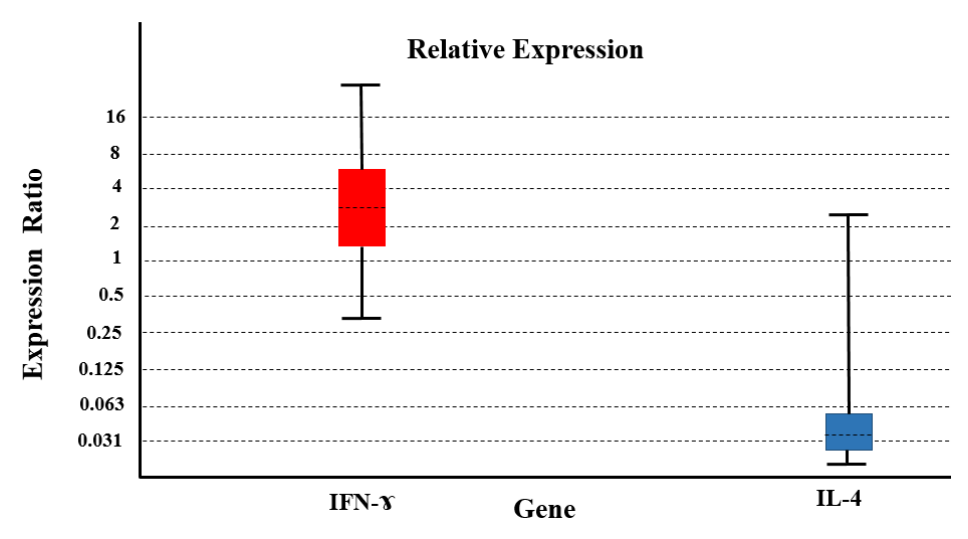
In this treatment, INF-γ expression was increased (by almost 3.2-fold) compared to the control group (P=0.03). However, IL-4 expression decreased compared to the control group (P=0.04) (Figure 1).
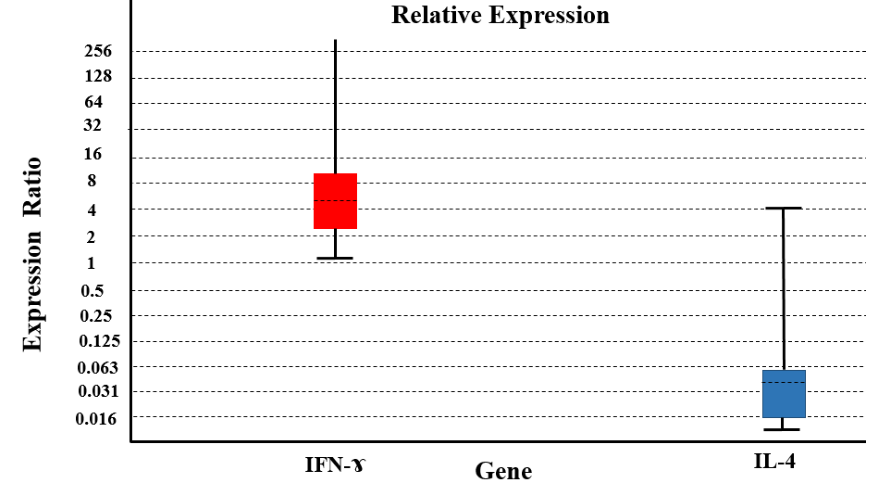
The expression level of INF-γ in this treatment showed an increase in expression (by almost 10.9 fold) compared to the control group (P=0.007), and the level of IL-4 expression compared to the control group showed a decrease in expression (P=0.03) (Figure 2).
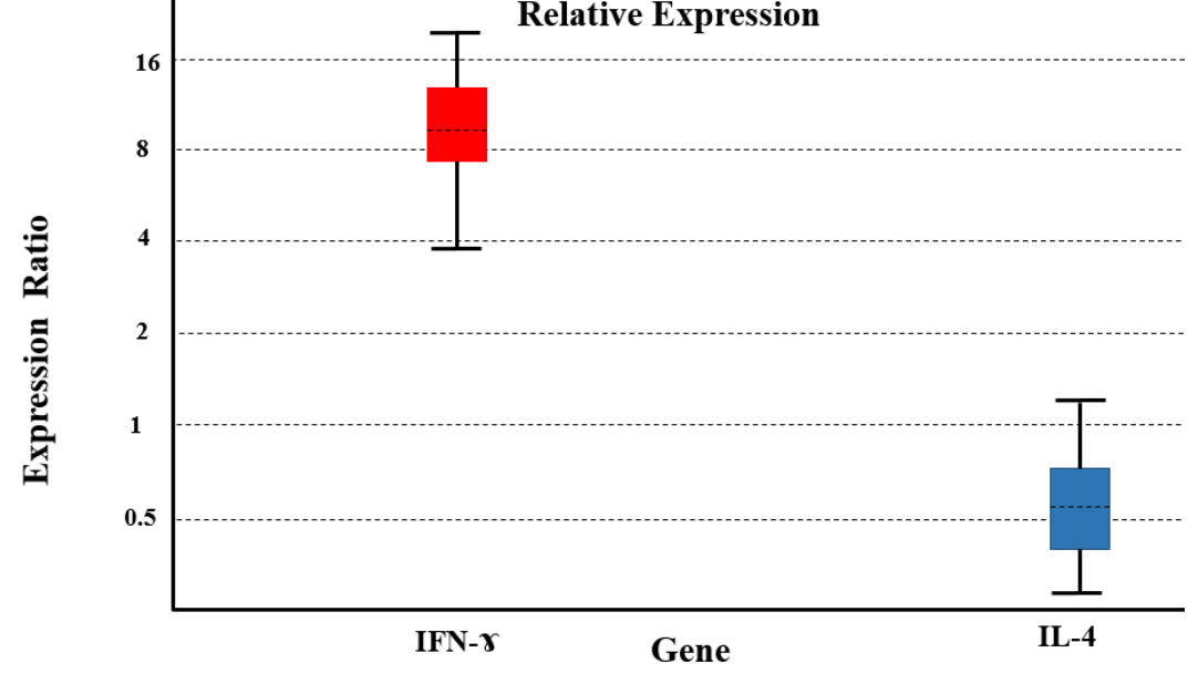
INF-γ expression was increased in this treatment (by almost 9-fold) compared to the control group (P = 0.008). But the expression level of IL-4 compared to the control group showed a decrease in expression (P=0.01) (Figure 3).
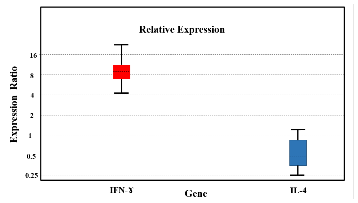
The expression level of INF-γ in this treatment showed an increase in expression (by almost 8.5-fold) compared to the control group (P= 0.009), but the expression of IL-4 levels did not change compared to the control group (P=0.06) (Figure 4).
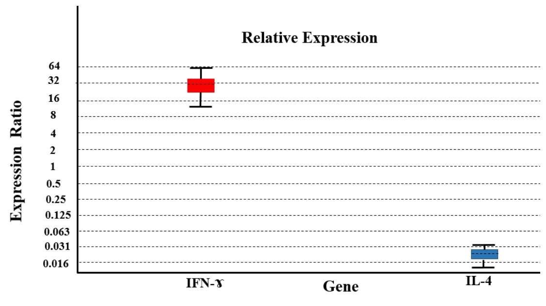
INF-γ expression was increased in this treatment (by almost 29-fold) compared to the control group (P=0.008). However, IL-4 expression relative to the control group decreased (P=0.002) (Figure 5).
The objective of this study was to examine the immune response induced by probiotic lactobacilli against gastroduodenal diseases, such as H. pylori infection. Immunity against H. pylori is characterized by a strong Th1 response, and the generation of an adaptive Th1 response against H. pylori infection requires innate immunity. For example, the neutrophil-activating protein of H. pylori induces interleukin IL-12 and IL-23 secretion [12]. T cells in the gastric mucosa are known to consist of Th1 cells that produce IFN-γ mostly [13]. The expression of IFN-γ and IL-12 was increased in gastric biopsy samples from patients infected with Helicobacter pylori [14]. The results of Vinagre et al. in 2018 showed that IL-4 and IL-10 concentrations in the stomachs of patients infected with H. pylori were significantly lower than in uninfected patients. Their results also showed that the concentrations of IFN-γ and IL-12 in patients infected with Helicobacter pylori were significantly higher than those in uninfected patients [15]. Many studies show that probiotics can be used as a complementary therapy for the management of H. pylori infection [4]. There is substantial evidence that certain probiotics play a significant role in regulating the immune system [16]. For example, it was reported that Lactobacillus can promote mononuclear cells to produce IFN-γ, IL-12, and IL-18. These organisms activate signaling pathways, such as NF-κB and STAT, in human macrophages [17]. Besides, probiotics can induce the production of T-helper 1-classified cytokines [18]. These studies suggest that certain probiotic strains may upregulate innate immune responses [19]. Kabir et al. showed that lactobacilli are a promising option for treating H. pylori, which may be due to their ability to inhibit pathogen binding. They also showed that mice fed a diet containing L. salivarius had no pathological problems in the stomach, as evidenced by the lack of IL-8 production by gastrointestinal epithelial cells [20].
Treatment of mice with L. reuteri and L. brevis increased the expression of inflammatory and Th1 cytokines such as IL-2, TNF-α, and IL-1β. However, conflicting results have been obtained regarding the role of probiotics in increasing the production of cytokines such as IFN-γ, IL-10, IL-1β, IL-6, TNF-α, and IgA production [21-25].
Incidentally, it has been shown that Lactobacillus species exhibit different abilities to produce proinflammatory cytokines in bone marrow-derived dendritic cells [26]. Koga has shown that L. gasseri prevents H. pylori-induced gastritis by inhibiting the bacterial type 4 secretory system, which reduces IL-8 production by gastrointestinal epithelial cells [27]. The use of two probiotic bacteria, L. paracasei and L. reuteri, reduces proinflammatory cytokines and intake of L. plantarum in mice that were deficient in IL-10 production, reduces mucosal IL-12 levels, and, consequently, reduces intestinal inflammation [28]. Activated macrophages can produce TNF-α and IFN-γ that have an important role in the immune defense against pathogens (viral and microbial) and induce immune responses [29-31]. Studies by Gill et al. showed that spleen cells in mice treated with L. rhamnosus and L. acidophilus produced significant amounts of IFN-γ [32]. The induction effect of L. acidophilus on IFN-γ production was demonstrated by mouse spleen macrophages [33,34]. A recent study by Ren et al. showed that mice that received the probiotic bacteria L. plantarum and L. salivarius, compared to the control group, showed an increase in the production of IL-10, IFN-γ, and TLR-2 [35]. Lactobacillus casei Strain Shirota can reduce the colonization of H. pylori and gastritis caused by this bacterium, and increase cellular immunity [36,37].
In this regard, our studies also showed that the expression level of IFN-γ in the group that was treated only with H. pylori increased 29 times compared to the control group, and IL-4 expression decreased; this confirms the superiority of the cellular immune response against this bacterium. The group that received the probiotic bacteria L. acidophilus ATCC4356 and L. rhamnosus PTCC1607 had an increase in IFN-γ expression level, which was 3.2 times in treatment with L. acidophilus and 9 times in treatment with L. rhamnosus. Therefore, the stimulatory effect of L. rhamnosus was greater, and it exerts the effect of strengthening the cellular immune response by helping stimulate T cell differentiation to Th1.
In the groups that received the combination of probiotics and H. pylori, the expression level of IFN-γ increased; in treatment with L. acidophilus plus H. pylori, it increased 10.9-fold, and in treatment with L. rhamnosus plus H. pylori, it increased 8.5-fold. Therefore, both probiotics have a stimulating effect and can help increase monocyte and macrophage activity, reduce inflammation, and strengthen the cellular immune response against H. pylori. It should be noted that the group that received only H. pylori showed a 29-fold increase in expression. However, the groups receiving the combination of probiotics and H. pylori increased IFN-γ expression, with an increase of 10.9 times in treatment with L. acidophilus and 8.5 times in treatment with L. rhamnosus, which was lower than the group that received only H. pylori. This may be due to the inhibitory effect of L. acidophilus and L. rhamnosus, which reduces the excitatory effect of H. pylori by preventing its binding to the gastric epithelium, as confirmed in similar studies [36]. The increase in expression in the groups that received the combination of probiotic and H. pylori is due to the combined effect of probiotic bacteria and H. pylori. Since no expression of the cytokine IL-4 gene was observed in any of the groups compared to the negative control group, and the changes were in the form of reduced expression or no change in expression, it can be stated that the above probiotics do not have the desired strengthening function in stimulating the humoral immune response in mice. In other words, the immunomodulatory effects of these probiotics are to help to induce a cellular immune response by aiding the stimulation of interferon-gamma production.
It can be concluded that the function of probiotic strains is different; each probiotic strain is a unique organism with its own characteristics, and care must be taken in selecting the strains to ensure effectiveness. What is clear in this study is that two probiotics, L. acidophilus ATCC4356 and L. rhamnosus PTCC1607, stimulate the production of interferon-gamma to increase the phagocytic activity of macrophages and also facilitate the differentiation of T cells to Th1. Th1 cells produce several cytokines that ultimately reduce proinflammatory cytokines such as IL-8 and IL-1, thereby directing the innate and acquired immune responses and keeping the body on standby.
The authors declare no conflict of interest.
Not applicable. No generative AI was used for producing this article.
All authors participated in the presentation of the original idea and design, search for sources and review of articles, initial writing or revision of the article, and all with the final approval of the present article, take responsibility for the accuracy of the content.
This project has been approved by the Ethics Committee of Al-Zahra University, Iran, with ID 5263.
Not applicable.