
Dental implants have become the gold standard in everyday oral rehabilitation, offering predictable clinical outcomes and improving the quality of life for individuals with missing teeth [1]. By restoring function, aesthetics, and overall oral health more effectively than traditional prosthetic solutions, they have become the preferred choice in modern dental practice [2-4].
Tooth loss is a common concern among postmenopausal women, due to a combination of age-related changes, systemic health conditions, and the cumulative effects of periodontal disease [5,6]. While dental implants are a valid and effective modality for restoring oral function and aesthetics [7], postmenopausal women present distinct biological and clinical challenges. Estrogen deficiency after menopause is linked to decreased bone mineral density, altered bone remodeling, and a heightened risk of osteoporosis—all factors that may affect osseointegration and long-term implant stability [8-10]. Furthermore, the higher prevalence of comorbidities, polypharmacy, and immune function changes in this population requires an individualized and comprehensive approach to treatment planning [11-13].
A growing body of literature has investigated implant survival rates, peri-implant bone loss, and complication profiles in postmenopausal women, mainly in subjects with osteoporosis [14-17]. Despite these advances, inconsistencies remain regarding the extent to which hormonal changes and altered bone metabolism influence clinical outcomes. A clearer understanding of these biological factors is essential for refining surgical protocols, selecting implant designs optimized for compromised bone quality, and implementing adjunctive strategies such as hormone replacement therapy or bone augmentation procedures.
Given the high prevalence of osteoporosis and the increasing demand for implant-based rehabilitation in this population segment, the relationship between osteoporosis treatments, bone metabolism, and implant placement is the focal point in current research.
This review synthesizes current evidence on the biological mechanisms that might influence implant therapy in postmenopausal women. It evaluates reported clinical outcomes in postmenopausal women and proposes practical treatment guidelines to improve prognosis. By integrating insights from basic science with clinical data, this study aims to provide a comprehensive, evidence-based framework for clinicians involved in the implant rehabilitation of postmenopausal women.
Menopause is characterized by a significant decline in circulating estrogen, a hormone that plays a central role in maintaining skeletal homeostasis [18-20]. Estrogen exerts anti-resorptive effects by downregulating osteoclast differentiation through inhibition of the receptor activator of nuclear factor kappa-Β ligand (RANKL) pathway and by promoting osteoprotegerin production, thereby reducing osteoclastic bone resorption [21,22]. It also enhances osteoblastic activity and facilitates calcium absorption via modulation of vitamin D metabolism [23,24].
In the context of estrogen deficiency, this tightly regulated balance is disrupted. Osteoclast lifespan is prolonged, osteoblast activity declines, and calcium absorption efficiency is reduced due to the decrease in estradiol levels, collectively resulting in a negative bone balance [25,26]. These systemic alterations lead not only to generalized skeletal demineralization, manifesting as postmenopausal osteoporosis, but also to localized effects within the oral cavity. The alveolar bone mirrors the skeletal bone, but given its high turnover rate, it is particularly vulnerable to accelerated resorption [23]. This localized bone loss contributes to periodontal breakdown, decreased tooth support, and impaired healing capacity, which can compromise the stability and longevity of dental implants and other rehabilitative interventions [27,28]. Thus, estrogen deficiency represents a shared mechanistic link between systemic osteoporosis and oral bone loss, highlighting the importance of considering hormonal status in both medical and dental management of postmenopausal women.
Osteoporosis is prevalent in postmenopausal women and is defined by reduced bone mineral density (BMD) and microarchitectural deterioration, leading to increased fracture susceptibility [29]. Several studies have reported that systemic osteoporosis is associated with decreased mandibular bone cortical width and trabecular density, potentially influencing implant site quality and the predictability of osseointegration [30,31]. However, the relationship between systemic BMD and alveolar bone support is still debated. Evidence regarding the relationship between systemic osteoporosis and oral bone loss remains inconsistent. Wang and McCauley reported that osteoporosis and periodontal disease may share common inflammatory and bone remodeling pathways, suggesting a biological association between systemic and oral skeletal deterioration [32]. Similarly, Erdem et al. demonstrated a positive association between reduced bone mineral density and compromised oral health parameters in postmenopausal women [33].
Conversely, Albandar et al. emphasized that local factors—including periodontal inflammation, mechanical loading, oral hygiene status, and microbiological composition—may independently influence alveolar bone loss regardless of systemic skeletal status [34]. Additional studies have similarly suggested that oral bone remodeling is multifactorial and may not directly reflect systemic bone mineral density [35–39].
In summary, menopause-induced estrogen deficiency disturbs the finely balanced process of bone remodeling, and these systemic alterations are reflected in the alveolar bone (Figure 1). Therefore, estrogen deficiency is a central driver of both systemic and oral bone deterioration, underscoring the need for integrated medical and dental strategies in the management of postmenopausal women.
In postmenopausal women, where estrogen deficiency already compromises bone remodeling and increases the risk of osteoporosis, host genetic variability may further amplify susceptibility to peri-implant bone loss [8,9]. Genetic determinants, therefore, help account for the heterogeneity of implant outcomes observed within this patient population [40,41]. Genetic factors shape inflammatory signaling and bone turnover and exert a direct influence on osseointegration, which is already challenged by the systemic hormonal environment of menopause [42].
Recent studies have highlighted the role of matrix metalloproteinase-8 (MMP-8) gene variants in peri-implant disease susceptibility [43,44]. A 2025 cross-sectional investigation demonstrated that the –799 C/T promoter polymorphism in MMP-8 was significantly associated with peri-implantitis, with carriers of the T allele exhibiting nearly a three-fold higher risk (OR = 3.04; 95% CI: 1.31–7.10; p = 0.010) compared with C allele carriers [42]. These findings are consistent with the established collagenolytic activity of MMP-8 in connective tissue degradation [42,43]. Notably, in the postmenopausal setting—where collagen metabolism and wound healing are already impaired—such polymorphisms may further compromise peri-implant tissue stability and long-term osseointegration [44].
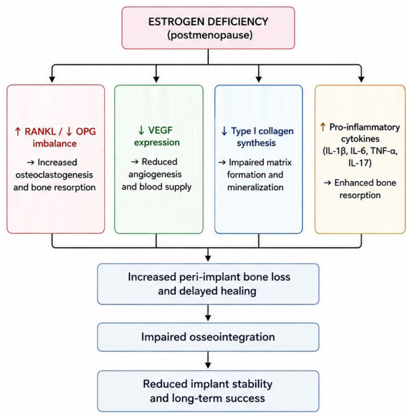
Legend: Estrogen deficiency initiates multiple biological mechanisms that negatively affect peri-implant bone healing and osseointegration. Reduced estrogen levels increase receptor activator of nuclear factor of kappa-light-chain-enhancer of activated B cells (RANKL) expression and decrease osteoprotegerin (OPG) production, thereby promoting osteoclast differentiation and bone resorption. Simultaneously, estrogen deficiency reduces vascular endothelial growth factor (VEGF) expression, impairing angiogenesis and early healing. Decreased type I collagen synthesis and altered extracellular matrix mineralization further compromise bone quality. Elevated levels of pro-inflammatory cytokines, including interleukin (IL)-1β, IL6, tumor necrosis factor-α (TNF-α), and IL-17, further stimulate osteoclastogenesis and peri-implant bone loss, collectively resulting in impaired osseointegration and reduced implant stability.
Complementary evidence concerns interleukin-17A (IL-17A), a cytokine with well-documented pro-inflammatory and bone-resorptive properties [45]. A 2024 case-control study reported that individuals carrying the AA genotype of the rs2275913 SNP had a 6.9-fold increased risk of peri-implantitis compared with healthy controls and implant-success patients (OR = 6.9; 95% CI: 1.71–27.46; p = 0.0065) [46]. A recent systematic review of the IL-17/IL-23 axis confirmed these findings, with approximately one-third of included studies confirming an association between rs2275913 and peri-implant inflammatory conditions [47]. In postmenopausal women, where estrogen deficiency promotes osteoclastogenesis and inflammatory bone loss, IL-17A polymorphisms may act concomitantly to accelerate peri-implant bone resorption, thereby impairing osseointegration [48].
‑ ‑ ‑ ‑ ‑ Moreover, genetic variants in the interleukin 1 gene cluster—including IL 1A, IL 1B, and the IL 1 receptor antagonist (IL 1RN) gene— ‑ represent central regulators of peri implant inflammatory responses [49]. Though recent data showed no significant difference in IL ‑ 1A, IL ‑ 1B, or IL ‑ 1RN polymorphism frequencies in peri ‑ implantitis patients versus controls [50], earlier evidence remains highly ‑ ‑ relevant: a classic study revealed that carriers of the IL 1RN allele 2 (VNTR in intron 2) had a significantly higher risk of peri implantitis (OR ≈ 3.0; 95% CI: 1.2–7.6; p = 0.02) [51]. In postmenopausal women—already predisposed to inflammatory bone loss—the absence or impaired function of IL ‑ 1RN may amplify the effects of IL ‑ 1A and IL ‑ 1B, acting in concert to accelerate bone resorption and disrupt osseointegration.
Together, all these findings underscore the genetic support of peri-implant disease in postmenopausal women and reinforce the rationale for incorporating host genomics into the framework of personalized implant therapy [52].
Despite emerging evidence supporting the role of genetic polymorphisms in peri-implant inflammation and bone remodeling, current findings remain insufficient for routine clinical implementation. Larger prospective studies are needed before genomic biomarkers can be integrated into personalized implant treatment protocols.
Osseointegration is defined by the Glossary of Prosthodontic Terms – 9th edition (GPT-9) as the apparent direct attachment or connection of osseous tissue to an inert, alloplastic material without the interposition of fibrous connective tissue [53]. This process requires adequate bone volume and quality for primary stability, as well as a favorable local environment to achieve long-term stability.
Estrogen deficiency poses significant challenges to osseointegration through several interconnected biological mechanisms. At the vascular level, reduced estrogen impairs the expression of vascular endothelial growth factor (VEGF), compromising neovascularization at the implant site and diminishing the oxygen and nutrient supply essential for early bone healing [54]. At the matrix level, decreased estrogen leads to reduced synthesis of type I collagen and impaired mineralization of the osteoid matrix, resulting in a less competent bone substrate for implant anchorage [55]. Furthermore, the disrupted RANKL/OPG ratio characteristic of estrogen deficiency accelerates osteoclastic resorption at the peri-implant interface, directly undermining the remodeling process necessary for long-term osseointegration [21,22].
Evidence from animal models supports these observations. Ovariectomized rat and rabbit models consistently demonstrate a significant reduction in bone-to-implant contact (BIC), decreased peri-implant bone volume fraction, and delayed cortical healing compared with intact controls [56–58]. Du et al. demonstrated that estrogen deficiency not only reduced BIC values but also altered the transcriptional profile of peri-implant tissue by downregulating genes associated with osteoblast differentiation and extracellular matrix organization [56]. Latimer et al. further confirmed impaired extraction socket healing and reduced implant stability in aged ovariectomized models, suggesting a cumulative effect of aging and hormonal deficiency [58].
Collectively, these findings suggest that estrogen deficiency negatively influences peri-implant healing through impaired angiogenesis, altered matrix formation, and enhanced bone resorption, potentially reducing the biological predictability of osseointegration in postmenopausal women.
| Study | Study design | Population | Follow-up | Implant survival | Main findings | References |
|---|---|---|---|---|---|---|
| Toy and Uslu (2020) | Retrospective | Postmenopausal women | 5 years | 94.7% | No significant reduction in implant success | [15] |
| Liapaki et al. (2022) | Retrospective pilot | Osteopenia/osteoporosis | 3 years | 95% | Slightly increased marginal bone loss | [16] |
| Alsaadi et al. (2021) | Retrospective | Osteoporotic PM women | Variable | Similar to controls | Menopause alone not associated with failure | [60] |
| Lemos et al. (2023) | Systematic review/meta-analysis | Osteoporosis patients | Multiple studies | Comparable overall survival | Greater marginal bone loss observed | [61] |
| Shibli et al. (2025) | Systematic review | Osteoporosis patients | Multiple studies | Comparable survival | Osteoporosis should not be considered an absolute contraindication | [17] |
While biological concerns regarding osseointegration in estrogen-deficient states are well founded, clinical evidence presents a more nuanced picture. Several retrospective studies and systematic reviews have reported implant survival rates in postmenopausal women comparable to those observed in the general population, suggesting that menopause itself should not be considered a contraindication for implant therapy [59–61]. The main clinical studies evaluating implant survival and peri-implant outcomes in postmenopausal women are summarized in Table 1.
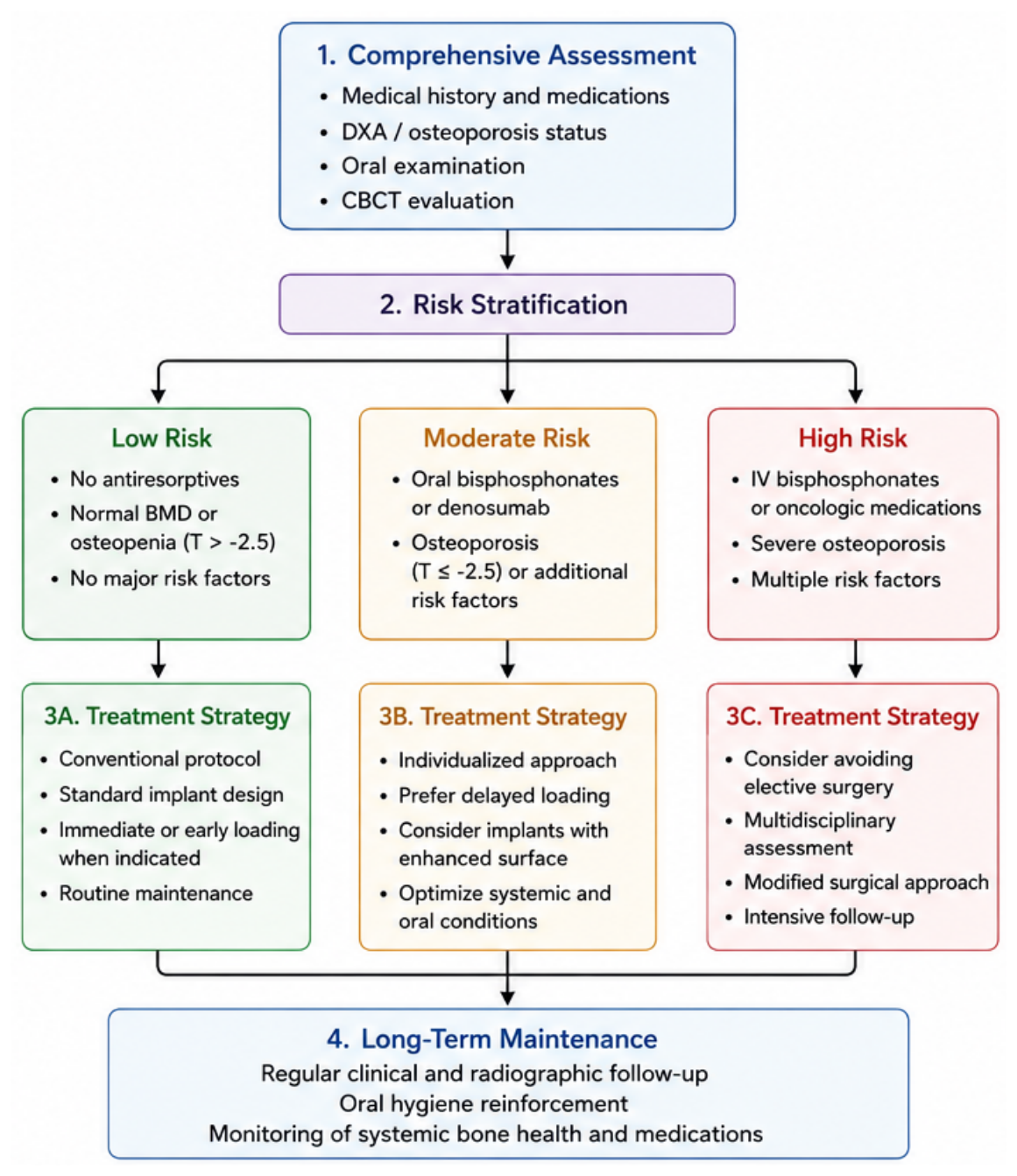
Legend: Proposed evidence-based clinical algorithm for treatment planning in postmenopausal women undergoing implant rehabilitation. The process integrates skeletal evaluation, medication history, risk stratification, and individualized implant strategies in order to maximize implant success and minimize complications such as peri-implant bone loss and medication-related osteonecrosis of the jaw (MRONJ). DXA, dual-energy X- ray absorptiometry; CBCT, cone-beam computed tomography; BMD, bone mineral density; IV, intravenous.
Analysis of currently available studies suggests that menopause alone does not significantly impair implant survival; however, osteoporosis and associated systemic conditions may influence marginal bone remodelling and long-term peri-implant stability [62].
Lemos et al. demonstrated that although implant survival rates were similar between osteoporotic and healthy patients, osteoporotic individuals exhibited greater marginal bone loss [61]. Furthermore, Sachelarie et al. reported that the coexistence of osteoporosis and diabetes adversely affected implant stability, suggesting a cumulative effect of systemic conditions [63].
Variability among studies may be explained by differences in patient selection, medication exposure, osteoporosis severity, implant protocols, and duration of follow-up. Collectively, these findings indicate that individualized risk assessment remains essential before treatment planning. A proposed clinical algorithm for risk assessment and treatment planning in postmenopausal women undergoing implant rehabilitation is illustrated in Figure 2.
Overall, the currently available evidence suggests that implant survival in postmenopausal women is influenced less by menopause itself and more by associated biological and systemic factors, including osteoporosis severity, medication exposure, and comorbidities. Therefore, successful implant therapy requires integration of biological understanding with individualized clinical risk assessment.
Bisphosphonates, administered either orally or intravenously, remain the primary therapy for osteoporosis due to their proven efficacy in reducing fracture risk [64,65]. However, their use has been linked to the development of medication-related osteonecrosis of the jaw (MRONJ), previously known as bisphosphonate-related osteonecrosis of the jaw (BRONJ), a rare but serious complication most frequently triggered by surgical dental procedures such as extractions or implant placement [66–68].
Ruggiero et al., in the updated AAOMS position paper, identified medication dosage, treatment duration, route of administration, and concomitant systemic risk factors as major determinants of MRONJ development [67]. Coropciuc et al. demonstrated that the risk of MRONJ significantly increased following invasive dental procedures in patients receiving antiresorptive therapy [69]. Furthermore, Migliorati et al. emphasized that high-dose intravenous bisphosphonate regimens prescribed for oncologic indications represent the highest-risk category for osteonecrosis development [70].
Among antiresorptive agents used specifically for osteoporosis management, denosumab—a fully human monoclonal antibody targeting RANKL—represents a widely prescribed alternative to bisphosphonates, with demonstrated efficacy in reducing vertebral, non-vertebral, and hip fractures in postmenopausal women [71,72]. However, similar to bisphosphonates, denosumab has been associated with MRONJ, particularly following invasive dental procedures such as extractions or implant placement [73,74].
The reported incidence of denosumab-associated MRONJ in osteoporosis patients remains low (approximately 0.04–0.3%), but risk increases with treatment duration and previous exposure to bisphosphonates [73]. A unique clinical consideration with denosumab is the risk of rebound bone resorption after discontinuation, leading to rapid bone mineral density loss and increased fracture risk. Therefore, current recommendations suggest scheduling elective implant surgery approximately six months after the last denosumab administration and delaying the next dose by at least three months following surgery, always in consultation with the prescribing physician [74,75].
Regarding implant survival, current evidence remains limited. Salabert et al. described successful implant-based rehabilitation after resolution of denosumab-associated MRONJ, indicating that implant placement may remain feasible in selected cases after complete healing and multidisciplinary assessment [74].
Anabolic agents present a contrasting therapeutic profile. Teriparatide (PTH 1–34), a recombinant parathyroid hormone analogue, stimulates osteoblast activity and promotes new bone formation [76]. Studies suggest beneficial effects on peri-implant bone healing and graft maturation, with successful use reported even in patients with MRONJ [77,78]. However, its routine use in implant dentistry remains insufficiently validated [79,80].
Abaloparatide, a synthetic PTHrP analogue, has demonstrated enhanced peri-implant bone formation in preclinical studies; however, evidence from human implant studies remains insufficient for routine clinical recommendation [81].
It is important to distinguish postmenopausal patients treated for osteoporosis from oncologic patients receiving bone-modifying agents, as the clinical context, medication dosage, and MRONJ risk profile differ substantially. Several medications used in oncology have been associated with MRONJ, including tyrosine kinase inhibitors such as sunitinib, anti-angiogenic monoclonal antibodies such as bevacizumab, fusion proteins such as aflibercept, mTOR inhibitors such as everolimus, and radiopharmaceutical agents such as radium-223 [82–87].
Unlike antiresorptive-associated osteonecrosis, the pathophysiology in these patients frequently involves impaired angiogenesis and reduced vascular supply in addition to altered bone remodeling. Consequently, implant placement in oncologic patients receiving these medications requires extensive multidisciplinary consultation and should generally be avoided during active treatment. The main osteoporosis-related and oncologic medications, their mechanisms of action, associated MRONJ risk, and implant-related considerations are summarized in Table 2.
| Drug class | Examples | Mechanism | MRONJ risk | Implant considerations | References |
|---|---|---|---|---|---|
| Bisphosphonates | Alendronate, zoledronate | Inhibit osteoclast-mediated bone resorption | Low in osteoporosis doses; higher with IV use, oncologic doses, treatment >2–4 years | Assess duration, route, dose, systemic risk factors; medical consultation recommended; consider drug holiday only in selected high-risk cases | [64–70] |
| Denosumab | Denosumab | RANKL inhibition, suppression of osteoclast activation | Low to moderate in osteoporosis patients; increases with duration and prior bisphosphonate exposure | Plan elective surgery near the end of the 6-month dosing interval; delay next dose until mucosal and bone healing, in consultation with physician | [71–75] |
| Teriparatide | Teriparatide, PTH 1–34 | Stimulates osteoblast activity and bone formation | Low; may support bone healing | Potential adjunct for bone regeneration and MRONJ healing; not validated for routine implant use; limited treatment duration | [76–80] |
| Abaloparatide | Abaloparatide | PTHrP analogue; stimulates osteogenesis | Low; evidence mainly preclinical | May enhance peri-implant bone formation in experimental models; insufficient human clinical evidence | [81] |
| SERMs | Raloxifene | Modulate estrogen receptors and reduce bone resorption | Low; isolated cases reported | Careful assessment, especially if combined with antiresorptives | [88,89] |
| Immunosuppressive agents | Corticosteroids, methotrexate | Impair immune response, wound healing, and bone turnover | Contributing risk factor | Optimize systemic condition before surgery; increased caution for healing and infection risk | [90,91] |
| Tyrosine kinase inhibitors | Sunitinib | Inhibit angiogenesis and tumor signaling pathways | Moderate; mainly oncologic population | Oncologic consultation mandatory; elective implant placement generally deferred during active therapy | [83,84] |
| Anti-angiogenic monoclonal antibodies | Bevacizumab | VEGF inhibition and impaired angiogenesis | Moderate; oncologic population | High caution; impaired vascularization may compromise healing; avoid elective surgery during active treatment | [82,85–87] |
| Fusion proteins | Aflibercept | VEGF ligand binding and angiogenesis inhibition | Moderate; oncologic population | Caution required; possible impairment of bone and soft tissue healing | [83] |
| mTOR inhibitors | Everolimus | mTOR inhibition; impaired bone remodeling and immune response | Moderate; reported mainly in oncology | Avoid elective implant placement during active oncologic therapy; multidisciplinary decision required | [82] |
| Radiopharmaceuticals | Radium-223 | Cytotoxic effect on bone cells | Moderate, especially when combined with antiresorptives | Oncologic consultation required; risk increases with combined bone-modifying therapy | [82,85–87] |
Taken together, available evidence highlights the importance of differentiating osteoporosis-related therapies from oncologic medications when assessing implant candidacy and MRONJ risk. Implant planning in postmenopausal women requires comprehensive medication history-taking, individualized risk stratification, and close collaboration with physicians to balance treatment benefits against possible complications.
Preoperative assessment in postmenopausal women planned for dental implant therapy must extend beyond the standard evaluation protocol applicable to the general population, incorporating a systematic appraisal of hormonal status, skeletal health, and pharmacological history.
A comprehensive medical history should document menopausal onset, duration of estrogen deficiency, current or previous hormone replacement therapy (HRT), and any diagnosis of osteopenia or osteoporosis confirmed by dual-energy X-ray absorptiometry (DXA) [92]. T-score values should be reviewed carefully, as osteopenia (−1 to −2.5) and osteoporosis (<−2.5) may influence surgical planning, implant design selection, and loading protocols [29,30].
Pharmacological history requires particular attention, with systematic identification of antiresorptive or anabolic therapies, including bisphosphonates, denosumab, SERMs, and teriparatide. Current AAOMS recommendations indicate that patients receiving oral bisphosphonates for less than four years and without additional risk factors generally do not require modification of therapy prior to implant placement. However, prolonged treatment duration or concomitant corticosteroid use requires individualized evaluation [67].
Oral examination should identify and eliminate active periodontal disease, residual infection foci, dental caries, and mucosal pathology before implant surgery. Menopause-associated changes such as reduced salivary flow, mucosal atrophy, altered microbiota, and impaired healing capacity may negatively influence peri-implant tissue stability [13,37,38].
Cone-beam computed tomography (CBCT) should be considered mandatory for evaluating bone volume, cortical thickness, bone density, and anatomical limitations. Patients with low-density bone may require modifications of surgical and prosthetic planning.
Patient counseling should include discussion of expected outcomes, possible complications, treatment limitations, and the importance of long-term maintenance.
The primary objective of surgical management in postmenopausal women is to maximize implant primary stability while minimizing tissue trauma and preserving vascular supply.
Implant selection should consider reduced bone density and altered trabecular architecture frequently associated with postmenopausal osteoporosis [93]. Evidence suggests that moderately rough implant surfaces (SLA or SLActive) improve bone-to-implant contact and accelerate osseointegration compared with machined surfaces [56,94].
Implant dimensions should also be adapted according to bone availability and density. Wider-diameter implants may improve stress distribution and primary stability in low-density bone; however, anatomical limitations should be carefully considered [93]. Similarly, short implants have demonstrated favorable clinical outcomes in severely resorbed ridges and may reduce the need for extensive bone augmentation procedures [95,96].
Loading strategies should be individualized according to primary stability. Conventional loading remains preferable in patients presenting with osteoporosis or reduced insertion torque values, whereas immediate loading protocols should be reserved for cases achieving adequate primary stability.
In patients presenting with low-density bone (type III–IV), under-preparation of the osteotomy site may improve primary stability without inducing excessive compression. A minimum insertion torque of 20–35 Ncm should be targeted for conventional loading protocols.
When severe ridge resorption is present, alternative approaches including short implants, tilted implants, zygomatic implants, or pterygoid implants may reduce the need for extensive bone augmentation procedures [95-97].
Collectively, individualized implant selection and minimally invasive surgical techniques may improve predictability and reduce complications in postmenopausal women.
Current evidence therefore suggests that implant success in postmenopausal women depends not only on systemic bone status but also on optimization of surgical variables, including implant macrodesign, surface characteristics, primary stability, and loading protocol selection.
Postoperative management should account for the possibility of delayed osseointegration and progressive systemic bone loss.
Conventional loading protocols following healing periods of three to six months are generally preferred over immediate loading in patients presenting with osteoporosis or compromised bone quality.
Final prosthetic design should ensure uniform distribution of occlusal forces and minimize cantilever loading to reduce peri-implant stress and marginal bone loss [98-100].
Clinical and radiographic follow-up should be performed at three, six, and twelve months after implant loading and annually thereafter. Evaluation should include peri-implant probing depth, bleeding on probing, implant mobility, and radiographic assessment of marginal bone loss.
According to implant success criteria proposed by Albrektsson et al., acceptable marginal bone loss should remain below 1.5 mm during the first year and below 0.2 mm annually thereafter [101].
Long-term maintenance remains essential because systemic bone changes may continue throughout the postmenopausal period.
Although estrogen deficiency introduces biological challenges to osseointegration through alterations in bone remodeling, angiogenesis, and inflammatory regulation, menopause itself should not be considered a contraindication to dental implant therapy.
Based on currently available evidence, successful implant rehabilitation in postmenopausal women should follow a structured clinical approach:
(1) comprehensive evaluation of skeletal status, including osteoporosis screening and medication history;
(2) identification of antiresorptive or other medications associated with medication-related osteonecrosis of the jaw (MRONJ);
(3) individualized risk stratification according to systemic conditions and bone quality;
(4) selection of implant designs and surgical protocols adapted for compromised bone conditions, including optimization of primary stability and loading strategies;
(5) implementation of long-term supportive care through regular clinical and radiographic follow-up.
Collectively, current evidence suggests that implant prognosis in postmenopausal women depends primarily on individualized treatment planning rather than menopause itself. Integration of systemic assessment, evidence-based surgical strategies, and individualized long-term maintenance protocols may improve treatment predictability and minimize complications.
The authors declare no conflict of interest. No generative AI-produced text was inserted in this manuscript, and no image was previously published in another journal or is under consideration for publication elsewhere. This research received no external funding.
Conceptualization, V.T., and D.A.S.; methodology, C.V.P.; software, D.A.S.; validation, V.T., C.V.P., and C.R.C.; formal analysis, V.T.; investigation, C.R.C.; resources, V.T.; data curation, D.A.S.; writing—original draft preparation, C.V.P.; writing—review and editing, C.R.C.; visualization, D.A.S.; supervision, V.T.; project administration, V.T.; funding acquisition, C.V.P. All authors have read and agreed to the published version of the manuscript.