
Non-melanoma skin cancers (NMSC) are among the most frequently diagnosed neoplasms worldwide [1]. According to the International Agency for Research on Cancer, NMSC ranks 5th in terms of global cancer incidence, with Europe reporting the second-highest mortality rates [2]. While non-melanoma skin cancers include a broad spectrum of cutaneous malignancies, the keratinocyte-derived neoplasms of basal cell carcinoma (BCC) and squamous cell carcinoma (SCC) account for approximately 99% of all NMSC [3].
Cutaneous BCC is the most common malignancy in the human population, with a steadily rising incidence that is projected to exceed the incidence of all other cancers combined [4] It constitutes almost 80% of NMSC cases, whereas SCC accounts for around 20%, with a reported BCC:SCC ratio of up to 10:1. While both BCC and SCC generally have a favourable prognosis, basal cell carcinoma is typically less aggressive and contributes minimally to NMSC-related mortality. In contrast, SCC accounts for a substantial proportion of deaths due to NMSC, with a reported metastatic rate of up to 9.9% [5].
The pathogenesis of these tumors is multifactorial, with risk factors including fair skin phenotype, UV radiation, genetic susceptibility, personal history of skin cancer, immunosuppression, and HPV infection. Furthermore, epidemiological data show that advanced age and male sex are independently associated with an increased incidence of NMSC [6].
BCC originates from basal cells of the epidermis and typically occurs in chronically sun-exposed areas. Accordingly, approximately 80% of these tumors are found on the skin of the head and neck [7]. Basal cell carcinoma is usually slow-growing and locally invasive, with limited metastatic potential. Although rare, metastatic BCC can involve lymph nodes, bone, or lungs [8]. According to the National Comprehensive Cancer Network, basal cell carcinomas located in the head and neck region are considered high-risk, regardless of size [9].
In contrast, SCC often arises from precursor lesions such as actinic keratosis or Bowen’s disease and may progress to an invasive carcinoma. Tumor progression may ultimately lead to metastatic disease in a minority of cases. Similar to BCC, SCC is strongly influenced by UV radiation and sun exposure, which shape its anatomic distribution. However, compared with BCC, a smaller proportion of SCC occurs in the head and neck region, with many tumors involving other sites such as the forearms, hands, and legs [10]. Several factors are considered when assessing recurrence and metastatic risk, including tumor size, depth of invasion, perineural involvement, and degree of histological differentiation; in addition, head and neck locations such as the lip and ear are generally considered at least high-risk sites [11].
For most NMSC, surgical removal is the first-line and often curative treatment. However, due to anatomic and functional constraints in the head and neck region, surgery can be challenging for both patient and clinician. The cephalic region is an area where function and aesthetics are of considerable concern, and the surgical plan often represents a compromise between cosmetic expectations and oncologic safety. As a result, narrower excision may be performed in some cases, potentially leading to positive surgical margins and a higher risk of local persistence/recurrence. In a 2025 retrospective study including 377 basal cell carcinomas, up to 37.2% of cases exhibited involved margins, largely due to constraints imposed by complex head and neck anatomy [12].
This study retrospectively analyzes a cohort of 117 patients with histopathologically confirmed head and neck NMSC treated surgically in a tertiary care center. The lesions included 113 basal cell carcinomas and 21 squamous cell carcinomas, including precursor lesions. Although classification remains controversial, some authors consider keratoacanthoma a premalignant lesion, whereas others regard it as a self-limited variant of SCC; therefore, histopathologically confirmed keratoacanthomas were included in the analysis. We describe the clinical and histopathological features of the tumors (including subtype and anatomic distribution) and summarize surgical management (excision technique, reconstruction, and margin status). We further explore factors associated with incomplete excision (R1) and with reconstruction choice following conventional excision.
We conducted a retrospective observational study, analyzing all primary head and neck NMSC treated from September 2025 to January 2026 in the Department of Dermatovenerology at the “Dr. Carol Davila” Central Military Emergency University Hospital of Bucharest, Romania. The study cohort included 117 patients who underwent resection of 134 head and neck NMSC; twelve patients underwent excision of more than one lesion. All cases were diagnosed based on histopathological evaluation of a single excision under local anesthetic (1% lidocaine). Excisional methods included conventional surgical excision and shave excision. In cases treated by shave excision, electrodesiccation of the lesion base was subsequently performed, preventing accurate histopathological assessment of maximum tumor depth; this limitation was encountered in eight cases. No incisional biopsies were performed, as current evidence supports primary excision as the preferred initial management for many NMSC [13]. Specimens were fixed in 10% neutral buffered formalin, embedded in paraffin, and stained with hematoxylin and eosin (H&E) for microscopic evaluation.
Inclusion criteria comprised patients with histologically confirmed head and neck NMSC who underwent surgical excision between September 2025 and January 2026, inclusive. The following variables were recorded for each patient: demographic data, clinical characteristics, histopathological parameters, and treatment-related data, as detailed in Table 1.
BCCs were classified histologically into six specified subtypes: superficial, nodular, micronodular, infiltrative, metatypical (basosquamous), and pigmented. No morpheaform or sclerosing BCC was identified during the study period. Some lesions (n=18) were reported as basal cell carcinoma, not otherwise specified (NOS), while others were classified as mixed, with coexisting histological patterns within the same lesion (e.g., nodular with adenoid and/or pigmented differentiation). The study also included squamous cell carcinomas and precursor lesions, including keratoacanthoma, actinic keratosis, and Bowen’s disease. While recent NCCN data suggest classifying SCC into two differentiation categories (well-to-moderately differentiated and poorly differentiated), in this study, squamous cell carcinomas were classified using the classic grading system: well differentiated (G1), moderately differentiated (G2), and poorly differentiated (G3) [14]. No anaplastic (undifferentiated, G4) cases were identified.
Due to the retrospective design, tumor dimensions were reconstructed from available clinical and histopathological records to provide a three-dimensional assessment. Tumor length and width were derived from clinical measurements, while maximum thickness was determined from histopathology reports.
The exclusion criteria included histologically unconfirmed NMSC, NMSC located outside the head and neck region, cutaneous melanomas, skin cancers treated by methods other than surgical excision and NMSC excised during periods other than the one specified above.
The primary outcome was resection status for conventionally excised lesions, categorized as complete (R0) or incomplete (R1), with R1 cases further classified by margin involvement (lateral, deep, or both). Secondary outcomes included the reconstruction method after conventional excision (direct closure, local flap, skin graft, or secondary intention) and flap subtype among flap reconstructions. Because prospective follow-up was not available within this study period, post-excision recurrence could not be assessed; a history of prior skin cancer excision within the previous 24 months was recorded as a separate baseline variable.
Categorical variables were summarized as counts and percentages, and continuous variables as means and ranges, as reported in the Results section and Tables. Associations between selected predictors and outcomes were explored using appropriate hypothesis tests for categorical data (e.g., chi-square), with multiple-testing control where indicated by false discovery rate (FDR) reporting.
| Category | Variable | Type | Definition |
|---|---|---|---|
| Demographic | Age | Continuous | Age (years) |
| Sex | Categorical | Male/Female | |
| Area of residence | Categorical | Urban/Rural | |
| Clinical | Tumor location | Categorical | The specific anatomical site of the head and neck (e.g nasal, cheek, auricular) |
| Evolution time | Continuous | Duration of lesion prior to excision (months) | |
| Tumor size | Continuous | Tumor length, width and maximum depth | |
| Temporal | Month of excision | Categorical | Month in which the lesion was surgically excised (September to January) |
| Histopathological | Tumor type | Categorical | BCC/SCC/Bowen/AK/Keratoachantoma |
| Histological subtype | Categorical | Superficial, nodular, infiltrative etc. (for BCC)/G1, G2, G3 (for SCC) | |
| pT stage | Ordinal | Pathological tumor stageing according to AJCC 8th edition (just for BCCs) | |
| Depth of invasion | Categorical | Deepest layer involved: dermis (papillary/reticular), hypodermis, muscle or lamina propria (for mucosal, lip lesions) | |
| Perineural invasion | Binary | Present/Absent | |
| Perivascular invasion | Binary | Present/Absent | |
| Ulceration | Binary | Present/Absent | |
| Desmoplastic reaction | Categorical | Absent/Minimal/Moderate/Important | |
| Necrosis | Binary | Present/Absent | |
| Solar elastosis | Categorical | Absent/Grade 1/Grade 2/Grade 3 | |
| Surgical | Surgical procedure | Categorical | Conventional excision/Excision-shave |
| Reconstruction method (for conventional excisions) | Categorical | Direct closure/Local flap/Skin graft/Secondary intention | |
| Flap subtype technique (for flap cases only) | Categorical | Advancement, rotation, transposition | |
| Surgical margins | Continuous | Lateral and deep margins (mm) | |
| Resection status | Binary | Complete (R0)/Incomplete (R1) | |
| Margin involvement (for R1 cases only) | Categorical | Lateral/Deep/Both | |
| Reintervention in the past | Binary | Yes/No |
BCC = basal cell carcinoma; SCC = squamous cell carcinoma; AK = actinic keratosis; G1=well-differentiated SCC; G2= moderately differentiated SCC; G3= poorly differentiated SCC; AJCC = American Joint Committee of Cancer.
In the performed study, the diagnostic and staging criteria of the American Joint Committee of Cancer (AJCC) were available only for a subset of cases, predominantly BCCs [15]. Given the retrospective design, staging data were limited to the pathological tumor component (pT), as nodal and metastatic status require clinical and imaging correlation that were not consistently documented for all cases.
The surgical procedures were performed by 7 doctors. The case cohort collected was divided into two classes: clean (R0) and positive (R1) margins, the latter being subdivided in turn according to the margin touched by the cancer (lateral, deep, or both).
The reconstruction methods were also recorded, being classified as direct closure, local flap, skin graft, or healing by secondary intention. For cases managed using local flaps, the flap’s subtype (such as advancement, rotation, or transposition) was documented, as well as the specific design modifications (M-plasty, A-T plasty, S-plasty, or V-Y plasty). During the research time period, only one case required reconstruction using a full-thickness skin graft. Thus, the defect involving the nose and adjacent cheek was covered by a right laterocervical skin patch. In the same way, a single case in the cohort was managed by secondary intention healing. After the excision of an auricular SCC with suspected cartilage involvement, the defect was intentionally left open, followed by the application of topical 5-fluorouracil.
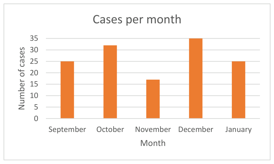
The study included 117 Caucasian patients who underwent resection of 134 primary NMSC located in the head and neck region. Of these, 75 (64.1%) were male and 42 (35.9%) were female, with a slight predominance of urban residents (n=65, 55.6%) compared with rural residents (n=52, 44.4%); this difference was not statistically significant. Patient age ranged from 39 to 92 years, with a mean age of 70.24 years. The most represented age group was 70–79 years (n=48, 41%), followed by 60–69 years (n=24, 20.5%). The remaining groups were 80–89 years (n=18, 15.4%), 50–59 years (n=15, 12.8%), 40–49 years (n=7, 6%), and 90–92 years (n=4, 3.4%); one patient (0.9%) was younger than 40 years. The month with the highest number of operated NMSC cases was December (n= 35/117), while November had the fewest (n=17/117) (Figure 1).
During the study period, 104 patients (88.9%) underwent surgical removal of a single lesion, whereas 13 patients (11.1%) underwent excision of two or more tumors (31 lesions in total). Two lesions were excised in 10/117 patients (8.5%); four lesions were excised in 2/117 patients (1.7%); and three lesions were excised in 1/117 patients (0.9%).
Out of the 134 lesions, 113 (84.3%) were basal cell carcinomas and 21 (15.7%) were squamous cell carcinomas, including precursor lesions. Among BCCs, 36.3% (n=41/113) exhibited mixed histological patterns, whereas 63.7% (n=72/113) showed a single morphological pattern; 18 lesions (16.0%) were reported as BCC, NOS. The most frequent specified subtype was nodular (n=32/113, 28.3%), followed by superficial (n=8/113, 7.1%), infiltrative (n=5/113, 4.4%), metatypical (n=4/113, 3.5%), micronodular (n=3/113, 2.7%), and pigmented (n=2/113, 1.8%). Pathological staging according to AJCC was available for the BCCs included in this cohort and showed a predominance of early-stage tumors: pT1 (n=104, 92%), pT2 (n=8, 7%), and pT3 (n=1, 1%); no pT4 lesion was recorded.
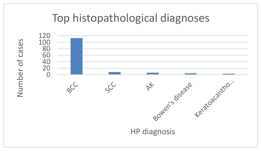
Regarding the histological distribution of SCC-related lesions, 8/21 (38.1%) were invasive squamous cell carcinomas, while the remaining 13 lesions (61.9%) were categorized as precursor or related lesions, including actinic keratosis (n=6/21, 28.6%), SCC in situ (Bowen’s disease, n=4/21, 19.0%), and keratoacanthoma (n=3/21, 14.3%). Among invasive SCCs (n=8), most were well differentiated (G1, n=5/8, 62.5%), followed by moderately differentiated (G2, n=2/8, 25.0%), with one poorly differentiated case (G3, n=1/8, 12.5%). The distribution of the primary histopathological diagnoses identified in the study cohort is illustrated in Figure 2.
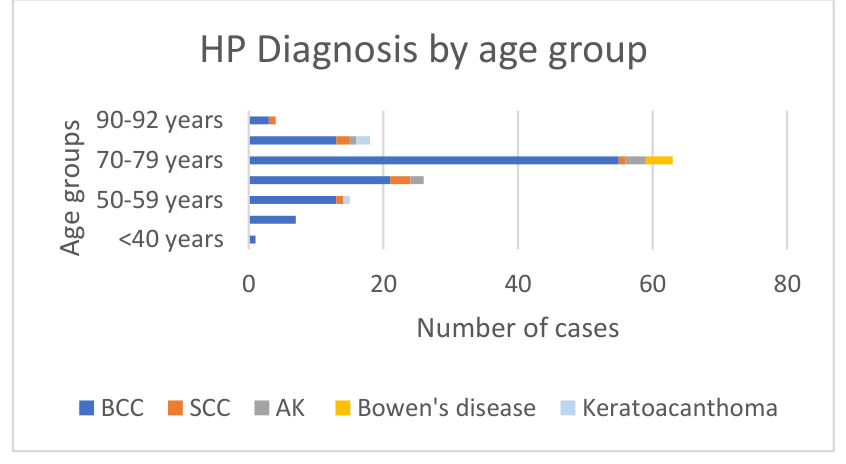
When stratified by age group, statistics demonstrates the predominance of basal cell carcinoma across all age categories, particulary in patients aged 70-79 years. SCC were less frequent, but were identified across most age categories (except <50 years) (Figure 3).
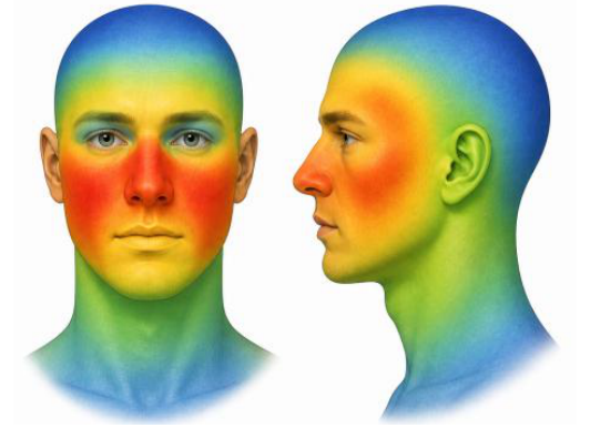
The most frequent anatomical site involved was the cheek area (n=42, 31.3%), followed by the nose (n=40, 29.8%). Within the nasal region, the nasal pyramid (n=31/40) was more commonly affected than the nasal alae (n=9/40). The ear and periauricular area were affected in 12 cases (9.0%), the temporal region in 10 cases (7.4%), and the lips in 8 cases (6.0%), with the upper lip (n=5/8) more frequently involved than the lower lip (n=3/8). The scalp, forehead, and periocular region showed equal involvement (6 cases each, 4.5% each), while the neck was least frequently affected (n=4, 3.0%). Regarding laterality, 42 lesions were located on the left side of the face (31.3%) and 41 on the right side (30.6%); 31 lesions involved the midline (23.1%), and laterality was not specified for 20 lesions (15.0%). The anatomical distribution of lesions across facial regions is highlighted by the “heatmap” representation shown in Figure 4, where warmer colors (red–orange) indicate areas with higher case density (nose and cheeks), while cooler colors (green–blue) represent regions with lower frequency, such as the scalp and neck.
The mean maximum tumor diameter was 10.0 mm (range: 1–23 mm), and the mean maximum depth was 1.9 mm (range: 0.2–10 mm).
Lesion evolution time before surgery varied considerably: most lesions (n=112/134, 83.6%) had been present for more than one year, while 22/134 (16.4%) had an onset within one year. Additional histological characteristics, including depth of invasion, perineural and perivascular involvement, ulceration, desmoplastic reaction, necrosis, and solar elastosis, are presented in Table 2.
Regarding the surgical approach, most lesions underwent conventional excision (n=126/134, 94.0%), while 8 lesions (6.0%) were treated by shave excision. Wound management after conventional excision was implemented primarily by direct closure in 94 lesions (74.6%), followed by local flaps in 30 cases (23.8%). More complex techniques were required in a small subset of cases: a full-thickness skin graft (harvested from the right laterocervical area) was used in one patient (0.8%) for reconstruction of a defect involving the nasal pyramid and part of the right cheek; secondary intention healing was also used in one patient (0.8%) for an auricular defect. Among flap reconstructions, 66.7% (n=20/30) were transposition flaps, 20.0% (n=6/30) were advancement flaps, and 13.3% (n=4/30) were mixed rotation-advancement flaps.
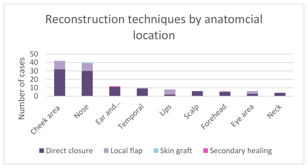
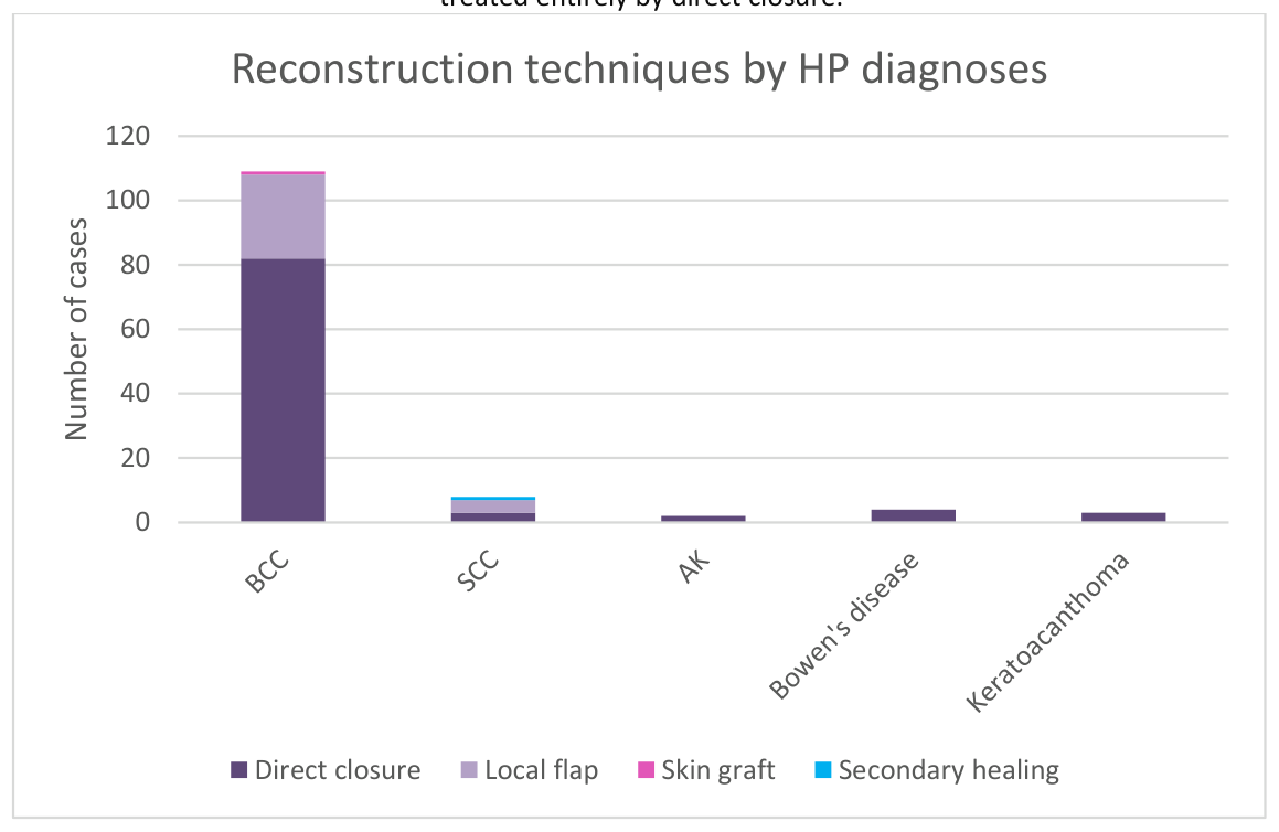
As illustrated in Figure 5, direct closure represented the predominant reconstructive approach across most anatomical regions, while local flaps were used more frequently in regions requiring greater reconstructive complexity, particularly the lips (n=6/8) and nasal area (n=3/6). When analyzed according to histopathological diagnosis, direct closure remained the predominant reconstructive technique across most lesion types, particularly in basal cell carcinoma (n=82/113). In contrast, SCC cases more frequently required complex reconstructive procedures, including local flaps (n=4/8) and secondary intention healing (n=1/8). The only skin graft case was observed in a BCC lesion (Figure 6).
The average lateral surgical margin was 3.2 mm, while the average deep margin was 1.4 mm. Histological evaluation of conventionally excised specimens showed that 92% (n=116/126) had complete margins (R0) and 8% (n=10/126) had positive margins (R1). Among R1 cases, lateral margins were most commonly involved (n=6/10, 60%), followed by deep margins (n=3/10, 30%); in one case (n=1/10, 10%), both lateral and deep margins were involved. Positive margins were observed in the nose (n=2/10, 20%), scalp (n=2/10, 20%), auricular (n=2/10, 20%), and periocular region (n=2/10, 20%). The temporal and cheek areas presented one case each with positive margins (n=1/10, 10%). No positive margins were recorded on the forehead, lips, or neck.
| Variable | Values |
|---|---|
| Mean Age | 70.24 |
| Gender | |
| Male | 75 (64.1%) |
| Female | 42 (35.9%) |
| Residence | |
| Urban | 65 (55.6%) |
| Rural | 52 (44.4%) |
| Tumor location | |
| Cheek area | 42 (31.3%) |
| Nose | 40 (29.8%) |
| Ear and periauricular area | 12 (9%) |
| Temporal | 10 (7.4%) |
| Upper lip | 5 (3.7%) |
| Lower lip | 3 (2.2%) |
| Scalp | 6 (4.5%) |
| Forehead | 6 (4.5%) |
| Eye area | 6 (4.5%) |
| Neck | 4 (3%) |
| Affected side | |
| Left | 42 (31.3%) |
| Right | 41 (30.6%) |
| Midline | 31 (23.1%) |
| Not specified | 20 (15%) |
| Evolution time of the tumor before surgery | |
| <12 months | 22 (16.4%) |
| >12 months | 112 (83.6%) |
| Tumor size | |
| Average length | 10 mm |
| Average depth | 1.9 mm |
| Tumor type | |
| BCC | 113 (84.3%) |
| SCC | 8 (6%) |
| AK | 6 (4.5%) |
| Bowen’s disease | 4 (3%) |
| Keratoacanthoma | 3 (2.2%) |
| BCC Histological Subtypes | |
| Nodular | 32 (28.3%) |
| Superficial | 8 (7%) |
| Infiltrative | 5 (4.4%) |
| Metatypical | 4 (3.5%) |
| Micronodular | 3 (2.7%) |
| Pigmented | 2 (1.8%) |
| NOS | 18 (16%) |
| Mixed histology | 41 (36.3%) |
| Pt category (just for BCCs) | |
| Pt1 | 104 (92%) |
| Pt2 | 8 (7%) |
| Pt3 | 1 (1%) |
| Pt4 | 0 |
| SCC Differentiation Grade (G) | |
| G1 | 4 (50%) |
| G2 | 2 (25%) |
| G3 | 2 (25%) |
| Depth of invasion | |
| Papillary dermis | 13 (9.7%) |
| Reticular dermis | 76 (56.7%) |
| Hypodermis | 13 (9.7%) |
| Muscle | 5 (3.7%) |
| Lamina propria (for mucosal lesions) | 3 (2.2%) |
| Not specified | 24 (18%) |
| Perineural invasion | |
| Yes | 5 (3.7%) |
| No | 125 (93.3%) |
| Not specified | 4 (3%) |
| Perivascular invasion | 0 |
| Ulceration | |
| Yes | 78 (58.2%) |
| No | 56 (41.8%) |
| Desmoplastic reaction | |
| Absent | 82 (61.2%) |
| Minimal | 11 (8.2%) |
| Moderate | 32 (23.9%) |
| Important | 9 (6.7%) |
| Necrosis | |
| Yes | 7 (5.2%) |
| No | 127 (94.8%) |
| Solar elastosis | |
| Absent | 23 (17.2%) |
| Grade 1 | 10 (7.5%) |
| Grade 2 | 39 (29.1%) |
| Grade 3 | 57 (42.5%) |
| Not specified | 5 (3.7%) |
| Surgical procedure | |
| Conventional excision | 126 (94%) |
| Shave-excision | 8 (6%) |
| Reconstruction method | |
| Direct closure | 94 (74.6%) |
| Local flap | 30 (23.8%) |
| Skin graft | 1 (0.8%) |
| Secondary intention | 1 (0.8%) |
| Flap subtype technique | |
| Advancement | 6 (20%) |
| Rotation | 0 |
| Transposition | 20 (66.7%) |
| Mixed (rotation-advancement) | 4 (13.3%) |
| Surgical margins (mm) | |
| Average size of lateral surgical margins | 3.2 mm |
| Average size of deep surgical margins | 1.4 mm |
| Resection status | |
| Complete (R0)/Clean margins | 116 (92%) |
| Incomplete (R1)/Positive margins | 10 (8%) |
| Margin involvement (for R1) | |
| Lateral | 6 (60%) |
| Deep | 3 (30%) |
| Both | 1 (10%) |
| Reintervention in the past 2 years | |
| Yes | 7 (6%) |
| No | 110 (94%) |
BCC= basal cell carcinoma; SCC= squamous cell carcinoma; AK= actinic keratosis; NOS= not otherwise specified; G1=well-differentiated SCC; G2= moderately differentiated SCC; G3= poorly differentiated SCC.
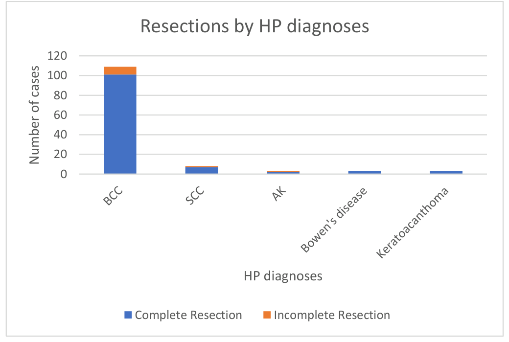
Regarding the relationship between resection status and histopathological diagnosis, basal cell carcinoma accounted for the majority of complete excisions (n=101/109). Although the remaining histopathological subtypes were represented by fewer cases, complete excision remained the predominant outcome across all lesion types, including SCC (n=7/8), AK (n=2/3), Bowen’s disease (n=3/3) and keratoacanthoma (n=3/3) (Figure 7).
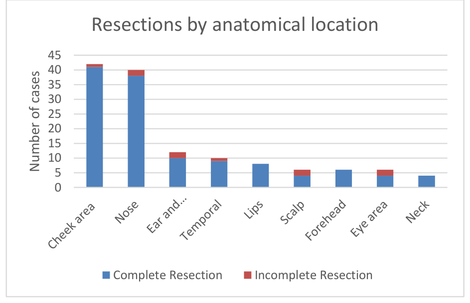
When analyzed according to anatomical site, the cheek and nasal regions accounted for the highest number of complete resections (n=42 and n=38, respectively). Incomplete resections were observed more frequently in anatomically complex areas, particularly the nose, scalp, and periocular/periauricular areas, all of which exhibited 2 cases each of positive edges (Figure 8).
Given the retrospective design and the short study period, prospective post-excision follow-up was not available to assess recurrence after the interventions included in this cohort. We therefore extracted baseline information from medical records regarding prior skin cancer surgery within the 24 months preceding the index procedure. Seven patients (6%) had undergone at least one previous surgical intervention for another skin cancer in the prior two years, while 110 patients (94%) had no such history documented.
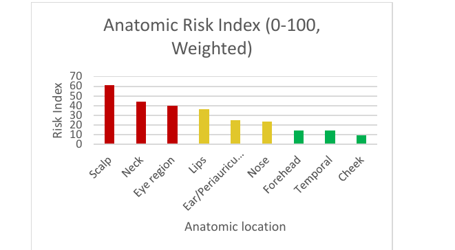
The burden of adverse histopathological indicators varied considerably according to anatomical site, as shown in Table 3. While the cheek area showed the highest case burden with relatively lower-risk characteristics, the scalp, neck, and nasal regions exhibited higher frequencies of incomplete excision, deep invasion, ulceration, and perineural involvement, suggesting a more unfavorable histopathological profile. To further integrate these adverse histopathological features into a unified anatomical vulnerability model, a weighted composite risk index was calculated for each region (Table 4). The scalp region demonstrated the highest composite risk score (61.2), followed by the neck (44.0) and eye region (40.0), classifying these areas as high-risk anatomical sites. These findings are further illustrated in Figure 9, which provide a visual representation of the weighted anatomical risk distribution using a “traffic light” classification system.
| Location Group | Cases | % Total | % Incomplete | % Mixed | % Deep Invasion | % Perineural Involvement | % Ulceration |
|---|---|---|---|---|---|---|---|
| Cheek area | 37 | 31.4 | 5.4 | 0.0 | 2.7 | 2.7 | 51.4 |
| Nose | 34 | 28.8 | 11.8 | 8.8 | 20.6 | 0.0 | 70.6 |
| Forehead | 16 | 13.6 | 12.5 | 0.0 | 6.2 | 6.2 | 43.8 |
| Lips | 8 | 6.8 | 0.0 | 0.0 | 62.5 | 0.0 | 62.5 |
| Temporal | 7 | 5.9 | 14.3 | 0.0 | 0.0 | 0.0 | 85.7 |
| Ear/Periauricular region | 6 | 5.1 | 0.0 | 33.3 | 16.7 | 0.0 | 100.0 |
| Scalp | 5 | 4.2 | 40.0 | 0.0 | 40.0 | 60.0 | 60.0 |
| Neck | 4 | 3.4 | 25.0 | 50.0 | 50.0 | 0.0 | 100.0 |
| Eye region | 1 | 0.8 | 100.0 | 0.0 | 0.0 | 0.0 | 0.0 |
| Location | N | Margin % | Deep Invasion % | Perineural % | Ulceration % | Risk Index | Traffic Light |
|---|---|---|---|---|---|---|---|
| Scalp | 5 | 40.0 | 40.0 | 60.0 | 60.0 | 61.2 | RED |
| Neck | 4 | 25.0 | 50.0 | 0.0 | 100.0 | 44.0 | RED |
| Eye region | 1 | 100.0 | 0.0 | 0.0 | 0.0 | 40.0 | RED |
| Lips | 4 | 0.0 | 62.5 | 0.0 | 62.5 | 36.2 | AMBER |
| Ear/Periauricular region | 6 | 16.7 | 16.7 | 0.0 | 100.0 | 24.7 | AMBER |
| Nose | 34 | 16.2 | 20.6 | 0.0 | 70.6 | 23.4 | AMBER |
| Forehead | 16 | 12.5 | 6.2 | 0.0 | 43.8 | 14.4 | GREEN |
| Temporal | 7 | 14.3 | 0.0 | 0.0 | 85.7 | 14.3 | GREEN |
| Cheek | 37 | 5.4 | 2.7 | 0.0 | 51.4 | 9.5 | GREEN |
Univariate analyses identified several variables significantly associated with excision completeness. Using chi square tests with rare categories collapsed into an “OTHER” group, statistically significant associations were observed for surgical procedure, tumor stage, resection margin status (lateral and deep), solar elastosis, desmoplastic reaction, and depth of invasion. All associations remained significant after correction for multiple testing using the false discovery rate (FDR). The strongest association was observed for surgical procedure, followed by tumor stage and resection margin characteristics. Histopathological features reflecting tumor aggressiveness and tissue response—specifically solar elastosis, desmoplastic reaction, and deeper tissue invasion—were also significantly associated with excision completeness. These findings indicate that procedural factors, tumor extend, and pathological indicators of infiltrative growth are each related to the likelihood of achieving complete excision at a univariate level, Table 5.
| Variable | Test | p-value | FDR q-value |
|---|---|---|---|
| Procedure | Chi-square (rare → OTHER) | 1.79 × 10⁻⁹ | 4.12 × 10⁻⁸ |
| Tumor stage | Chi-square (rare → OTHER) | 2.07 × 10⁻⁶ | 1.62 × 10⁻⁵ |
| Resection margins (lateral and deep, mm) | Chi-square (rare → OTHER) | 2.11 × 10⁻⁶ | 1.62 × 10⁻⁵ |
| Solar elastosis | Chi-square (rare → OTHER) | 5.83 × 10⁻⁴ | 3.35 × 10⁻³ |
| Desmoplastic reaction | Chi-square (rare → OTHER) | 3.74 × 10⁻³ | 1.72 × 10⁻² |
| Depth of invasion | Chi-square (rare → OTHER) | 4.77 × 10⁻³ | 1.83 × 10⁻² |
In multivariable logistic regression analysis evaluating independent predictors of complete excision, none of the examined clinical or histopathological variables demonstrated a statistically significant association after simultaneous adjustment. Patient age showed no meaningful effect on excision completeness (odds ratio [OR] ≈1.0), while indicators of tumor aggressiveness—including deep invasion into the hypodermis or muscle and advanced pathologic stage (pT ≥ 2)—were associated with lower odds of complete excision, although these associations did not reach statistical significance. Similarly, histopathological features such as increasing solar elastosis grade and the presence of ulceration were not independently associated with completeness of excision. The absence of statistically significant predictors in the adjusted model contrasts with the univariate findings and likely reflects collinearity among tumor stage, depth of invasion, and related pathological characteristics, as well as limited power due to the relatively small number of incomplete excisions. Overall, these results suggest that while surgical and pathological factors are associated with excision completeness at a univariate level, no single variable independently predicts complete excision when considered within a multivariable framework (Table 6).
| Predictor | OR | 95% CI |
|---|---|---|
| Age (per 1 year) | 0.99 | 0.94 – 1.04 |
| Deep invasion (hypodermis/muscle) | 0.6 | 0.15 – 2.40 |
| pT ≥ 2 | 0.44 | 0.09 – 2.18 |
| Solar elastosis (per grade) | 1.17 | 0.70 – 1.95 |
| Ulceration (YES vs NO) | 1.04 | 0.33 – 3.29 |
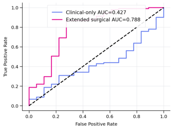
Receiver operating characteristic (ROC) analysis demonstrated superior predictive performance of the extended surgical model compared to the clinical-only model (Figure 10). The clinical-only model achieved an AUC of 0.427, indicating limited discriminatory ability, whereas the extended surgical model demonstrated substantially improved predictive accuracy, with an AUC of 0.788. Calibration analysis further demonstrated improved agreement between predicted and observed outcomes for the extended surgical model, particularly for complete and incomplete resection categories. As illustrated in Figure 11, the extended surgical model showed better calibration performance compared to the clinical-only model across most outcome categories. Prediction of mixed-pattern lesions remained limited in both models, likely reflecting the low number and heterogeneity of these cases. Overall, the inclusion of extended surgical and histopathological parameters improved both discrimination and calibration performance, supporting their added predictive value in resection outcome assessment.
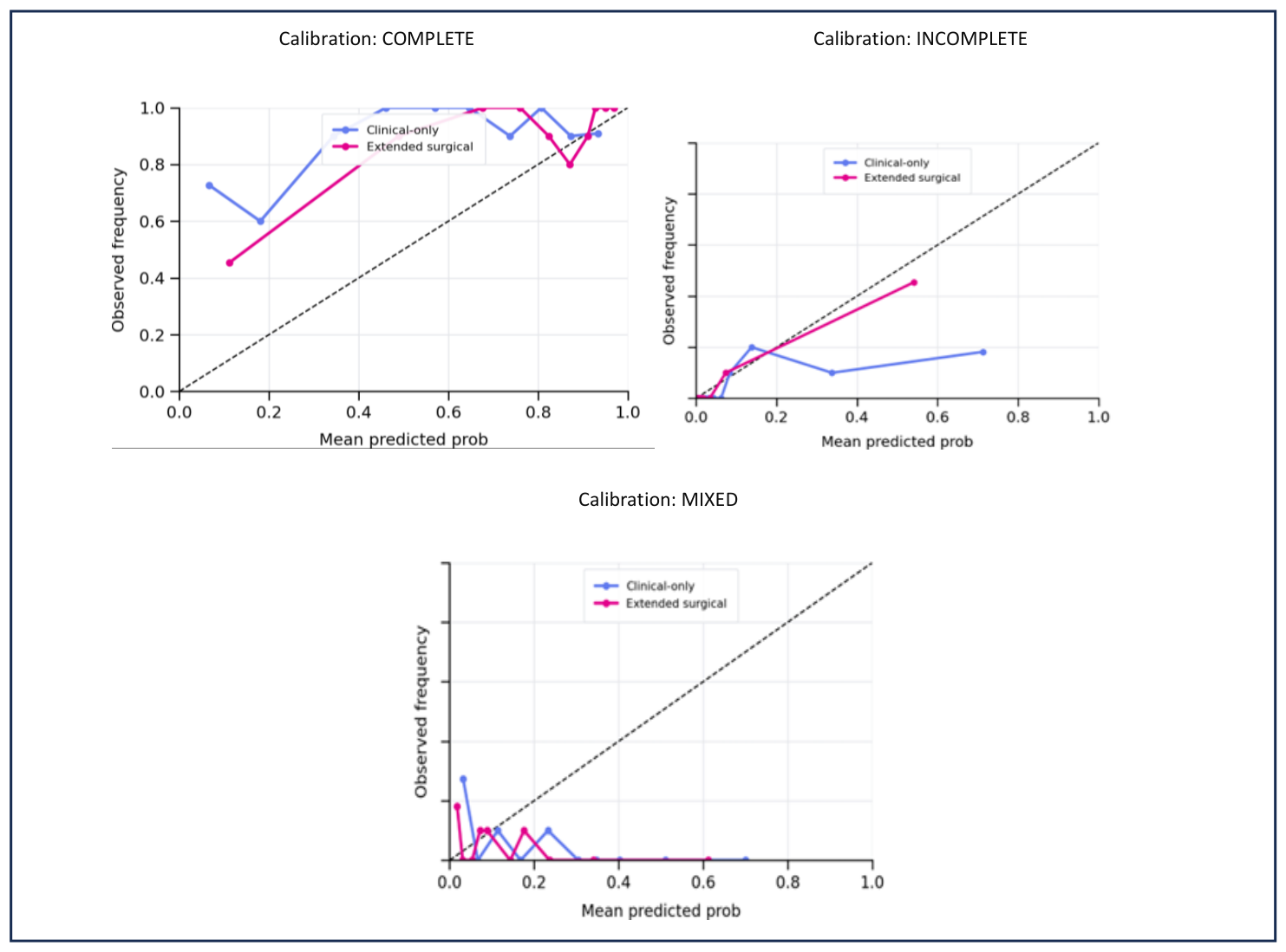
Table 7 summarizes 5 fold out of fold cross validation performance for a three class classification task (COMPLETE, INCOMPLETE, MIXED), comparing a clinical only model with an extended surgical feature set. The evaluation includes 102 cases, with marked class imbalance (91 COMPLETE, 6 INCOMPLETE, 5 MIXED), which limits the stability of estimates. Macro AUC (one vs rest) reflects average discrimination across classes and improves from 0.560 (clinical only) to 0.687 (extended). Log loss assesses the quality/calibration of predicted probabilities and improves substantially from 0.918 to 0.446, suggesting better probabilistic predictions with surgical features.
| Metric | Clinical-only | Extended surgical |
|---|---|---|
| N | 102 | 102 |
| Class counts | {‘COMPLETE’: 91, ‘INCOMPLETE’: 6, ‘MIXED’: 5} | {‘COMPLETE’: 91, ‘INCOMPLETE’: 6, ‘MIXED’: 5} |
| Macro AUC (OVR) | 0.560 | 0.687 |
| Log loss | 0.918 | 0.446 |
Table 8 is a confusion matrix for the clinical-only model’s 5 fold out of fold predictions in the three class task (COMPLETE, INCOMPLETE, MIXED). Rows are the true class and columns are the predicted class; values are the number of cases. Correct predictions are on the diagonal: 63 COMPLETE→COMPLETE, 2 INCOMPLETE→INCOMPLETE, and 0 MIXED→MIXED. The main errors are that many true COMPLETE cases are predicted as INCOMPLETE (11) or MIXED (17), and most true INCOMPLETE cases are predicted as MIXED (4). All true MIXED cases are misclassified (3 as COMPLETE, 2 as INCOMPLETE), indicating poor recognition of the MIXED class.
| True Label | Predicted Complete | Predicted Incomplete | Predicted Mixed |
|---|---|---|---|
| True Complete | 63 | 11 | 17 |
| True Incomplete | 0 | 2 | 4 |
| True Mixed | 3 | 2 | 0 |
Table 9 is also a confusion matrix for the same three-class task (COMPLETE, INCOMPLETE, MIXED), but presented in a more compact “alternate” layout. Like Table 8, rows are the true labels and columns are the predicted labels, so the diagonal cells are correct classifications. In Table 9, the model correctly predicts 84/91 COMPLETE cases, 5/6 INCOMPLETE cases, and 0/5 MIXED cases. The main errors are misclassifying COMPLETE as MIXED (6) and a small number of COMPLETE as INCOMPLETE (1), plus misclassifying MIXED as COMPLETE (4) or INCOMPLETE (1). Overall, MIXED is not learned well.
| True Label | Predicted Complete | Predicted Incomplete | Predicted Mixed |
|---|---|---|---|
| True Complete | 84 | 1 | 6 |
| True Incomplete | 0 | 5 | 1 |
| True Mixed | 4 | 1 | 0 |
In this single-center retrospective cohort of head and neck NMSC, basal cell carcinoma predominated and lesions clustered in chronically sun-exposed anatomic regions, particularly the cheek and nose. This distribution is consistent with the well-established role of cumulative UV exposure in keratinocyte carcinoma pathogenesis and with prior reports showing a high burden of disease in the facial “H-zone”, where complex anatomy may constrain excision margins.
Among conventionally excised lesions, the proportion of positive margins (R1) was relatively low (8%). Lateral margin involvement was more frequent than deep margin involvement, which may reflect the practical tendency to preserve functionally and cosmetically sensitive tissue laterally in the head and neck. Although the number of R1 cases was small, positive margins appeared concentrated in high-risk sites (e.g., nose, periocular, auricular, scalp), supporting the view that anatomic constraints and tumor subclinical spread can increase incomplete excision risk in these areas. In clinical practice, these findings highlight the importance of careful preoperative risk stratification and consideration of margin-controlled techniques when feasible in high-risk locations.[16]
Although conventional surgery remains the cornerstone of treatment, advanced imaging modalities such as PET-CT have shown value in assessing tumor extent and vascular involvement in other malignancies, highlighting the growing role of functional imaging in oncologic decision-making. [17]
When interpreted against published data, the 8% rate of positive margins in our conventionally excised cohort appears lower than rates reported in several head and neck BCC series focused on higher-risk lesions, where incomplete excision has been reported around 15–22% overall and may approach ~30–37% in particularly challenging facial sites such as the nose, periorbital region, ear, and scalp. In cutaneous SCC, a systematic review has estimated an overall incomplete excision rate of approximately 13%, with head and neck location repeatedly identified as a risk factor. Differences in case mix (including inclusion of precursor lesions), surgeon specialty, excision technique, and pathology processing may account for substantial between-study variability; nevertheless, these external estimates provide a useful benchmark suggesting that even in tertiary practice, incomplete excision remains a clinically relevant outcome that warrants ongoing audit and process optimization.[18]
The predominance of lateral margin involvement in our R1 cases may be explained by the competing priorities of oncologic clearance and tissue preservation in cosmetically and functionally critical facial units. In routine wide local excision, the “tightest” margin is often the lateral margin because it determines final defect size and reconstructive complexity; by contrast, the deep plane is frequently taken down to a relatively fixed anatomic layer. In addition, certain BCC growth patterns can extend subclinically beyond visible tumor borders, increasing the risk of lateral positivity despite apparently adequate clinical margins. From a practical standpoint, these observations support a workflow in which tumors in high-risk sites or with suspected aggressive behavior are considered early for margin-controlled approaches (e.g., Mohs micrographic surgery or staged excision when available), and where conventional excision is performed, clear specimen orientation and explicit reporting of lateral versus deep margin involvement are emphasized to facilitate timely and anatomically targeted re-excision.[19]
For head and neck lesions—particularly in high-risk facial subunits (perinasal, periocular, auricular, and scalp)—preoperative counseling should address the possibility of positive margins and the potential need for staged management (re-excision or margin-controlled surgery where available). When conventional excision is selected, careful clinical border assessment (including dermoscopy when appropriate), documentation of planned margins, and close communication with pathology regarding margin orientation may reduce avoidable R1 resections. Finally, because reconstruction choices can limit re-excision options, surgeons should consider a reconstruction approach that preserves tissue and defers complex flap reconstruction when margin uncertainty is high, or alternatively plan for margin-controlled approaches in lesions with higher pretest risk.[20]
A specific implication for head and neck surgery is the interaction between margin uncertainty and reconstruction timing. Complex local flaps can distort anatomic planes and may complicate subsequent margin-directed re-excision if an R1 result is returned. For lesions with higher pretest risk of incomplete excision (high-risk sites, suspected aggressive subtype, larger diameter, deeper invasion, or perineural involvement), a pragmatic strategy is to either pursue a margin-controlled technique up-front, or adopt a staged approach with temporary defect management (or simpler closure) until histologic clearance is confirmed, followed by definitive reconstruction when appropriate. Embedding this decision-making into preoperative counseling may reduce patient distress when additional procedures are needed and may help preserve reconstructive options.[21]
Direct closure was the most common reconstructive approach, while local flaps accounted for approximately one quarter of reconstructions. This pattern is expected in a cohort with predominantly small-to-moderate tumor sizes and emphasizes the value of anticipating closure options during surgical planning. In particular, transposition flaps were the most frequently used flap subtype, reflecting their versatility for common facial defects. Systematically recording reconstruction methods, as done in this study, can support local audits and may help identify anatomic regions where more advanced reconstruction is more likely to be required.
Future work should prioritize prospective follow-up to capture recurrence and the downstream management of R1 cases (re-excision, observation, adjuvant therapy where relevant), since margin status is a surrogate outcome and may not translate directly into clinically meaningful events in all patients. Larger multi-center datasets would also allow more robust multivariable modeling with adequate event counts and could separate analyses for BCC versus invasive SCC, while treating precursor lesions (AK, Bowen’s disease, keratoacanthoma) as a distinct category or sensitivity analysis. Finally, standardizing the recording of planned clinical margins, specimen orientation, and pathology reporting within institutional pathways would support both quality improvement and external comparability across studies.[22]
This study has several limitations. First, its retrospective, single-center design and short accrual window (September 2025–January 2026) may limit generalizability and can amplify local referral patterns, seasonal effects, and institutional practice preferences. In addition, selection and referral bias are possible: complex head and neck tumors may be overrepresented in a tertiary service, whereas very high-risk lesions may be preferentially referred for margin-controlled surgery in other settings, which could influence observed R1 rates.
Second, the analysis was performed at the lesion level, and some patients contributed multiple tumors; without explicit methods to account for within-patient clustering, independence assumptions may be violated. Procedures were performed by multiple clinicians, and variability in margin selection, specimen orientation, and reconstructive approach may confound associations. Margin status itself is subject to routine histopathology constraints (orientation, inking, sectioning) and incomplete documentation may lead to misclassification of lateral versus deep involvement or underestimation of true positive margins. Tumor dimensions were reconstructed from clinical measurements and pathology reports, which may introduce measurement error and reduce comparability across cases.
Third, the cohort included invasive SCC as well as SCC precursor lesions and keratoacanthoma, which broadens clinical scope but complicates comparisons with studies restricted to invasive tumors. AJCC staging and documentation of nodal/metastatic assessment were not uniformly available, limiting risk stratification. Prospective follow-up was not available to evaluate recurrence after the index excision; prior surgery history within 24 months is not equivalent to post-treatment recurrence. Finally, the small number of R1 events creates substantial class imbalance and limits statistical power; any multiple testing or predictive metrics presented should be regarded as exploratory and hypothesis-generating until supported by a prespecified analysis plan, fuller methodological reporting, and external validation.
Head and neck NMSC in this cohort occurred predominantly in elderly patients and was most frequently located on the cheek and nose. Conventional excision achieved complete margins in most cases, with positive margins occurring in a small subset and tending to involve high-risk anatomic sites. Direct closure was the most common reconstruction, while local flaps—predominantly transposition flaps—were required in roughly one quarter of conventionally excised cases. Prospective follow-up and larger multicenter datasets are needed to better quantify risk factors for incomplete excision and to evaluate prediction approaches and outcomes, including recurrence.
The authors declare no conflict of interest. This research received no external funding.
Authors’ contribution: Conceptualization, M.M., M.D. and A.C.; methodology, R.C. and G.M.; software, A.Z.C.A. and D.V.; validation, M.M., C.S. and A.C.; formal analysis, M.D. and R.C.; investigation, M.M. and C.S.; resources, G.M. and A.Z.C.A.; data curation, M.D. and D.V.; writing—original draft preparation, M.M. and M.D.; writing—review and editing, M.M. and R.C.; visualization, G.M. and D.O.C.; supervision, C.S. and D.O.C.; project administration, A.C. and D.V.; funding acquisition, A.Z.C.A. and D.O.C. All authors have read and agreed to the published version of the manuscript.
The study was conducted in accordance with the Declaration of Helsinki and approved by the Local Ethics Committee of the Dr. Carol Davila University Emergency Central Military Hospital, Bucharest (registration number).
Not applicable, because it is a retrospective study.