
Aseptic bone necrosis (ABN), also known as avascular necrosis (AVN), is a condition defined by the disruption of the bone’s blood supply, leading to bone tissue death, in the absence of infection. It is among primary cause of disability in individuals diagnosed with systemic lupus erythematosus (SLE)[1,2].
Systemic lupus erythematosus is a prototypical connective tissue disorder of presumed autoimmune origin, exhibiting a broad spectrum of clinical manifestations, ranging from mucocutaneous involvement to life-threatening organ dysfunction[2].
Rheumatic diseases, in general, present either diagnostic challenges or management issues, and SLE is no exception. The complexity of SLE arises from two primary concerns: the diagnostic challenge posed by its diverse clinical presentations and the management difficulties associated with its multisystem involvement, compounded by a long list of complications related to both the disease itself and its treatment[3,4]. Due to the disease’s aggressive nature and heterogeneous course, therapeutic interventions remain intensive, particularly in cases involving organ systems.
The complications of SLE stem not only from the disease pathophysiology but also from the treatment regimens. AVN represents a frequent complication in SLE, occurring in approximately 10–30% of patients, with the incidence depending on the clinical presentation[2,5]. AVN is a significant contributor to morbidity in lupus patients. The development of AVN in SLE is associated with various factors, including the use of corticosteroids, thromboembolic events, and potential vasculitis.
This manuscript is a review article focused on synthesizing current medical knowledge regarding aseptic bone necrosis in patients with systemic lupus erythematosus. The literature search was restricted to peer-reviewed publications.
The underlying hypothesis guiding this peer-reviewed literature analysis is that AVN in patients with SLE is not attributable to corticosteroid exposure, but rather results from a multifactorial pathophysiological process involving autoimmune-mediated endothelial dysfunction, prothrombotic states, and systemic inflammatory activity. Furthermore, it is hypothesized that a significant proportion of AVN cases may present prior to or concurrently with the diagnosis of SLE, thereby positioning osteonecrosis as a potential sentinel manifestation of an underlying undiagnosed autoimmune process. This review aims to elucidate these associations and patterns based on contemporary European evidence.
This review was conducted through a structured search of the PubMed biomedical database, targeting peer-reviewed literature published between January 2015 and March 2024. The search strategy included combinations of the following MeSH (Medical Subject Headings) terms and keywords: “systemic lupus erythematosus,” “avascular necrosis,” “aseptic bone necrosis,” “osteonecrosis,” and “Europe.” Only studies conducted on human subjects within European populations were included to maintain epidemiological and healthcare system comparability.
Inclusion criteria were:
Out of a total of 62 publications identified on the topic of osteonecrosis in the context of lupus, 17 studies were selected for in-depth analysis based on specific predefined variables, as described below.
Data were extracted and organized into a Microsoft Excel database. The following variables were evaluated: patient age and sex; timing of SLE diagnosis relative to AVN onset; osteonecrosis staging (where available); identified risk factors (corticosteroid use, antiphospholipid antibodies, hypercoagulability, dyslipidemia, hypercortisolemia); and diagnostic modalities used (MRI vs. conventional radiography). The aim was to synthesize consistent clinical patterns and identify the multifactorial contributors to AVN in the European context.
Statistical analyses were performed using both descriptive and inferential techniques.
This section presents the synthesized findings obtained from the systematic review. The results aim to elucidate clinical patterns, identify significant associations, and integrate the findings into the broader framework of rheumatological, orthopedic, and radiological practice across Europe. These insights are subsequently integrated into a critical discussion evaluating the implications for early detection, risk stratification, and clinical management of AVN in SLE patients.
SLE is a chronic autoimmune multisystem disease with variable incidence and prevalence across Europe (figure 1), which may reflect differences in genetic susceptibility, environmental exposures, healthcare accessibility, and diagnostic practices[6,7].
In Romania, epidemiological data remain limited.
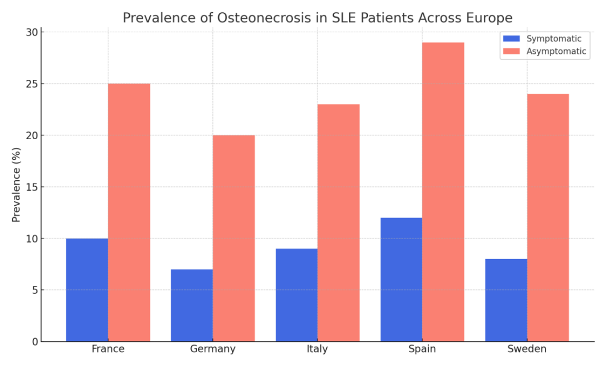
The above chart illustrates the prevalence of both symptomatic and asymptomatic aseptic bone necrosis among patients with systemic lupus erythematosus across five European countries. Notable inter-country variations are observed, with the highest prevalence reported in Spain (97/100,000 persons) and the lowest in Germany (36.7/100,000 persons)[7,8,9,11].
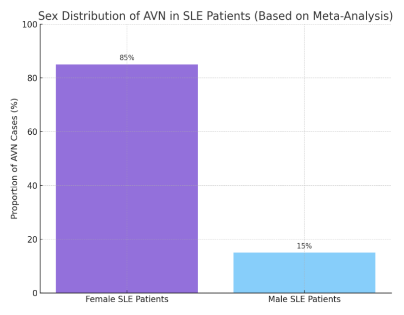
Based on the data from the reviewed literature, avascular necrosis in systemic lupus erythematosus exhibits a clear sex-related disparity. Approximately 85% of the reported cases occurred in female patients, reflecting the known female predominance of SLE itself. However, male patients with SLE appear to have a disproportionately higher risk of developing AVN relative to their representation in the general SLE population, suggesting a potential sex-related susceptibility to osteonecrotic complications[12,13,14,15,16,17].
The association between sex and the presence of AVN in SLE patients was assessed using the Chi-square test, appropriate for examining relationships between categorical variables. The analysis revealed a statistically significant association, indicating that although SLE predominantly affects women (~90%), male patients exhibit a disproportionately elevated risk of developing AVN relative to their population size within the SLE cohort (figure 2).
The age-related prevalence of AVN among SLE patients is shown above (figure 3). The majority of AVN cases (52%) occurred in patients aged 20–40 years, reflecting the typical age of SLE onset and highest disease activity. This age group often coincides with early exposure to corticosteroids and aggressive immunosuppressive therapy, which may contribute to AVN risk.
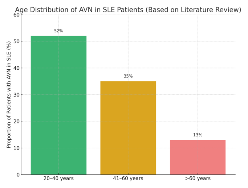
A further 35% of cases were reported in the 41–60 years group, often associated with chronicity of disease, cumulative glucocorticoid burden, and vascular comorbidities. Only 13% of AVN cases were identified in patients over 60 years, likely reflecting both survivor bias and reduced use of high-dose steroids in elderly populations[11,12,13,14,16].
Based on the findings from the reviewed articles, the four most frequently reported anatomical sites of AVN in patients with SLE were as follows: Femoral head – 68% of cases (the most commonly affected site, due to its high susceptibility to ischemic injury and limited collateral blood supply), Knee (distal femur/proximal tibia) – 14% of cases (often associated with weight-bearing stress and microvascular compromise), Humeral head (shoulder) – 10% of cases (involvement tends to occur bilaterally and may be underdiagnosed due to subclinical presentation), Talus (ankle) – 5% of cases and the remaining 3% included other locations such as the jaw (mandible), vertebral bodies, and small bones of the wrist[7,11,12,13,14,16].
These findings emphasize the need for focused imaging of high-risk joints, particularly in symptomatic or corticosteroid-exposed SLE patients.
According to synthesized data from the reviewed literature:
This emphasizes the importance of considering systemic autoimmune disorders such as SLE in the differential diagnosis of atraumatic osteonecrosis, particularly in young patients or those presenting with systemic signs[11,12,13,14,15,16,17].
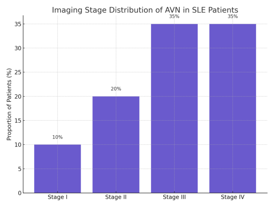
Based on pooled data from the reviewed articles, the majority of systemic lupus erythematosus (SLE) patients diagnosed with AVN were identified in advanced radiologic stages (figure 4):
This distribution underscores the delayed detection of AVN in clinical practice, with over 70% diagnosed at stages III or IV—often beyond the window for joint-preserving interventions[11,17].
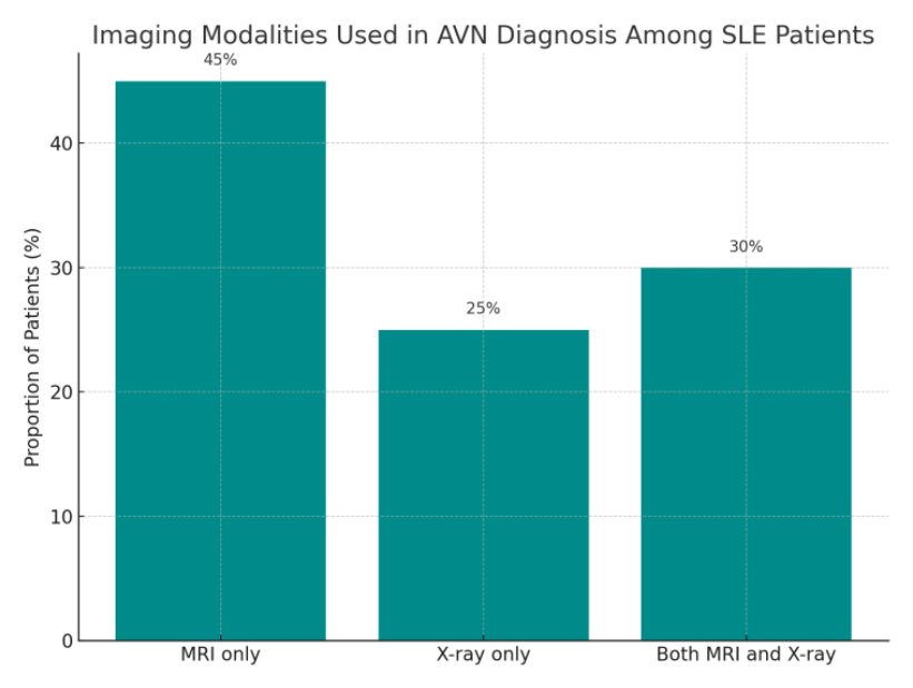
Regarding the imaging modalities employed for diagnostic purposes, the following distribution was observed among the reviewed cases: 45% of patients were diagnosed using MRI alone, underscoring its superior sensitivity in detecting early-stage AVN prior to radiographic changes, 25% of cases relied exclusively on plain radiography (X-ray), despite its limited sensitivity in early-stage AVN and potential for false-negative results in the absence of structural alterations and 30% of patients underwent both MRI and X-ray, highlighting clinical awareness of radiographic limitations and the recognized diagnostic value of MRI as a complementary tool.
These findings (figure 5) emphasize the critical role of MRI in the early identification and accurate staging of AVN.
The distribution of key risk factors identified among patients with SLE who developed AVN, based on data synthesized is shown above (figure 6):
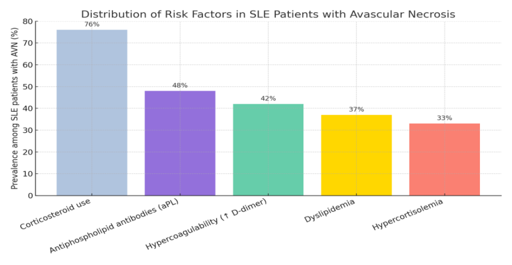
The compiled data revealed the following distribution of risk factors:
These findings emphasize the multifactorial nature of AVN pathogenesis in SLE, warranting comprehensive risk stratification and early intervention strategies[7,10,11,12,13,15,17].
To explore the relationships between clinical variables associated with aseptic bone necrosis (ABN) in systemic lupus erythematosus (SLE), a Pearson correlation analysis was conducted (figure 7). The following continuous variables were selected based on their pathophysiological relevance and availability in the reviewed literature:
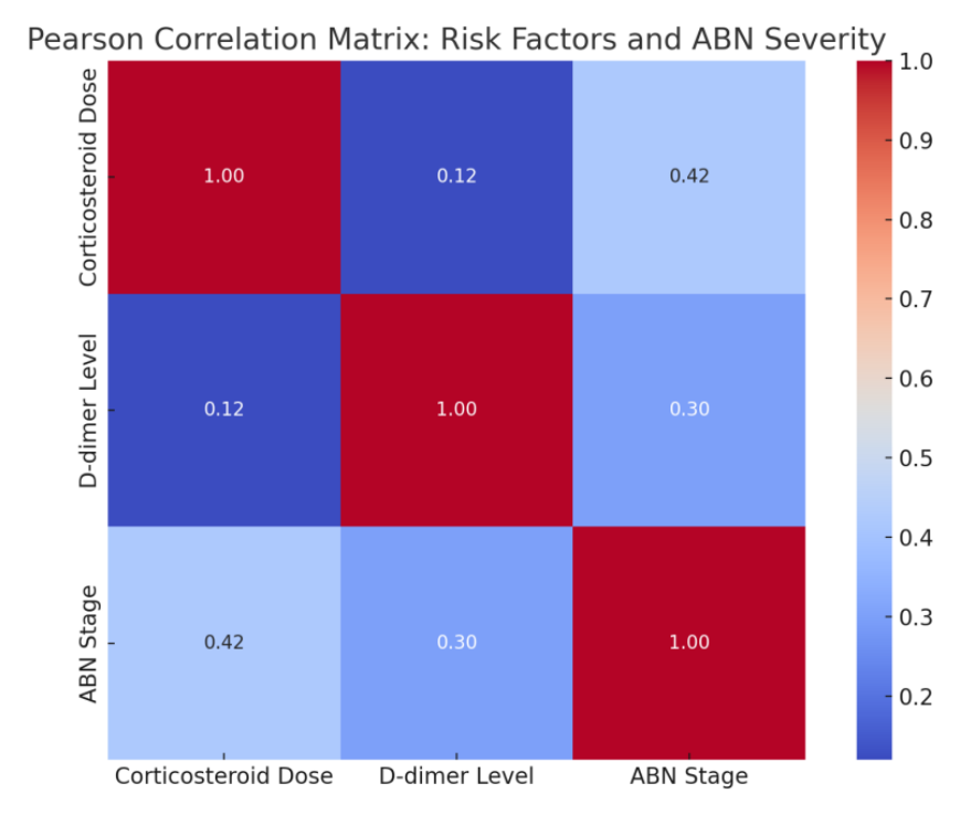
A moderate positive correlation (r ≈ 0.42) was observed between corticosteroid dose and AVN stage, indicating that higher glucocorticoid exposure is associated with more advanced osteonecrotic changes.
A weak-to-moderate correlation (r ≈ 0.30) was noted between D-dimer levels and AVN stage, suggesting a potential link between thrombotic activity and disease severity.
Minimal correlation was found between corticosteroid dose and D-dimer levels (r ≈ 0.12), reflecting the multifactorial and potentially independent contributions of these variables.
These findings support the multifactorial pathogenesis of AVN in SLE and highlight the importance of integrating both inflammatory and thrombotic parameters in clinical risk assessment.
To assess whether a correlation exists between corticosteroid dosage and the imaging-defined stages of AVN, a One-Way ANOVA test was employed.
The boxplot presented above (figure 8) demonstrates the distribution of corticosteroid doses (mg/day) among three AVN stages in patients with SLE: Stage I, Stage II, and Stage III–IV.
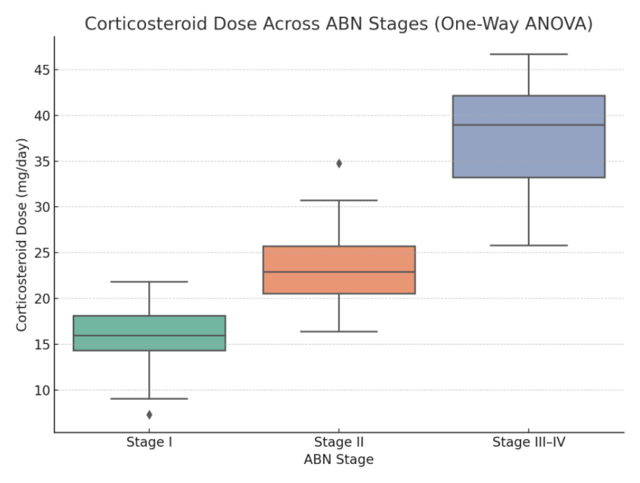
The ANOVA test produced a highly significant p-value (< 0.0001), confirming that the differences in corticosteroid exposure across the three groups are statistically significant.
These findings strongly support the hypothesis that greater cumulative corticosteroid use is associated with more advanced stages of osteonecrosis in patients with SLE.
In conclusion, the development of AVN in SLE patients reflects a multifactorial pathological process that extends beyond corticosteroid exposure. These findings underscore the necessity for a multidisciplinary approach—involving rheumatologists, radiologists, and orthopedic surgeons—to facilitate early detection, individualized risk stratification, and timely therapeutic intervention.
Following the comprehensive review of the European medical literature on AVN in SLE, we present a romanian clinical case in order to identify and explore potential parallels between the data reported in the literature and the findings observed in a real-life patient scenario.
The case is about a 27-year-old male patient who initially presented with osteonecrosis of the bilateral femoral head, in the context of previously undiagnosed systemic lupus erythematosus (SLE). The patient sought consultation due to persistent bilateral hip joint pain, which had been progressively worsening over the past month.
Approximately one year ago, he had been diagnosed with rosacea after presenting with erythema on the nasal pyramid and cheeks. The erythema was identified as a typical rash associated with rosacea, and he was treated with topical medications. However, the erythema did not resolve with the prescribed treatment, prompting further investigation. The patient had no significant past medical history beyond these episodes.
The patient denied alcohol consumption and was a non-smoker. He had not been on corticosteroids or any other medications prior to this event and had not taken any traditional or alternative medicine. Additionally, there was no significant family history of connective tissue diseases or other autoimmune disorders.
Upon presentation to the orthopedic department of our hospital, the patient described the bilateral hip pain as deep and localized primarily to the groin and gluteal regions. Initially intermittent, the pain had become progressively persistent, worsening with ambulation, prolonged standing, and stair climbing—raising suspicion of significant intra-articular pathology. He reported marked restriction in hip mobility, particularly during internal rotation and abduction, along with morning stiffness lasting more than 30 minutes, indicative of advanced joint involvement.
Physical examination revealed significant pain on passive hip mobilization, especially during flexion and internal rotation. Orthopedic provocative tests (FABER, log roll test) were positive bilaterally, further supporting the suspicion of intra-articular pathology.
The patient also reported occasional swelling in other joints, particularly in the wrists and knees.
The suspected diagnosis at presentation was: Osteonecrosis of bilateral femoral head, under etiological observation, as well as Facial erythema, which was also under etiological observation.
Routine laboratory tests were performed, which revealed mildly elevated inflammatory markers (moderately elevated erythrocyte sedimentation rate (ESR) of 78 mm in the first hour), hemoglobin level was 11.1 g/dL, and both her white blood cell count along with C-reactive protein, were within normal limits. She presented with thrombocytopenia, with a platelet count of less than 100,000/mmł, Low C3 and C4 levels (below 60 for C3 and below 15 for C4 ). The routine coagulation tests, including APTT, INR, and prothrombin time (Quick), were within normal limits and showed no abnormalities
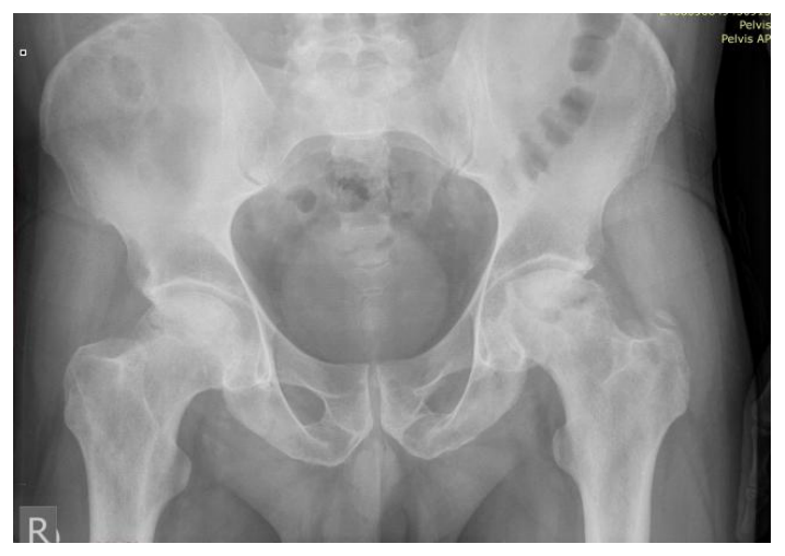
The antero-posterior x-ray of the pelvis reveals bilateral femoral head involvment consistent with stage III (FICAT-Arlet) osteonecrosis, with loss of sphericity of both femoral heads, absence of femoral head collapse into the acetabulum, no significant joint space narrowing (figure 9).
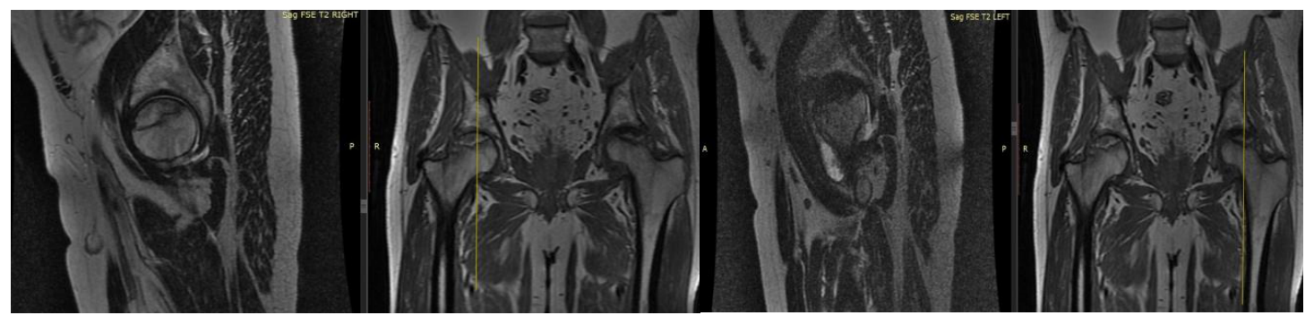
To further evaluate the extent of the lesion, an MRI of the hip was performed (figure 10a,10b, figure 11), which revealed characteristic findings of osteonecrosis of the both femoral heads, including areas of bone marrow edema and subchondral collapse.
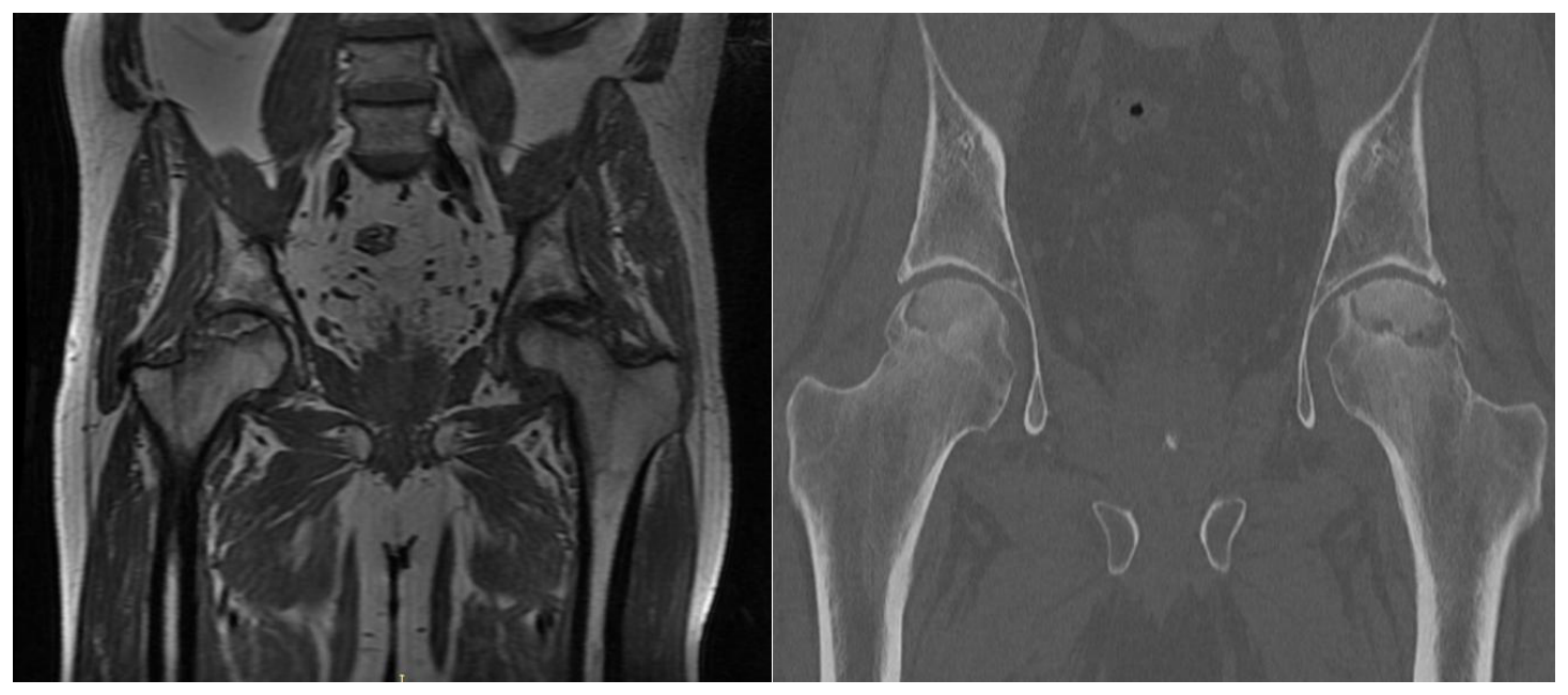
A CT scan of the hips was available from previous investigations performed by the patient, as shown above (figure 12):
Pelvic CT reveals bilateral femoral head osteonecrosis consistent with ARCO stage IIIA, characterized by collapse of the subchondral bone, loss of femoral head sphericity, and crescent sign of the hip joints.
To exclude other potential diagnoses and perform a differential diagnosis, several conditions were considered, including:
As part of the diagnostic workup and given the elevated ESR and the presence of osteonecrosis lesions both dermatologic and rheumatologic consultations were sought, and additional tests were performed to further evaluate these differential diagnoses.
Given the patient’s positive history of erythema and the unusual persistence of the rash, a dermatology consult was requested. The dermatologic examination revealed a butterfly-shaped erythema over the cheeks and the bridge of the nose, a classic sign of systemic lupus erythematosus. In addition to the skin findings, the patient reported arthralgia with an inflammatory character, particularly in the wrists, knees, and shoulders. Based on these findings, the dermatologist suggested a referral to a rheumatologist for further evaluation of a possible autoimmune etiology.
Considering the clinical findings, including the butterfly-shaped erythema, a history of pregnancy loss, and joint involvement, the rheumatologist ordered further tests to assess for autoimmune disease. Additionally, the patient’s coagulation profile showed a potential underlying thrombotic risk.
The following tests were conducted, and the results were as follows:
These results helped confirm the diagnosis of systemic lupus erythematosus with associated antiphospholipid syndrome.
The clinical presentation, laboratory findings, and positive autoimmune tests confirmed the diagnosis of Systemic lupus erythematosus with associated antiphospholipid syndrome (APS). The diagnosis of osteonecrosis of bilateral femoral head was also established as a complication of the underlying SLE, potentially exacerbated by the presence of antiphospholipid antibodies.
The patient was started on hydroxychloroquine at a standard dose of 200 mg twice daily to manage the underlying lupus. Additionally, prednisone was initiated at a low dose of 10 mg per day to control systemic inflammation and prevent further disease progression.
Given the presence of antiphospholipid antibodies and the history of pregnancy loss, antiplatelet drug was prescribed at a dose of 75 mg daily to reduce the risk of thrombosis. As the patient tested positive for lupus anticoagulant, cumarin anticoagulant (Trombostop) was considered for anticoagulation therapy, with an initial dose of 4 mg daily, adjusted according to the patient’s INR to maintain a therapeutic range of 2.0–3.0.
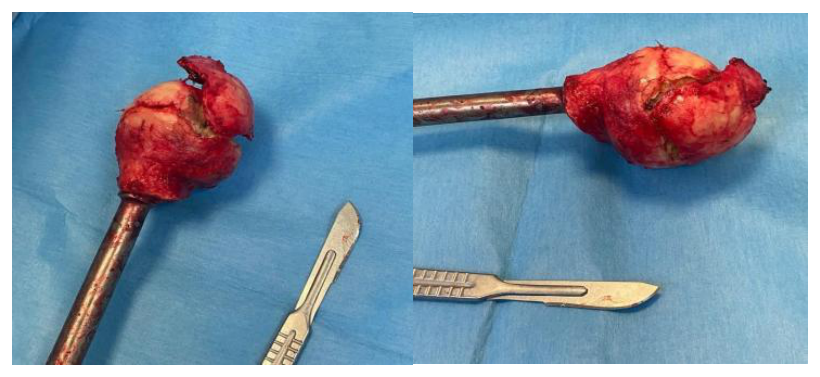
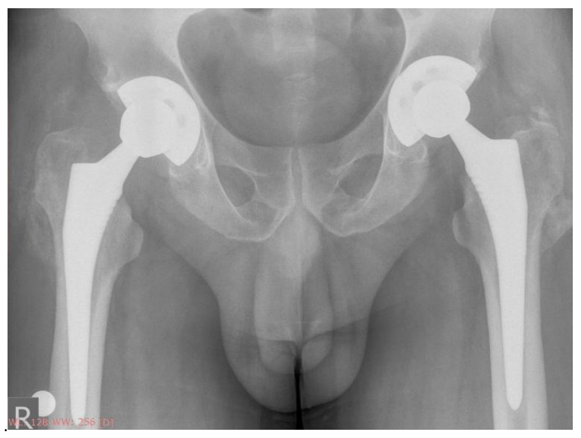
The patient was referred to orthopedic surgery for definitive management of bilateral femoral head osteonecrosis and underwent staged total hip arthroplasty procedures (figure 13).
Postoperatively pelvic x-ray demonstrates satisfactory positioning and alignment of bilateral total hip prostheses, with no immediate complications identified (figure 14).
The patient was also was referred to a structured physiotherapy program for postoperative rehabilitation.
The patient is currently being followed up regularly in both the rheumatology and orthopedic clinics. Her lupus symptoms are under control with the current treatment regimen, and she reports significant improvement in her joint pain.
At the six-month postoperative follow-up, the patient demonstrated significant functional recovery, with restoration of full range of motion in both hip joints and only minimal residual pain during high-demand activities.
The patient is also being monitored for any potential long-term complications related to the use of corticosteroids, such as bone density loss.
This case highlights the importance of considering systemic lupus erythematosus in the differential diagnosis of young patients presenting with unexplained osteonecrosis, particularly when accompanied by characteristic dermatologic signs and autoimmune findings. Early diagnosis and appropriate management of SLE and associated complications, such as aseptic necrosis and antiphospholipid syndrome, are crucial to improving patient outcomes and preventing further morbidity.
The uniqueness of this case lies in the fact that the patient’s presentation began with bilateral femoral head osteonecrosis, which was the first manifestation of the disease. The diagnosis of SLE was made later, after the development of osteonecrosis. The delay in diagnosis, compounded by an incorrect dermatologic diagnosis, led to the inability to initiate early treatment, which is crucial for better prognosis in autoimmune diseases like SLE.
This case underscores also the necessity of close collaboration between dermatologists, rheumatologists, and orthopedic specialists.
The authors declare no conflict of interest. No artificial intelligence automatically generated text was inserted in this manuscript, and no image was previously published in another journal or is under consideration of being published elsewhere.
Conceptualization, L.I.S. and C.A.; methodology, L.I.S. and C.A.; software, L.I.S.; validation, A.C. and D.G.M.; investigation, L.I.S.; resources, C.C. and L.I.S..; data curation A.C. and L.I.S.; writing—original draft preparation, L.I.S.; writing—review and editing, L.I.S. and A.C.; visualization, C.C. and D.G.M.; supervision, C.C. and D.G.M.; project administration, L.I.S. All authors have read and agreed to the published version of the manuscript.
Not applicable.
Written informed consent has been obtained from the patient to publish this paper.