
Knee osteoarthritis (KOA) is a chronic, degenerative disease that affects not only the bone and cartilage but also all periarticular soft tissues such as menisci, ligaments, synovial tissue, tendons, and muscles. Osteoarthritis most commonly affects the knee, followed by the hip joint. Pain dominates the symptomatology, leading to a progressive limitation of joint mobility and an increase in patient morbidity. In the context of an increase in life expectancy and an ageing population, knee osteoarthritis is becoming a public health problem both through the complications caused by the disease and through the high costs associated with care and treatment. Approximately 240 million people worldwide suffer from osteoarthritis, which makes understanding the pathogenic mechanisms, identifying risk factors and identifying disease-modifying drugs a priority[1]. For patients with a significantly impaired quality of life, total knee arthroplasty is recommended. The identification of effective biomarkers is essential for the early diagnosis and treatment of knee osteoarthritis. Recent studies have shown that osteoarthritis is not just a disease induced by mechanical destruction of articular cartilage, and the main pathogenic mechanisms involved are the immune system and inflammatory mechanisms[2-4]. Imaging, pathological, and clinical results suggest that synovial alterations manifest prior to the pathological deterioration of cartilage, while synovitis, characterized by the recruitment of immune cells, is present throughout the entire developmental course of osteoarthritis[5,6]. Transcriptomic investigations in osteoarthritis have recently advanced due to the introduction of RNA sequencing. RNA sequencing has been used to elucidate significant molecular differences between groups of patients with knee osteoarthritis. In patients with KOA who experience significant pain, transcriptomic analysis of synovial tissue from those undergoing total knee arthroplasty reveals differential regulation of 32 genes
(mRNA), two long non-coding RNAs (lncRNAs) and 35 micro-RNAs (miRNAs). This regulation appears independently of radiological severity or gender[7]. Fisch et al. have identified a series of transcription factors, prioritising eight as high-priority candidates for therapeutic intervention. Among these, JUN, JUND, FOS and FOSL2 encode different subunits of AP-1. The role of AP-1 in chondrocytes requires further elucidation. Studies suggest that IL-1 promotes the synthesis of MMP13 proteases via activation of JUN/FOS heterodimers[8-10]. The expression of growth and differentiation factor 5 (GDF5) poses a significant risk for the development of osteoarthritis of the knee.
Murakami T et al. showed that GDF5 expression was amplified in post-injury articular cartilage of the medial meniscus and was increased in human articular cartilage of patients with KOA, according to the findings obtained via immunohistochemistry and microarray analysis[11].
Zhang Q. et al. identified a diagnostic model based on the expression of four genes, which shows significant predictive performance in knee osteoarthritis. The identified genes are implicated in the TNF signalling pathway and the IL-17 signalling pathway, along with their roles in the response to steroid hormones, glucocorticoids and corticosteroids. Notably, four of these genes- MYC, JUN, DUSP1, and NFKBIA- are specifically expressed in the immune system.
Clinical studies have demonstrated a consistent change in these four genes in synovial tissue obtained from patients with KOA[12].
Gene expression analysis allows the comprehension of the pathophysiological mechanisms, the identification of potential therapeutic targets, and the detection of patients at risk of adverse outcomes requiring total knee arthroplasty. Genetic studies have prompted a trend to classify knee osteoarthritis from a molecular perspective, rather than merely on osteocartilaginous lesions.
A total of 29 patients were selected who presented to the orthopedics and traumatology department of the University Emergency Hospital of Bucharest with knee pain and were divided into two groups. Group 1 comprised 26 patients, aged between 59 and 81 years, who were diagnosed with stage four KOA according to the Kellgren and Lawrence (KL) classification and were scheduled for total knee arthroplasty. Group 2 consisted of three patients who constituted the control group. These patients, aged between 30 and 45 years, did not manifest any radiological or MRI signs of knee osteoarthritis. They required arthroscopic surgery for pathologies associated with the meniscus or anterior cruciate ligament. The study was conducted from January 1, 2023, to March 31, 2024. The inclusion criteria for group 1 comprised patients aged over 45 with knee pain and radiographic evidence of KOA. The exclusion criteria included patients with recent trauma, signs of active osteoarticular infection, a lack of radiological changes in KOA demanding a differential diagnosis, individuals with known rheumatological conditions, and those for whom complete required data could not be obtained.
The main objective of this study is to analyze gene expression in suprapatellar synovial tissue using the RT-PCR technique in patients with knee osteoarthritis. Five genes with the potential to show expression changes in synovial tissue were selected, according to the data available to date in articles retrieved from PubMed/MEDLINE (https://pubmed.ncbi.nlm.nih.gov) and EMBASE (https://www.embase.com), focusing on studies published between January 2005 and January 2023. The five selected genes are represented by GDF5, MYC, JUN, DUSP1, and NFKBIA.
Synovial tissue harvesting in patients who required total knee arthroplasty was done through an anterior internal parapatellar approach and harvesting from the suprapatellar region of synovial tissue using a cold scalpel, in order not to alter the quality of the sample. In the case of the control group, synovial tissue harvesting was done during arthroscopic surgery, using a grasper and harvesting fragments of synovial tissue with a diameter of approximately 1 centimeter, then immersed in RNA preservation fluid. Fresh synovial tissue harvested during the surgical procedure was immediately immersed in RNAprotect Tissue stabilization reagent (Qiagen, Hilden, Germany) to prevent RNA degradation, and the sample was stored at -20°C until further processing. For each sample, approximately 30 mg of synovial tissue was cut into cubes of approximately 3 mm. Total RNA extraction from synovial tissue was performed using the RNeasy Fibrous Tissue Mini kit (Qiagen), which uses silica membrane column purification technology. The stabilised fragment of synovial tissue was placed into a homogenisation tube containing Buffer RLT (Qiagen), which was supplemented with 14.3 M β-mercaptoethanol. Tissue homogenization and complete mechanical disaggregation were performed using a TissueLyser II (Qiagen) with stainless steel beads at optimal parameters (2 × 2 minutes at 20 Hz) to ensure complete tissue lysis. RNA concentration and purity were determined spectrophotometrically using the Infinite 200 PRO multimodal plate reader (Tecan Group Ltd., Switzerland) with the NanoQuant plate. The absorbance ratio at 260 and 280 nm was between 2.0 and 2.2, indicating adequate purity of the isolated RNA. Each PCR reaction was performed in triplicate for each gene used in the experiment. Tissue homogenisation and complete mechanical disaggregation were performed using a TissueLyser II (Qiagen) with stainless steel beads at optimal parameters (2 × 2 minutes at 20 Hz) to ensure complete tissue lysis. The NanoQuant plate was utilized to determine the RNA concentration and purity spectrophotometrically using the Infinite 200 PRO multimodal plate reader (Tecan Group Ltd., Switzerland). The isolated RNA was of sufficient purity, as evidenced by the absorbance ratio of 2.0 to 2.2 at 260 and 280 nm. Each gene utilized in the experiment was subjected to a triplicate PCR reaction. A separate plate was designated for each gene, and all reactions for the samples included in the experiment were conducted on the same plate to ensure consistency and comparability in the results. The amplification efficacy of each gene was determined by utilizing the standard curve, which was generated from serial dilutions (1:10) of the amplified product. The efficacy of the dilutions was automatically determined by the LightCycler® 480 software through linear regression, utilizing the Ct values and log10 of the initial concentration. Efficiency values of over 96% were observed for all genes examined, with the exception of the GDF5 gene, which had an efficiency value of 91%. Two reference genes were employed to analyze the expression of the genes of interest (NFKBIA, DUSP1, JUN, MYC, GDF5): β-glucuronidase (GUSB) and β-actin (ACTB). These genes were chosen based on their expression stability under varying experimental conditions. The technical replicates of each gene were tested in triplicate for each sample. For each sample and gene, the average Ct value from the three replicates was calculated to provide a robust estimate and reduce technical variability. The variability among the replicates was minimal, with the maximum difference between the Ct values obtained in the replicates not exceeding 0.5 cycles, thereby confirming the reproducibility of the measurements. Relative gene expression levels were determined using the E-ΔCt analysis method. To compare gene expression between the disease group (osteoarthritis) and the healthy control group (control), the relative expression values E-ΔCt were used. These values represent the expression of the genes of interest normalized to the 2 reference genes (GUSB and ACTB), in each sample, without using a separate calibrator. Thus, E-ΔCt allows for direct comparison of gene expression between groups (Disease vs Control).
Statistical analysis was performed using IBM SPSS Statistics 27, MedCalc Software Ltd. (Version 23.2.1), Excel 2019 MSO (Version 2503) and GraphPad Prism version 10.4.1.
Differences in gene expression were identified using the nonparametric Mann-Whitney U test, where p-values ≤ 0.05 were considered significant. The Pearson coefficient (r) was used to measure the strength and direction of the linear relationship between genes. The value of the coefficient varies between 0 and +1, with a value of 0 indicating the absence of a linear relationship between the analyzed genes. To quantify the magnitude of rise or decrease in a gene’s expression on a logarithmic scale, hence aiding in the understanding of significant variations, we used log10 fold change. To effectively visualize the expression of genes of interest and to discern significant changes in gene expression, as well as to detect patterns and clusters, we employed a heatmap.
Study approval was obtained from the local ethics committee of the University Emergency Hospital of Bucharest, and informed consent was obtained from all participants, ethical approval code 40525 / 2023.
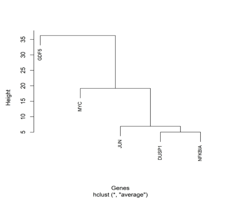
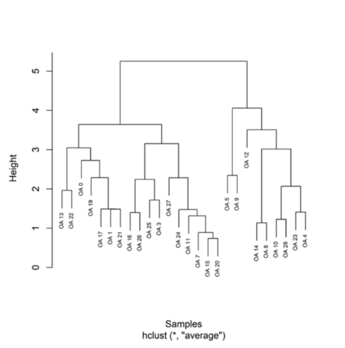
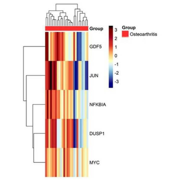
Dendrogram analysis (Fig. 1) revealed a clear separation of the investigated genes (JUN, NFKBIA, DUSP1, MYC, GDF5) into two major groups: (i) a core formed by JUN, DUSP1 and NFKBIA, which exhibited high correlations among them, and (ii) MYC and GDF5, of which GDF5 showing the most distinct expression profile. The dendrogram of the samples (Fig. 2) indicated the clustering of osteoarthritis patients into several subclusters, with pairs or groups of extremely similar samples. DUSP 1 has been identified as overexpressed in the synovial tissue of the patients with KOA. DUSP1 may indirectly regulate JUN/AP-1 activity by functioning as a phosphatase on MAPKs, which activate JUN through phosphorylation.
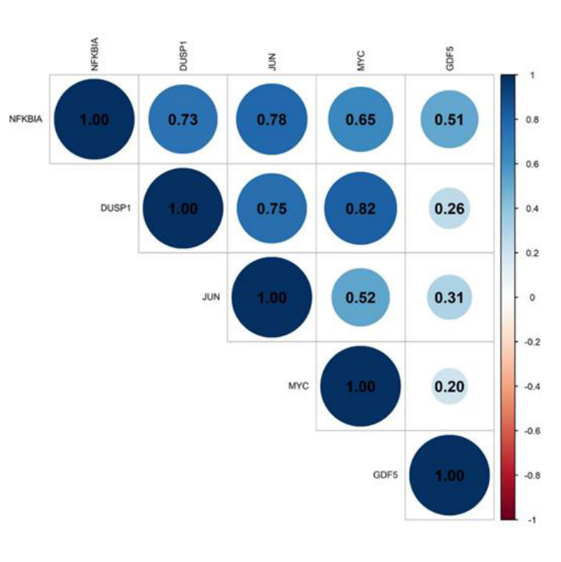
The Pearson correlation plot (Fig. 3) showed strong associations between: DUSP1–MYC (r=0.82), NFKBIA–JUN (r=0.78), DUSP1–JUN (r=0.75) and NFKBIA–DUSP1 (r=0.73). Correlations with GDF5 were weak (r=0.20–0.51), confirming the distinct profile of this gene. These findings indicate a coordinated transcriptional mechanism linking the MAPK/AP-1 and NF-κB pathways. At the same time, the positive correlation between NFKBIA and DUSP1 rather suggests a chronic inflammatory process that is self-regulating.
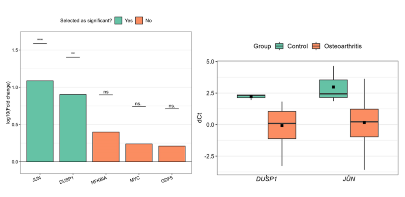
The dCt represents the discrepancy between the Ct value of the target gene and the Ct value of the reference gene. The gene’s expression is reduced when the dCt value is higher.
JUN and DUSP1 exhibit substantial fold alterations, with JUN exhibiting a greater change than DUSP1. The absence of substantial fold changes in MYC and GDF5 between the two groups implies that these genes may not be as pertinent to osteoarthritis as JUN and DUSP1.
The dCt values of DUSP1 and JUN are both higher in the control group, whereas their expression is substantially lower in the osteoarthritis group. This suggests that the expression of these genes in the synovial tissue of patients with osteoarthritis who require total knee arthroplasty has increased.
The utility of the heatmap, besides the visual representation of the results, is to observe large differences in expression between conditions to identify patterns and clusters (genes that behave similarly). The heatmap (Fig. 5) revealed clear blocks of co-regulation: JUN, DUSP1 and NFKBIA demonstrated similar patterns, MYC showed an intermediate expression, while GDF5 presented a distinct profile, characterized by considerable variations between samples.
The functions of several immune cells are essential in the onset and advancement of osteoarthritis. The timely use of diagnostic and therapeutic biomarkers could greatly improve the prognosis for osteoarthritic patients; however, there are currently few molecular candidates that meet the standards for high sensitivity and specificity.
In this study, we examined the alterations in gene expression and immune cell infiltration within osteoarthritic synovial tissue in comparison to normal control. The analysis of gene expression for JUN, DUSP1, NFKBIA, GDF5, and MYC demonstrated heterogeneous expression patterns in the synovial tissue of patients suffering from knee osteoarthritis relative to those in the control group without any signs of arthritis.
One study indicates that synovitis exacerbates pain and contributes to cartilage degradation; for every 0.1 mm of lost cartilage over a 24-month period, a Western Ontario and McMaster Universities Osteoarthritis Index (WOMAC) pain subscale score of 0.32 (95% CI: 0.21–0.44) is obtained[13].
The role of JUN in knee osteoarthritis appears to be complex and varies from case to case. Recent research suggests that elevated levels of JUN in the synovium contribute to inflammation, angiogenesis, and synovial proliferation. This process may occur through the activation of the MAPK/JNK pathways, which stimulate pro-inflammatory or degradative genes. The overexpression of JUN in the patients enrolled in the study, who are part of the group of patients with stage IV knee osteoarthritis according to Kellgren and Lawrence, is consistent with available data in previously published literature[14].
Moreover, DUSP 1 has also been found to be overexpressed in the synovial tissue of patients with KOA. Due to its role as a phosphatase on MAPKs (which activate JUN by phosphorylation), DUSP1 may indirectly regulate JUN/AP-1 activity. Thus, under conditions where DUSP1 is low, JUN activation may be more intense, favoring pro-inflammatory/degenerative phenotypes. The concomitant overexpression of JUN and DUSP1 in the synovium of patients with advanced gonarthrosis (KL IV) suggests a chronic activation of the MAPK–AP-1 pathways, alongside an anti-inflammatory compensatory feedback response through DUSP1 induction. However, the increased level of DUSP1 appears insufficient to restore synovial homeostasis, allowing for the progressive perpetuation of inflammation and contributes to the progressive degradation of the joint tissue.
Pearson correlation analysis demonstrated robust positive correlations between DUSP1–MYC (r = 0.82), NFKBIA–JUN (r = 0.78), and NFKBIA–DUSP1 (r = 0.73). These results indicate a synchronized transcriptional program that connects the MAPK/AP-1 and NF-κB pathways. The connection between DUSP1 and MYC suggests that MAPK-dependent proliferative and metabolic signaling is intertwined with feedback inhibitory mechanisms within the same cellular environment. The concurrent activation of NF-κB and AP1, two critical transcriptional pathways implicated in osteoarthritic inflammation, is reflected in the positive correlation between NFKBIA and JUN. Additionally, the correlation between NFKBIA and DUSP1 underscores the concurrent activation of feedback inhibitors—IκBα for NF-κB and MKP-1 for MAPK—which implies a chronic, self-regulating inflammation rather than an acute activation..
This expression pattern indicates a dysregulated inflammatory milieu in the end-stage synovium of osteoarthritis that is highly active. It is distinguished by the co-expression of feedback regulators (DUSP1, NFKBIA) and pro-inflammatory transcription factors (JUN, MYC). This co-activation is likely indicative of an adaptive response that is designed to reduce the excessive signaling of MAPK and NFκB. Nevertheless, this response does not achieve homeostasis; rather, it exacerbates the persistence of synovitis and tissue remodeling that are indicative of advanced KOA[12, 16].
The inconsistent expression and weak correlations of GDF5 with inflammatory markers (JUN, DUSP1, MYC, and NFKBIA) corroborate the unique profile of this gene. In contrast to genes associated with MAPK/NF-B signaling and the inflammatory response, GDF5 is involved in the processes of chondrogenic differentiation and tissue regeneration. In the synovium of patients with advanced KOA (KL IV), where chronic inflammation is predominant and regenerative processes are fragmented, GDF5 expression is heterogeneous and lacks functional coherence.
This indicates the loss of or dysregulation of local anabolic potential. Consequently, the lack of significant correlations supports the idea that GDF5 belongs to a distinct regulatory axis, separate from the proinflammatory JUN–MYC–DUSP1–NFKBIA networks. Furthermore, its role in advanced OA is likely residual or compensatory, not significantly influencing the inflammatory synovial phenotype[16].
The promising results of gene expression analysis highlight the complex transcriptomic profile of synovial tissue in patients with stage IV KL KOA who required total knee arthroplasty after conservative treatments failed. The complex changes in synovial tissue highlight the proinflammatory status of synovial tissue, leading to damage to articular cartilage. The analysis of the expression of four genes indicates that compensatory mechanisms are either insufficient or exhausted due to the chronic proinflammatory status. Proinflammatory transcription factors (JUN, MYC) and feedback regulators (DUSP1, NFKBIA) require extensive clinical trials for the use of disease-modifying drugs. The limitations of the study are induced by the limited number of patients and the limited number of the control group. Conducting a study with a large number of participants across multiple centres is also necessary to validate the statistical data obtained.
The authors declare no conflict of interest.
This research received no external funding.
Carol Davila University of Medicine and Pharmacy, 050474 Bucharest, Romania
Authors’ contribution
“Conceptualization, S.A.I., and C.F.C.; methodology E.R; software, D.G.M.; validation, C.F.C., D.G.M., and A.C.; formal analysis, M.C.; investigation, B.C.; resources, M.I.G.P.; data curation, B.S.; writing—original draft preparation, S.A.I.; writing—review and editing, S.A.I.; visualization, D.G.M.; supervision, C.F.C.; project administration, E.R; funding acquisition, A.C. All authors have read and agreed to the published version of the manuscript
The study was conducted in accordance with the Declaration of Helsinki, and approved by the Institutional Review Board (or Ethics Committee) of University Emergency Hospital of Bucharest (protocol code 40525/2023.
Informed consent was obtained from all subjects involved in the study.