
Radiology has been closely linked to the degree of technological evolution and has experienced revolutionary developments in recent years. This review examines the implementation of Artificial Intelligence (AI) in radiology, examining its practical applications, associated challenges, and potential paths for progress.
Radiology is a medical field that, through several imaging technologies, including Magnetic Resonance Imaging (MRI), Computed Tomography (CT), Positron Emission Tomography (PET), ultrasound, and conventional radiology, contributes to the formation of medical diagnoses [1].
Challenges facing radiology today include a high workload that is not adapted to the existing workforce, corporatization, streamlining of workflow, and burnout. All of these contribute to the “perfect storm,” creating pressure on radiologists and the institutions in which they work [2].
In this context, AI can contribute to the evolution of radiology, from improving image acquisition to image interpretation with the help of advanced image analysis [1].
Artificial intelligence has a contribution in radiology through machine learning (ML) and deep learning (DL) algorithms that play a role in image analysis.
Artificial intelligence is the replication of human intelligence in machines programmed to simulate human thought and action, with the aim of creating intelligent systems that operate autonomously and have the ability to solve problems that would normally require human intelligence.
Machine learning algorithms have emerged in recent years and are based on the introduction of decision trees, support vector machines, and neural networks, with these algorithms forming the basis of image analysis.
Deep learning is a subset of ML that uses multilayer artificial neural networks, which allow DL algorithms to understand complex data, which is successful in visual recognition. The layers are learned from the data, with architecture inspired by the human brain [1, 3].
AI is crucial to radiology’s future in many aspects:
Through DL, AI can analyze large volumes of unstructured data, such as images, and learn patterns to automatically recognize features from the raw data. Through these technologies, and particularly through Convolutional Neural Networks (CNNs), AI has revolutionized the early identification of lesion patterns in CT scans [3].
Integrating AI into clinical workflows gives radiologists quicker access to advanced imaging, speeding up results and helping prioritize complex or urgent cases [1]. There are areas where the use of artificial intelligence tools has reduced the workflow of radiologists by about 34%, such as in the case of breast cancer screening [4]. One of the main problems that arises when implementing AI in radiology is the lack of standardization between platforms and the fact that there are multiple AI tools with different user interfaces, which ultimately leads to an inefficient workflow, as radiologists must access different systems to reach the information provided by AI. The existence of a centralized AI platform, which integrates multiple diagnoses, with a single interface, can facilitate the workflow [5].
The ability to make an accurate prediction of a patient’s prognosis is essential for timely interventions. The emergence of predictive analytics through artificial intelligence allows the analysis of a large volume of patient data, including demographic data, medical history, and previous tests, which, together with imaging, can be used to establish predictive models with greater accuracy [6].
The current approach to education emphasizes a series of theoretical notions and a practical case-based experience. However, the method has its limitations, exposing learners to a limited number of cases compared to real-world work situations, as well as limited access to expert radiologists in certain fields. This is an area where AI can have a transformative impact through its ability to analyze large volumes of data and provide real-time feedback to complement the knowledge acquired through traditional education. Also, the existence of work platforms with simulations of real cases, connected to large databases, can increase the quality of training for students and residents [7].
Another transformative element is the inclusion of interdisciplinary education; radiologists must go beyond the traditional focus and have experience in fields such as genomics, pathology, laboratory medicine, oncology, molecular medicine, and immunology. Thus, the radiologist can understand the complex data that modern AI systems introduce into analysis to generate multimodal results. Radiologists occupy a unique position at the intersection of diverse diagnostic fields and play a central role in the diagnosis and care of patients, which is why their expertise in placing imaging data in an integrative clinical context is essential. Interdisciplinary collaboration is the way to ensure that radiologists are active users of artificial intelligence. Another area that can be improved is communication skills, an area in which the existence of structured reports also helps interdisciplinary communication and the transmission of clear conclusions [8].
Radiology training programs should prepare radiologists to understand artificial intelligence and to be able to make decisions under uncertainty without being influenced by cognitive biases, a situation that training in risk assessment and probabilistic reasoning helps to improve. This shift involves more than simply adapting the curriculum; it involves an integrative mindset focused on continuing education and an education that emphasizes interdisciplinary collaboration. These are the values that can help radiologists adapt to the complexity of the modern data they face [8].
Correct interpretation of images, considering tumor heterogeneity and imaging characteristics, is a key factor in cancer diagnosis. By integrating AI into the oncology imaging workflow, multidimensional information about tumors can be extracted, which together can aid in tumor staging and stratification, molecular diagnosis, metastasis prediction and treatment response [9]. The main directions of focus for the inclusion of AI in oncology imaging include automatic image segmentation, lesion review, and the generation of standardized responses based on automated analysis. A significant aspect in oncology is the development of radiomics, but also the contribution of AI in radiotherapy.
Among the areas where artificial intelligence is successfully integrated into radiology is oncology, where techniques for automatic image segmentation, lesion review, generation of standardized responses based on automated analyses, and the development of radiomics have been successfully implemented.
DL significantly contributes to image segmentation and classification and therefore increases the speed and accuracy of diagnosis. CNNs contribute significantly to the ability of artificial AI to learn complex patterns, which are found in fields with visual components, such as radiology. This is possible due to the complex, layered architecture of these networks, which have demonstrated increased ability in object detection and segmentation. CNNs have shown a key role in segmenting brain tumors in MRI examinations. Other applications where CNN segmentation has been successfully applied in radiology are the individualization of lung nodules, also distinguishing between benign and malignant tumors in mammography [1, 10, 11].
Radiology exploits the most useful AI algorithms to automatically identify, locate, and recognize abnormalities found on medical images, whether they are tumors, fractures, or other types of injuries. Among these, DL and reinforcement learning help AI systems increase diagnostic accuracy, helping to halve the radiologist’s workload and medical decision-making time. DL models can identify small lesions more easily through resolution enhancement techniques such as interpolation and super-resolution. To increase the performance of AI analysis algorithms in medical object detection, image pre-processing procedures are applied. Among the image preprocessing techniques used in artificial intelligence for object detection in radiology, there are some important ones, such as spatial resampling, intensity normalization, image registration, data augmentation, noise reduction, and segmentation. Data augmentation techniques include rotating, scaling, and flipping images to create larger data sets for use in AI models. Radiological applications of AI object detection apply to both tumors and other types of lesions in a wide range of examinations, including X-ray and CT scans, but one of the most innovative applications is CNN-based segmentation in MRI imaging [12].
The existence of structured reports in radiology improves workflow efficiency by faster generation of medical reports, especially when there are variable templates for various clinical aspects. The European Society of Radiology (ESR) emphasizes the need to use structured medical reports to streamline workflow and reduce the potential for errors. The use of structured reports that include a standardized list of issues reduces the risk of error by guiding the radiologist through a system of reviewing key points. At the same time, a structured approach ensures better communication between specialties and ultimately contributes to better patient care. With multiple AI tools, there is a risk that radiologists will be overwhelmed, but AI algorithms for rapid image analysis and standardized reporting can create consistency and make these disparate AI tools work together more effectively [5].
Among AI techniques, ML and radiomics involve extracting predefined features from radiological images through a data characterization algorithm. These features encompass various aspects of tumors, such as intensity-based measurements, shape characteristics, volume, heterogeneity, vascularity, and effect on surrounding structures. The entire process aims to increase diagnostic and predictive capabilities [1, 9].
Technological progress in diagnostic imaging has improved the efficiency of radiotherapy by introducing artificial intelligence software that optimizes the segmentation of tumors and organs at risk, prognostic assessment, treatment planning and optimization, thus saving considerable time for the radiation oncologist [13].
An advantage and a significant improvement of the workflow in radiology services appeared with the programs for automatic analysis and segmentation of pulmonary nodules [14]. These programs have become routine and are permanently attached to PACS systems, identifying, measuring dimensions, analyzing the structure, density and location of pulmonary nodules with dimensions between 3-30 mm [14].
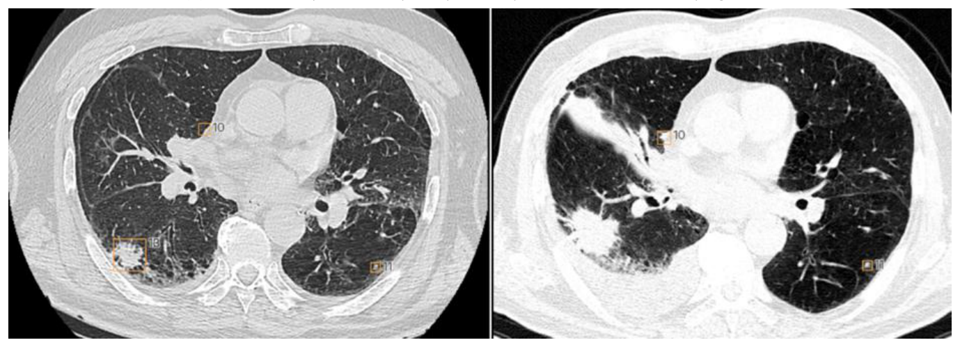
There are also limitations in the automated analysis of pulmonary nodules, among which the most significant are the false identification of vascular trajectories as nodular hyperdensities, and other limits in identifying lesions in areas with ventilation disorders, with motion artifacts or with fibronodular sequelae. However, automated analysis programs are continuously improving and shortening the times for preparing medical reports, increasing work efficiency, and becoming working tools for the redistribution of highly qualified human resources in other directions where they are still irreplaceable [15].
Another useful contribution is the automated comparative analysis of the evolution of pulmonary nodule sizes, in accordance with the intervals specified by specialist guidelines, such as that of the Fleischner Society, an aspect that contributes to establishing the most appropriate therapeutic and follow-up conduct [16]. Automated analysis generates medical reports that enter patients’ electronic records in parallel with the generation of a set of images with automatically marked lesions [15]. A concrete example in practice is that provided by CAD-type analyses in the identification of undetected tuberculous lesions.
In imaging departments serving emergency rooms in civilian or military field hospitals, cerebral hemorrhagic lesions, or hemorrhages in any part of the body, represent the most common cause of post-traumatic death. Rapid recognition of hemorrhages and the most appropriate therapeutic interventions are of paramount importance. The role of AI systems using ML is to optimize diagnosis in these situations through algorithms that recognize hemorrhagic lesions and assess their risk potential through CT and FAST ultrasound methods [17]. These programs use semiological notions of lesion recognition, such as Language Models (LM) for automatic 3D labelling to recognize hemorrhage subtypes based on brain segmentation, midline deviation, mass effect, cerebral edema, and hyperdensity values. Diagnostic models in neurotraumatology utilize various emergency diagnoses, including hemorrhage, ischemia, cerebral infarction, space-occupying lesions, mass effect, hydrocephalus, and midline deviation. Neurotraumatology lesion recognition models based on imaging data obtained through radiography, computed tomography, and MRI can also be used for the rest of the body, through 3D anatomical segmentation, 2D CNNs software, and Recurrent Neural Network (RNN) [18].
By using the Picture Archiving and Communication System (PACS) system, lesions with complex neurological substrates can be effectively detected and characterized through automatic algorithms that have entered routine analysis in some types of cerebral pathologies.
Automatic Cerebral Volumetry consists of an efficient automated analysis based on segmentation using the 3D T1 MPRAGE sequence. For reference, the images and measurement values are compared with those of a comparative database of patients from the same age groups for whom the brain volume is reported. Thus, this program provides cerebral volumetric assessment in case of suspected dementia, by evaluating the frontal, parietal and temporal lobes, and particularly the hippocampus. Brain volume assessment can also be extremely useful in multiple sclerosis and other degenerative diseases, such as Multiple Systemic Atrophy (MSA), Progressive Supranuclear Palsy (PSP), and Cortico-basal Degenerative Disease (CBD).
The program can also analyze the volume of specific brain areas such as the caudate nucleus, putamen, globus pallidus, cerebellar cortex, and midbrain [19].
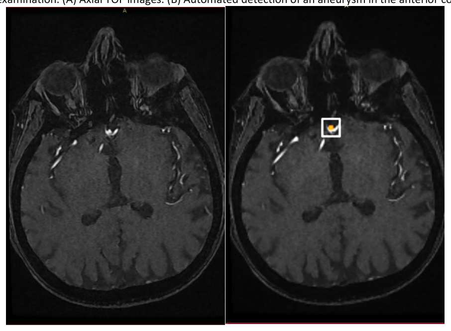
The Evaluation of Arterial Intracranial Vascular Lumen uses a Time-of-flight (TOF) acquisition for analysis before being processed with the Maximum Intensity Projection (MIP) program. The analysis program is attached to the PACS system and generates a medical report that includes measurements and imaging of the identified lesions. In the case of intracranial aneurysms, it is estimated that 10-15% of them are underdiagnosed, and these automated analysis programs can improve the anomaly detection rate with an accuracy of almost 100% [20].
The Characterization of a Demyelinating Lesion is based on the analysis of 3D Fluid-Attenuated Inversion Recovery (FLAIR) sequences that highlight the location of the lesions. The analysis includes classification of the lesions using various reference systems, such as the McDonald and Fazekas criteria.
The program can generate a medical report that includes data on the location and size of the lesions, the presence of new lesions compared to previous examinations, and key illustrative images that are included in the report. Thus, this type of analysis can be extremely useful in all demyelinating diseases, including multiple sclerosis [20].
Brain tumor analysis allows for the automated assessment of tissue structures associated with tumor presence in entities with different anatomopathological substrates. Automated tumor lesion analysis is performed after intravenous administration of a contrast agent, which evaluates the behavior and uptake of the contrast. Automated lesion analysis is applied to highlight and differentiate various lesions and tissue components, which can be especially useful in the characterization of glioblastoma-type lesions, metastases and other tumor types, such as meningiomas. Also, complex data analysis allows for the possibility of integration into radiotherapy and neurosurgery programs [21].
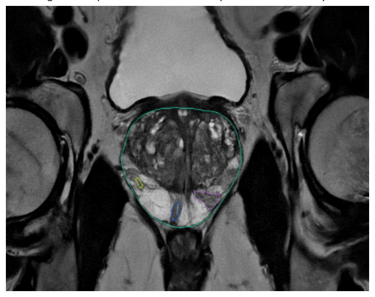
AI has become a commonly used tool for the diagnosis of prostate cancer, useful for both radiologists and urologists in multiparametric MRI examinations. The speed and efficiency of prostate cancer diagnosis and staging are among the greatest achievements for AI in prostate MRI evaluation [22].
Prostate cancer staging is performed using the PIRADS classification system and is integrated into the PACS system. The AI-generated medical report provides PIRADS classification, prostate volume, and detailed multiparametric analysis of prostate lesions, including nodule identification and categorization. In addition, when available, previous examinations can be evaluated comparatively. Medical reports also include appropriate graphical representations and dimensional assessments, as well as mapping of prostate lesions [22, 23].
When the patient’s PSA analysis is also available, this aspect can be represented in tables and associated with the presentation of prostatic density in the workflow report. This medical report can also be effectively used for image-guided biopsy, depending on the representation and arrangement of the most representative lesions [22, 23].
The most useful evaluation method is the one that uses the Area Under the Curve (AUC) indicator in binary detection. The method is derived from the Receiver Operating Characteristic (ROC) curve, which uses sensitivity and specificity as parameters to state whether a lesion is present or not [17]. In the ideal mode, the optimal value of AUC is 1. Between 1 and 0.5 AUC has the appearance of a random model and below 0.5, it cannot be brought into discussion. At the current time, the interpretation in an efficient model has AUC values close to 0.9. The evaluation of the AUC indicator has, in turn, references and standards related to diagnosis established at the level of junior and senior radiologists with expertise. This level of over 0.9 is more easily achieved in lesions defined by few parameters and with clear values, such as hemorrhages, fractures, pneumothorax, lesions from mammographic examinations, while multiparametric defined lesions, such as prostatic or hepatic ones, require a senior radiologist-type standard of expertise. In the radiological evaluation of polytrauma in emergency services, high AUC values, above 0.9, and short processing time are required [25].
The legal and social implications of artificial intelligence technologies in radiology need to be seriously assessed. One concern is the lack of transparency behind the decision-making process through artificial intelligence. Another key aspect in the implementation of artificial intelligence is the lack of a legislative system that regulates the level of involvement of artificial intelligence. Medical liability in case of malpractice is attributed to the radiologist. AI tools must meet rigorous validation and approval standards to establish their safety. Once approved, they must be continuously monitored to ensure that the results they provide are dependable. The implementation of AI systems requires adherence to established standards, appropriate regulatory approval, and awareness among radiologists and medical practitioners regarding their limitations, ensuring these factors are carefully considered [1, 26].
Despite the development of artificial intelligence, when implemented in real life, its performance varies for certain subgroups. The question arises whether the goal should be fairness to the individual or fairness to the group. It has been observed that artificial intelligence takes shortcuts in the learning process, which can lead to biased results towards certain subgroups, for example, when multiple factors, including age, gender, and race, are taken into account in the diagnosis [27].
The integration of artificial intelligence into clinical radiology faces several challenges. One of these is the existence of robust hardware capable of managing the substantial amounts of data generated by medical imaging systems, which implies investment in such infrastructure. On the other hand, this new hardware and software must be able to integrate with existing radiology systems. There is also a need for training of radiologists and for them to collaborate with artificial intelligence developers [1].
The future transformation of radiology includes the integration of Augmented/Virtual Reality (AR/VR) and AI. AR/VR technologies are expected to play a significant role in radiological practice and radiological education by improving the visualization of radiological images, thereby contributing to diagnosis and treatment planning. Also, through machine learning, AI improves its image analysis capabilities, thereby contributing to reducing diagnostic errors. A tool based on AI is Computer-Aided-Diagnosis (CAD), which can contribute to the development of an integrative diagnosis by incorporating radiological, anatomopathological and genomic information into the AI-assisted workflow [1].
Artificial intelligence is developing rapidly, and understanding its use in medical practice is mandatory. Radiology is transforming, and specialists must keep up with it, adopting these innovative technologies and being integrated into multidisciplinary clinical teams, with collaboration between healthcare providers being paramount to fully capitalize on the benefits of this scientific knowledge.
The authors declare no conflict of interest. This research work received no external funding. No AI tools were used to draft or edit the manuscript.
Authors’ contribution
Conceptualization, D.C., and A.S.C.; methodology, D.C., and A.S.C.; software, D.C., and A.S.C..; validation, D.C., and A.S.C.; formal analysis, D.C., and A.S.C.; investigation, D.C., and A.S.C.; resources, D.C., and A.S.C.; data curation, D.C; writing—original draft preparation, D.C., and A.S.C.; writing—review and editing, D.C., and A.S.C.; visualization, D.C.; supervision, D.C. All authors have read and agreed to the published version of the manuscript.
Patient consent for publication Not applicable.