
Diabetes mellitus (DM) encompasses a group of heterogeneous diseases characterized by hyperglycemia. The most prevalent forms of disease are Type 1 DM (T1DM) and Type 2 DM (T2DM). IDF has also just confirmed type 5 diabetes. It is reportedly characterized by severe insulin deficiency, mainly caused by undernutrition during childhood or adolescent years [1]. T1DM affects approximately 8.75 million individuals globally, whereas T2DM currently impacts about 6.28% of the world’s population, accounting for 80% of all diabetes cases. DM represents a significant health issue both in terms of prevalence and the impact of its chronic complications, which can lead to organ failure (e.g., kidneys, retina, heart) [2-4]. Projections indicate that these values are expected to increase significantly in the coming years [5].
T1DM and T2DM are multifactorial diseases. T1DM is typically diagnosed following extensive destruction of insulin-secreting pancreatic β cells (70-80%) by T lymphocytes, resulting in the presence of self-reactive autoantibodies (T1A-DM). Non-immune mechanisms account for 10-30% of T1DM cases (type 1B – T1B-DM subclass), which are classified as idiopathic T1DM [6-8]. Idiopathic T1DM does not have an autoimmune background. Although it can present with insulin deficiency, this subclass of DM does not have a clear trigger. Fulminant T1DM represents another subclass that is also non-immune-mediated. It manifests by fast β-cell death, which may be caused by environmental and genetic backgrounds [9]. Altered insulin secretion, often paired with insulin resistance, represents the main mechanism involved in T2DM [10].
The predisposition for DM remains only partially understood. With some exceptions, a complex network of genetic and epigenetic factors can predispose individuals to T1DM and T2DM [11-15]. Since 2004-2005, exosomal microRNAs (miRNAs) have been included in the list of factors associated with DM and its chronic complications [16-17].
Small RNA molecules (e.g., microRNAs -miRNAs; small interfering RNAs -siRNAs) are synthesized in the nucleus and subsequently transported to the cytoplasm. The origin of the miRNA molecules is a primary transcript that contains hairpin-shaped structures [18]. The RNase Drosha recognizes and cuts these hairpin structures in RNA, resulting in a miRNA precursor (pre-miRNA) of about 70 nucleotides in length. These molecules exit the nucleus (by means of exportin-5 complex) and in the cytoplasm are recognized and cut at the loop part by the enzyme Dicer [19]. The double-stranded miRNA is then loaded into the Argonaute (Ago) protein complex, and a complete silencing complex is formed, and the miRNA is used as a template for target recognition [18].
These double-stranded RNA molecules are utilized by the RNA-Induced Silencing Complex (RISC) to regulate gene expression [19]. It has been reported that the structural similarity of these miRNA hairpins and anti-Tn-C aptamers indicates miRNA functionality beyond RISC, making them even more sophisticated regulators [20]. The RISC complex acts as a translational repressor or as an mRNA breaker, depending on the number of base pairs that form between base pairs. One miRNA can target multiple genes, and they often return to silence the primary transcript from which they were synthesized.
The silencing process has critical roles in the physiological processes (e.g., cell differentiation, organism development) [21]. The concentration of miRNA was estimated between 1000 copies/ 1000 mm3 cells and around 200 copies/cell (or 166pM). It has been reported in previous studies that the median half-life of miRNA is approximately 5 days, with some exceptions (e.g., miR-155 was reported to have lower stability) [20]. The temporal and spatial deregulation of miRNA expression and the silencing process can result in pathological consequences (e.g., by modulating the expression of pro-inflammatory and pro-apoptotic effectors), which impact disease onset, progression, and severity [18,22]. The possibility of repeated sampling has increased scientists’ interest in assessing the potential of miRNAs as biomarkers for malignant (e.g., lung adenocarcinoma, gastric or breast cancers) and non-malignant diseases (e.g., including DM) [23-27]. It should be taken into consideration that the multi-role of these molecules when comprising a panel for clinical usage. Thus, miR-21, miR-326 (e.g., glioblastoma), miR-34a, miR-2a (e.g., hepatocellular carcinoma), miR-155 (e.g., pancreatic ductal adenocarcinoma), miR-23b (e.g., melanoma, prostate cancer), miR-98 (e.g., squamous cell carcinoma; head and neck carcinoma), miR-186 (e.g., Hodgkin’s lymphoma), miR-223 (e.g., chronic myeloid leukemia) were associated with different malignant diseases. But some of these miRNAs were also associated with different nonmalignant diseases (e.g., miR-21- cardiomyocyte hypertrophy; immune response in sepsis; miR-326 and miR-186 – multiple sclerosis; miR-590- extrinsic cardiomyopathy, autoimmune myocarditis; miR-223 – hereditary neutrophilia) [28].
The study aims to perform a bibliometric analysis to evaluate research trends in the field and to identify and characterize miRNAs implicated in DM using curated bioinformatic resources.
This study was conducted in two main phases: an analysis of the scientific literature and the identification of miRNAs altered in DM based on records in specialized databases.
Bibliometric Analysis. A comprehensive literature search was performed using the Scopus database [29]. The search strategy targeted the Title and Abstract fields with the following keywords: (“hsa-microRNA” OR “hsa-miRNA” OR “microRNA” OR “miRNA”) AND (“type 1 diabetes” OR “type 2 diabetes” OR “T1D” OR “T2D”) AND (“human” OR “humans” OR “Homo sapiens”) NOT (“mouse” OR “mice” OR “rat” OR “rats” OR “murine” OR “animal model” OR “in vivo” OR “non-human” OR “rodent”). The initial search yielded 1730 articles. After excluding irrelevant subject areas (e.g., Engineering, Mathematics, Materials Science, Arts and Humanities, Social Sciences, Physics and Astronomy), 1,707 articles remained. Further filtering by document type (research articles only) reduced the number to 1085. Applying language (English) and publication stage filters resulted in a final dataset of 1051 articles. The literature analysis was conducted using the Bibliometrix R-package (version 4.4.0) and Biblioshiny (version 5.0), which enabled descriptive statistics, thematic mapping, and trend analysis [30]. All analyses were performed in R (version 2025.05.0, Build 496).
MiRBase [31] and TargetScanHuman 8.0 [32] were consulted to check for predicted target transcripts and conservation status of each miRNA. Diseases associated with miRNA were extracted from the Human ncRNA Gene Database [28].
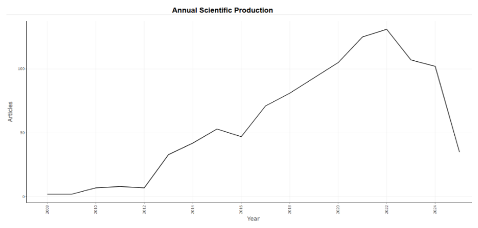
The timespan of the analyzed publications was 2008 to 2025. The dataset comprised 1051 documents published across 440 sources, involving a total of 6409 authors and citing 49120 references. The annual growth rate of scientific output was 18.34%. The average age of the documents was 5.34 years, and the mean number of citations per document was 28.12.
Interest in the publication of this data began between 2008 and 2012 (Figure 1). A high increase was noticed from then on until 2015, with a spike from 2016 to 2022.
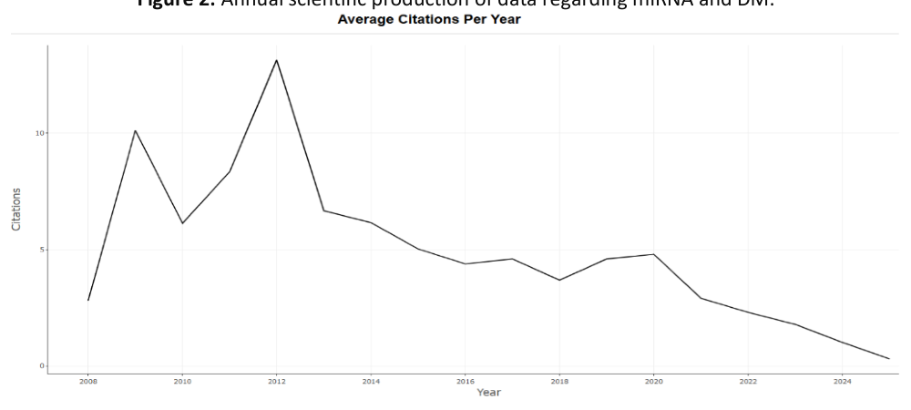
As for the average number of citations per year, it aligned with the wave of scientific production (Figure 2). The first spike was observed in 2009, followed by a second spike in 2012. A slight rise was noticed again, from 2018 to 2020.
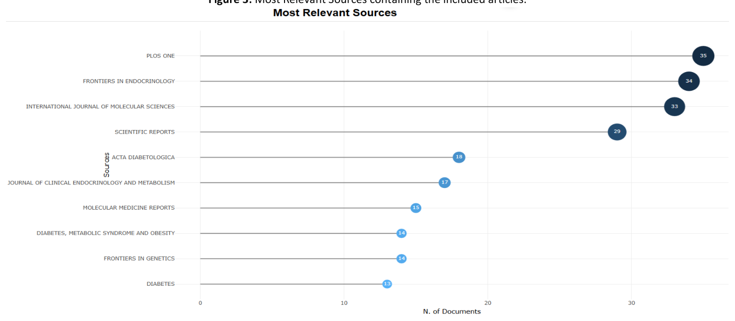
The 1051 articles were published in various sources. The majority of them can be found in PLOS One, Frontiers in Endocrinology, International Journal of Molecular Sciences, and Scientific Reports. Other sources included: Acta Diabetologica, Journal of Clinical Endocrinology and Metabolism, and Molecular Medicine Reports (Figure 3).
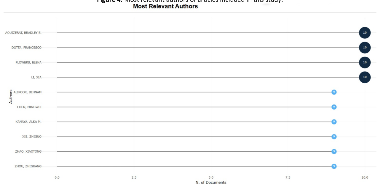
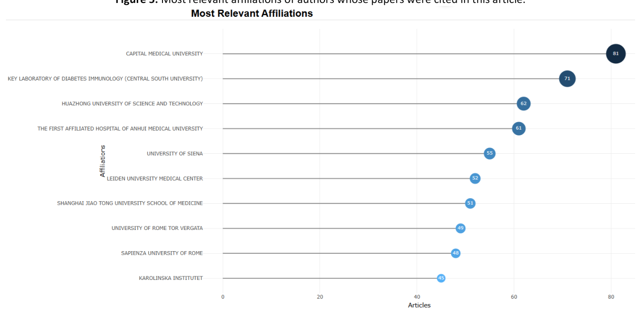
From the 6049 authors who participated in the research included in this paper, some of the most relevant are Dotta F., Flowers E., Li X., Chen M., and Zhao X. (Figure 4). Their affiliations include: Capital Medical University, Key Laboratory of Diabetes Immunology, University of Siena, and Huazhong University of Science and Technology (Figure 5).
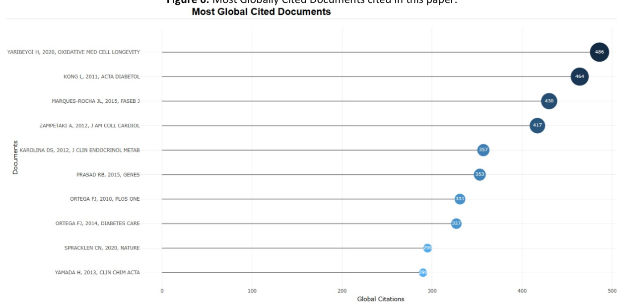
Speaking at a global level, the papers with the most citations included in this study were [2,33-36]. The sources in which the works of the aforementioned authors were published include, in order: Oxidative Medicine and Cellular Longevity, Acta Diabetologica, The FASEB Journal, Journal of the American College of Cardiology, and The Journal of Clinical Endocrinology & Metabolism (Figure 6).
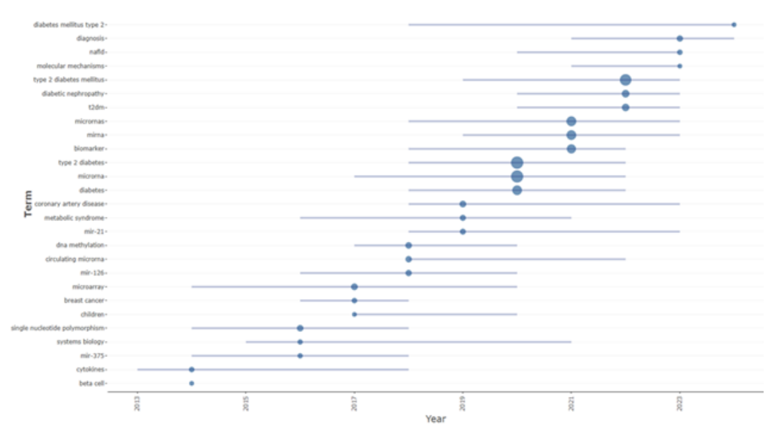
The trending topics identified in the reviewed articles highlight the evolving focus areas and emerging themes within the research field over time (Figure 7). Authors’ keywords pinpointed in articles that constitute the database change over the years. First time span consisted of keywords such as: “cytokines”, “miR-375”, “single nucleotide polymorphism”. In recent years, keywords such as “diagnosis”, “molecular mechanisms”, “biomarker”, and “type 2 diabetes mellitus” were employed.
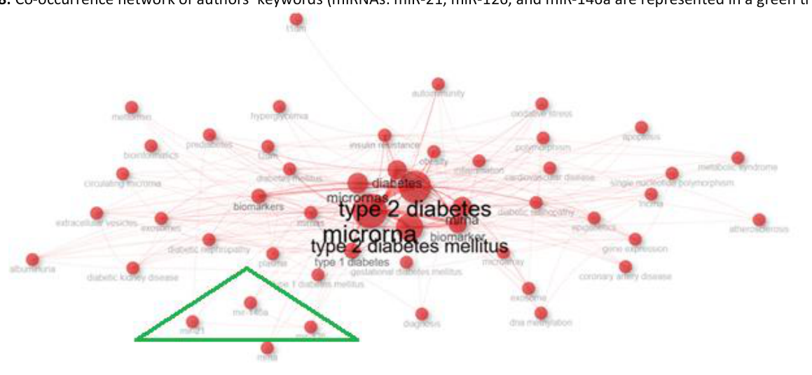
The co-occurrence network of authors’ keywords and the microRNA species miR-21, miR-126, and miR-146a highlights the relationship and thematic connections within the literature contained in the database of this paper (Figure 8).
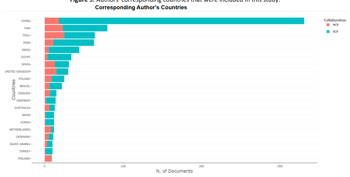
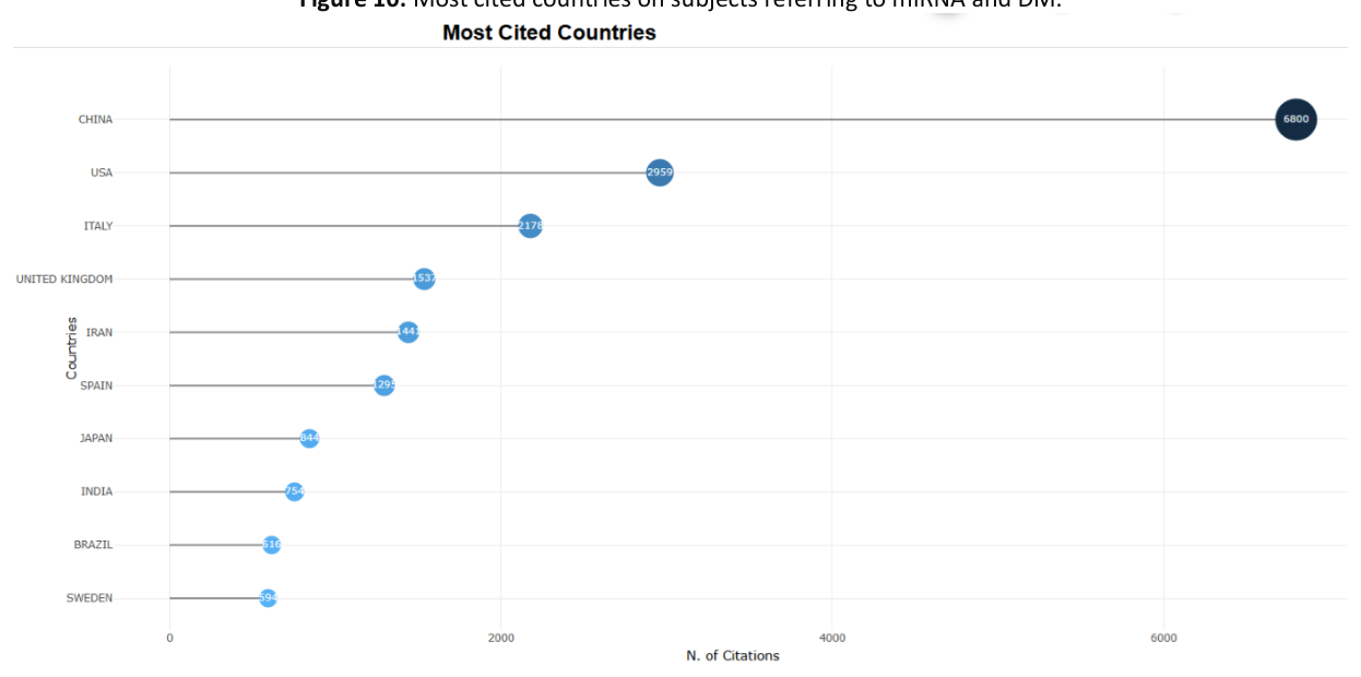
The distribution of corresponding authors by country mirrors the ranking of the most cited countries (Figure 9; Figure 10). Leading the list are countries such as China, the United States, and Italy, which also represent the most active contributors to DM research. In terms of citation impact, Italy is followed by the United Kingdom, Iran, Spain, and Japan.
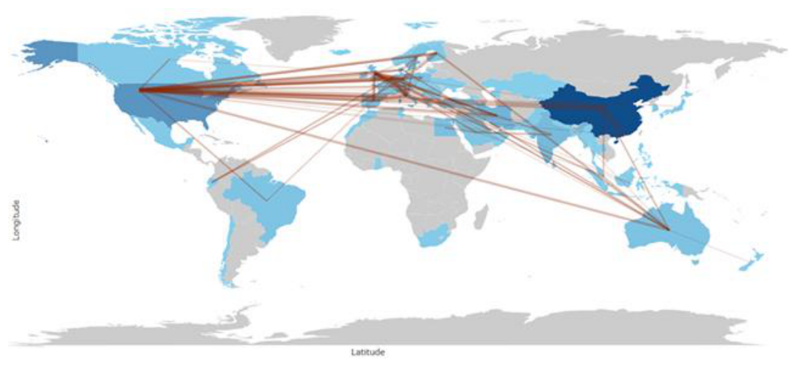
Visualization of international research collaborations by country is seen in Figure 11. Highlighting the global networks formed through co-authorship and cross-border scientific partnerships, its scope encompasses every continent.
Bioinformatic analysis can be conducted in order to predict possible miRNA targets. Examples of databases constructed for this purpose are miRBase [31], TargetScan [32], and miRecords [37]. For this research, miRbase [31] and TargetScanHuman v8.0 [32] were used for consulting target transcripts and conservation status for each miRNA. Target gene IDs can be found in Tables 1, 2, and 3. MiR ID was normalized according to at least two articles and two miR databases, according to that miR’s sequence.
| miR in T1DM | Target Gene/Mechanism in which they are involved | Referenes |
|---|---|---|
| miR-326 ↑ | TNFSF14, 15; CEP85 | Zheng et. al., 2017 |
| miR-23 ↑; miR-98 ↑; miR-590-5p ↑ | Trail, Faslg | |
| miR-186 ↑ | CXCL13; IRF8; STAT4 | [38] |
| miR-223 ↑ | HSP90B1; TP53; MCL1 | |
| miR-142-5p ↑ | HERPUD1 | [39] |
| miR-155 ↑ / ↓ | CASP3 | [39]; Katsarou et. al., 2017 |
| miR-100-5p ↓ | RAVER2; AP1AR | Ferraz et. al., 2022 |
| miR-181a-5p ↑ | ZNF; TNFSRF11B | Liu et. al., 2019 |
| miR-1275 ↑ | KCNC3; KIR2DL4 | [22] |
| miR-30b-3p ↑ | CCL28, 22; TAF8; NCR3 | Nizam et. al., 2024 |
| miR-25-3p ↑ | HIPK3; SNAPC1; RBM47 | Liu et. al., 2019 |
| miR-10a-5p ↑ | CRLF2; HOXA3 | Santos et. al., 2022 |
| miR-98 ↑ | IGDCC3; IGF2BP2; CCL3 | Khan et. al., 2020 |
| miR-22-3p ↓ | H3F3C; GADD45A | Kaur et. al., 2015 |
| miR-16-5p ↑ | TNFSF13B; BTLA | Gao et. al., 2020 |
| miR-574-3p ↓ | NKG7; IL6; ATPIF1; IFNLR1; SRF | Garcia-Contreras et. al., 2017 |
| miR-103a-3p ↑ | SPI1 1; AGO4 | Assmann et. al., 2018 |
| miR-454-3p ↑ | IRF1; IGF | Erener et. al., 2017 |
| miR-450a-2-3p ↑ | IFG2; IL6ST; NOX5; IL17A | Takahashi et. al., 2014 |
| miR-100-5p ↓ | INSM1; AGO2 | Hezova et. al., 2010 |
TargetScanHuman v8.0 was used to query each miRNA to check predicted target genes.
Some factors involved in the pathophysiology of T1DM (e.g., ILs, tumor necrosis factor) can interfere with the expression of these miRNAs and thereby modulate the efficiency of silencing processes. These observations complicated the relationship between T1DM and miRNAs.
MiRNAs can interfere with T2DM physiopathological mechanisms [40] (Table 3). Some of these molecules were associated with both T1DM and T2DM (Table 2).
Although there is significant interest in developing new biomarkers for DM, only a few miRNAs have been consistently associated with T2DM in at least two studies (Table 3).
| miR in T1DM & T2DM | Target Gene/Mechanism in which they are involved | Referenes |
|---|---|---|
| miR-21 ↑ | IL12A; FASLG; CCL1 | [41] |
| miR-34a ↑ | CDIP1; MSR1; PDCD6 | |
| miR-146a ↓ | INSR; ZNF; GRIN2B | |
| miR-29 ↑ | NFIA; ATAD2B | Massart et. al., 2017 |
| miR-142-3p ↑ | CASP3 | [39]; Zhu & Leung, 2015 |
| miR-24-3p ↑ | FASLG; BCL2L14 | Blanco et. al., 2023; Garavelli et. al., 2020 |
| miR-148a-3p ↑ | ATP6AP2; GADD45A | Grieco et. al., 2018; Ghoreishi et. al., 2022 |
| miR-150-5p ↓ | MYB; TADA1; AIFM2 | Kim et. al., 2019; Qiu et. al., 2021 |
| miR-210-5p ↑ | ERP29; GDF11 | Patra et. al., 2023 |
| miR-342-3p ↓ | OSER1; SPI | Jiang et. al., 2020 |
| miR-375 ↑ | INS; HOXA5; ATP1B1 | Higuchi et. al., 2015; Marchand et. al., 2016 |
| miR-101 ↑ | UBE2D1; APP | Santos et. al., 2019; Higuchi et. al., 2015 |
| miR-199a ↑ | PAWR; ACOX1 | Wang, et. al., 2018; Yang et. al., 2020 |
| miR-335-5p ↓ | CASP; IL17RD | Li & Zhang, 2021; Hezova et. al., 2010 |
| miR-20b-5p ↓ | PDCD; IRF9 | Katayama et. al., 2019; Hezova et. al., 2010 |
| miR-15b ↑ | CYP7A1; CASP | [42] |
| miR-186 | CRP | |
| miR-155 ↑ / ↓ | BCL2 | Corral-Fernández et. al., 2013; Katsarou et. al., 2017 |
TargetScanHuman v8.0 was used to query each miRNA to check predicted target genes.
| miR in T2DM | Target Gene/Mechanism in which they are involved | Authors & Year |
|---|---|---|
| miR-143 | IGFBP; FADS6; ITM2B | [43] |
| miR-142-3p | BCL2 | Zhu & Leung, 2015 |
| miR-126 | INSR; SCL7A5 | Zhu & Leung, 2015 |
| miR-375 | INS; VEGFA; HOXA5; ATP1B1 | Poy et. al., 2009; Chakraborty et. al., 2014 |
| miR-26a-5p | POLR3G; INS; BAX | Xu et. al., 2020; |
| miR-146a-5p | IGSF1; IRAK1; PSMA4 | Li et. al., 2012 |
| miR-1 ↓ | CYP7A1; XCR1; PRPF39 | Chakraborty et. al., 2014 |
| miR-16 ↓ | RRM2B; MRPS22; C8B; PDX1 | Bork-Jensen et. al., 2015 |
| miR-30e ↓ | GABRB1; IL1R1APL2; SOCS1 | Dieter et. al., 2019 |
| miR-9 ↑ | DSCC1; GLT1D1 | Al-Muhtaresh et. al., 2018 |
| miR-33a ↑ | TEX38; TLR4; PTPLAD1 | Saeidi et. al., 2023 |
| miR-320a ↑ | ETFA; TRIAP1; RGS9BP | Du et. al., 2021 |
| miR-148b ↑ | GADD45A; MMD; TGFA | Liang et. al., 2020 |
| miR-21 ↓ | IL12A; FASLG; GPR64 | [44] |
| miR-29a ↑ | MANEA; HOXA10; RTP3 | Massart et. al., 2017 |
| miR-34a ↑ | MSR1; PDCD6; GORAB | Shen et. al., 2017 |
| miR-103 | RNF38; TRIAP1; AGO4 | Rafiee et. al., 2024 |
| miR-15a ↓ | POLR3K; SLC22A9; RAVER2; | Al-Kafaji et. al., 2015 |
| miR-155 ↓ | H3F3A; ZNF652; FGF7 | Katsarou et. al., 2017 |
| miR-221 ↑ | GABRA1; KCNQ3; SOCS5 | Liu et. al., 2018 |
| miR-145 ↓ | ATP6V0B; ITGB8; CDC37L1 | He et. al., 2020 |
| miR-24 ↓ | SNN; BCL2L11; KCNK2 | Xiang, 2015 |
| miR-182 ↑ | TMEM50B; CHIC1; VLDLR | Krause et. al., 2024 |
| miR-192 ↑ | insulin-like growth factor 2; SLC44A1 | Jaeger et. al., 2018 |
| miR-194 ↑ | PRKAR1A; SLC10A7; BNIP2 | Jaeger et. al., 2018 |
| miR-223 ↑ | FBXW7; IL6ST; HSP90B1 | Sánchez-Ceinos et. al., 2021 |
| miR-130a ↑ | IGF1; TGFBR2; HIVEP2 | Ofori et. al., 2017 |
| miR-19a ↑ | QKI; | Yan et. al., 2020 |
| miR-26b ↑ | ID2; IGFBP3; SOCS1 | Xu et al., 2015 |
| miR-27a | FOXO1, FOXA2, STAT3 | [45] |
| miR-27b ↓ | GOLM1; GPAM | Ghoreishi et. al., 2022 |
| miR-28 ↑ / ↓ | TRAF3 | [45;46] |
TargetScanHuman v8.0 was used to query each miRNA to check predicted target genes.
The utility of miRNAs as biomarkers for screening or diagnosis of T1DM is of high interest. There have been some attempts to propose a panel of miRNAs for elaborating these tests, although normalization of miRNA levels is still necessary [47].
A systematic review published in 2017 mentioned 48 miRNAs that were investigate in tissues relevant for T1DM pathogenesis (e.g. serum, plasma, pancreatic tissue, blood cells); for 11 of them the expression level differ significantly in T1DM patients and control subjects (e.g. miR-21-5p, miR-24-3p, miR-100-5p, miR-146a-5p, miR-148a-3p, miR-150-5p, miR-181a-5p, miR-210-5p, miR-342-3p, miR-375 and miR-1275) [22]. There are certain miRNAs (e.g., miR-375) that directly influence specific pathways leading to diabetogenesis. These molecules target genes that modulate immune T cell activity. Human and mouse T lymphocytes have been shown to release exosomal miRNAs (e.g., miR-142-3p, miR-142-5p, and miR-155), which can predispose or support the apoptosis of pancreatic β cells [39]. Additionally, overexpression of some miRNAs can reduce insulin synthesis, insulin secretion, or their biological effects [48].
Cytotoxic attack in T1DM is mediated by proinflammatory cytokines, such as interleukin-1β (IL-1β) and tumor necrosis factor α (TNFα). These cytokines have been found to induce the synthesis of miR-21, miR-34a, and miR-146a in vitro, in human and mouse pancreatic cells [41].
miR-21 is predicted to target multiple transcripts (e.g., IL12- natural killer cell stimulatory factor and cytotoxic lymphocyte maturation factor, CXCL1- chemokine ligand 1). Increased levels of IL-12 could be predictors of T1DM. IL-12 plays a part in determining the levels of CD4+T cells and increasing the CD8+ T cells’ toxicity in DM [49]. It also has a role in activating the apoptotic pathway of the β cells. Existing literature proved the implication of the CXCR1/2 axis, CCL4-CCR5 axis, and CXCL10-CXCR3 axis in the inflammatory background of DM. These pathways were proven to be engaging more aggressive immune T-cells. This furthermore accentuates the β cell death [50].
A decrease in miR-155 expression has been shown to impair the function of regulatory T cells (Tregs), which exhibit an upregulated expression of suppressor of cytokine signaling 1 (SOCS1) upon maturation. Treg cells have a primary role in alleviating the aggressivity of CD4+ and CD8+ T cells. Therefore, their underdevelopment has been linked with one of the causes of T1DM, implicating the STAT3 mutations. The signal transducer was reported to recognize and enhance the miR-155 promoter. The development of Treg cells is also threatened by mutations in the FoxP3 gene, and its altered transcriptional activity has been linked with the presence of miR-155 [51].
The apoptotic genes (Trail, Faslg) are predicted to be targeted by miR-23, miR-98, and miR-590-5p. These miRNAs are overexpressed in CD8+ T cells of T1DM patients [52]. MiR-326 targets different genes (e.g., the gene coding the vitamin D receptor), which modulate the function of the immune system [44]. Expression levels of miR-326 were found to be increased in lymphocytes of T1DM patients who tested positive for the islet-specific autoantibodies IA-2A and GADA [53].
MiR-34a is known to inhibit macrophage polarization, thus impairing tissue homeostasis and repair. This miRNA is involved in the regulation of insulin signaling and glucose metabolism through the targeting of specific mRNAs, including those of sirtuin 1 (SIRT1) and vesicle-associated membrane protein 2 (VAMP2) [54]. Dysregulated miR-34a in cancer patients has been proven to lead to low survival chances. It targets multiple oncogenes and plays a part in apoptosis mediated by the p53 complex, the tumor suppressor gene [55].
In a cross-sectional study, Januszewski and his coworkers demonstrated increased levels of miR-186 and miR-223 in T1DM patients. MiR-186 is known for its role in apoptosis and chemokine and insulin signaling pathways [42]. MiR-186 is also known for being a tumor suppressor in prostate cancer. There was an increase in granulocytes in model mice with miR-223 deficiency, and its expression was highly decreased in patients with active nephritis. This miR-223, along with others, has been associated with multiple cellular processes: p53 pathway regulation, cell cycle regulation, tumor metabolism, and apoptosis [38].
The cumulative roles of miRNAs in T1DM are various, implicating many cellular and molecular pathways and mechanisms. Extensive research is needed to further validate the expression levels in vivo model organisms [47].
Certain miRNAs found in scientific publications or databases are associated with the pathogenesis of diabetes or with particular features of diabetic patients. Subjects with impaired glucose tolerance presented abnormal levels of miR-18b [45]. miR-143 has a role in precursor adipocyte differentiation and in predisposition for T2DM (e.g., by targeting the insulin-AKT pathway) [43]. MiR-21 targets mainly FOXO1, FOXA2, and STAT3 transcripts. Its levels were considered informative for gluconeogenesis [56], insulin resistance [57], and altered glucose tolerance [45]. Markers of low-grade inflammation in T2DM (e.g., IL6, TNFα) are also associated with changes in miRNAs. TNFa enhances the intracellular accumulation of miR-223, which targets genes involved in lipid and glucose metabolism, inflammation, and T2DM (e.g., IGF-IR, Hsp90, FOXO1) [58;59].
A randomized community-based study analyzed a sample of 1000 individuals. It was identified several downregulated miRNAs (e.g. miR-126, miR-15a, miR-29b, miR-223, miR-20b, miR-197) in the plasma samples from T2DM patients [46]. Upregulated (e.g., miR-140, miR-142, miR-222) or downregulated (e.g., miR-125b, miR-126, miR-130, miR-192, miR-195, miR-423-5p, miR-532-5p) miRNAs in T2DM patients were also identified in a cross-sectional, double-blinded study. It was observed that the expression levels of four miRNAs (e.g., miR-126, miR-140-5p, miR-195, miR-423-5p) demonstrated a strong ability to distinguish subjects with T2DM from those with normal fasting glucose levels [60]. MiR-126 has been reported by a considerable number of research groups to be a suitable biomarker for the assessment of T2DM progression [44, 46, 61]. MiR-186 expression levels were found to be statistically significantly associated with HbA1C levels in T2DM patients and with the risk of diabetic neuropathy [62].
There are numerous factors to consider when evaluating the potential of miRNAs as biomarkers for DM. For instance, the dynamics of miRNA concentration may be correlated with causes of dysregulated expression (e.g., number of cells releasing these small noncoding molecules or with miRNA overexpression in individual cells), miRNA half-life, and factors influencing their stability. Other factors can also significantly impact the significance of results. For example, an increase in adipose tissue volume can increase the levels of some miRNAs; these associations may influence the results, particularly in studies involving obese patients with T2DM [45]. The timing of miRNA testing poses a challenge, especially in presymptomatic stages of diabetes [63]. It has been hypothesized that dysregulated miRNAs may be present only in specific cells or tissues. Comprehensive research involving paired analyses of body fluids and biopsy tissues, along with validation studies, could be beneficial for future investigations [64]. There also remains a critical need for the standardization of miRNA analysis across different research platforms, tissues, and conditions to facilitate the development of clinically useful biomarkers [65].
The size of the sample pool influences the study’s power and the reliability of results. Studies with small sample sizes (e.g., fewer than 10 patients and 10 control subjects) may yield biased outcomes [66]. Consequently, findings from such studies may not be corroborated by research involving larger sample sizes. Therefore, the statistical significance and clinical relevance of some miRNA studies may be limited. Reliable statistics are needed against large sample pools and of different categories.
MiRNAs can target a large number of transcripts, and their levels in specific tissues change during the natural history of the disease. But these factors also increase the difficulty of identifying the relevant genes for diabetogenesis and validating the clinical use of specific miRNAs. Standardization in profiling techniques and validation of these biomarkers across large, diverse populations remain essential steps toward their clinical application.
This review underscores the emerging role of miRNA as one of the key molecular players in the pathogenesis, diagnosis, and potential treatment of DM. Numerous studies have identified distinct expression patterns of specific miRNAs (e.g., miR-21, miR-126, miR-143, miR-155, miR-223, and miR-375) that correlate with immune dysregulation, β-cell apoptosis, insulin resistance, and metabolic imbalance.
The authors declare no conflict of interest. This research work was carried out with the support of the Romanian Ministry of Education and Research, under the Core Program, project PN 23 01 03 03. The funders had no role in the design of the study; in the collection, analyses, or interpretation of data; in the writing of the manuscript; or in the decision to publish the results.
Conceptualization, X.I.P. and DC.; methodology, X.I.P., RN and DC; software, D.C. and X.P; validation, X.I.P., O.A.A.T, I.R, L.B., I.A.E. and S.N.; formal analysis, D.C.; investigation, D.C, X.I.P. and R.N; resources, D.C.; data curation, X.I.P., O.A.A.T, I.A.E, S.N, and R.N.; writing- X.I.P. and D.C; writing-review and editing, O.A.A.T, I.R, L.B., I.A.E, S.N. and R.N.; visualization, D.C.; supervision, D.C.; project administration, L.B.; funding acquisition, L.B. All authors have read and agreed to the published version of the manuscript.