
Gastrointestinal neuroendocrine tumors (NETs) are rare, slow-growing tumors characterized by specific histological, biological, and clinical features. These tumors have shown a significant increase in incidence and prevalence over recent decades, reaching an increase of over 100% in the past 40 years, due to advancements in diagnostic methods and increased medical awareness [1-6]. Gastrointestinal NETs represent an important subtype of neuroendocrine tumors, affecting various segments of the gastrointestinal tract, as follows: the small intestine (45%), rectum (20%), appendix (16%), colon (11%), and stomach (7%) [7].
One of the most notable characteristics of these tumors is their predominant setting in the small intestine, particularly in the terminal ileum, approximately 60 cm from the Bauhin valve. This makes them the most frequent malignancy of the small intestine [8]. Small intestinal neuroendocrine tumors are often diagnosed in advanced stages because clinical symptoms are frequently nonspecific and can include abdominal pain, intestinal obstruction, and symptoms caused by carcinoid syndrome [2]. Additionally, these tumors develop distant metastases more frequently than neuroendocrine tumors from other locations, further complicating their clinical management. Mesenteric lymph node metastases are found in over 80% of patients at the time of diagnosis, regardless of the primary tumor’s size [9-14]. Imaging techniques such as contrast-enhanced CT, MRI, and somatostatin receptor-based PET scans have improved detection, while immunohistochemical markers such as chromogranin A and synaptophysin aid in confirming the diagnosis.
Histologically, gastrointestinal neuroendocrine tumors are characterized by the presence of neuroendocrine cells that secrete various peptides and biogenic amines. These substances can cause diverse clinical manifestations, including diarrhea, flushing, and wheezing, within the context of carcinoid syndrome [15].
Biologically, the tumors exhibit variable expression of neuroendocrine markers such as chromogranin A and synaptophysin, which are used in the diagnosis and monitoring of the disease [7, 16-18].
Recent studies have highlighted the genetic and molecular diversity of gastrointestinal neuroendocrine tumors. Mutations in the MEN1, DAXX, and ATRX genes are frequently associated with these tumors. The identification of these mutations has allowed for a better understanding of the tumor pathogenesis and opened new perspectives for targeted therapies [8, 19-21].
The clinical management of patients with gastrointestinal neuroendocrine tumors involves a multidisciplinary approach, including advanced imaging, endoscopic techniques, surgery, and systemic therapies. Surgical intervention remains the treatment of choice for resectable tumors [22], while systemic therapies, including somatostatin analogs, mTOR inhibitors, and radionuclide therapies, are used for advanced or metastatic disease [23-29].
Gastrointestinal neuroendocrine tumors (NETs) represent a relatively rare group of malignancies, yet recent literature indicates a significant rise in their incidence worldwide. Small intestinal NETs account for approximately 45% of all gastrointestinal NETs, establishing these tumors as the most prevalent malignancy of the small bowel [1,7]. The estimated global annual incidence ranges between 3 to 6 cases per million inhabitants, with a clear upward trend observed over the last four decades, primarily attributed to improved diagnostic methods and increased physician awareness [2,4].
A comprehensive analysis of epidemiological data from the Surveillance, Epidemiology, and End Results (SEER) database (1977–2016) by Liu et al. (2023) demonstrated that gastrointestinal NETs have exhibited a dramatic increase of over 100% in diagnosed cases during recent decades [2]. This significant upward trend reflects advances in diagnostic imaging modalities and improved endoscopic techniques, facilitating earlier and more accurate diagnosis [2,7].
In line with findings reported by Taal and Visser (2004), small intestinal NETs show notable geographical variation, yet consistently demonstrate an annual incidence ranging from 3 to 6 cases per million inhabitants globally [4]. This consistency suggests a common biological behavior and potentially shared risk factors influencing the occurrence of these tumors [4-5].
Epidemiological patterns also reveal that the diagnosis of small bowel NETs most commonly occurs between the ages of 50 and 70 years, highlighting the importance of clinical suspicion within this age group when nonspecific gastrointestinal symptoms are present. The age distribution emphasizes the need for targeted diagnostic vigilance in middle-aged and older patients presenting with chronic abdominal complaints without an obvious etiology [6-7,12].
In addition, several retrospective studies have documented the high likelihood of delayed diagnosis due to nonspecific symptomatology, often resulting in advanced disease at initial presentation, with a high frequency of mesenteric lymph node involvement and distant metastasis at the time of diagnosis [6-7].
Therefore, awareness of these epidemiological trends, along with improved understanding of the disease’s clinical behavior, is crucial for clinicians to achieve earlier diagnosis and intervention, ultimately enhancing patient outcomes.
The clinical presentation of gastrointestinal neuroendocrine tumors (NETs), especially those of the small intestine, is highly variable and nonspecific, frequently contributing to significant delays in diagnosis. Commonly reported symptoms include chronic abdominal pain, often intermittent and vague, unexplained weight loss, changes in bowel habits such as chronic diarrhea or intermittent constipation, and episodes of intestinal obstruction due to either tumor mass or mesenteric fibrosis [7,25,27].
In certain instances, especially when tumors exhibit secretory activity (functional NETs), clinical presentation may include symptoms associated with the classic carcinoid syndrome. This syndrome occurs predominantly when liver metastases are present, allowing serotonin and other vasoactive substances to enter systemic circulation. Characteristic features include episodic flushing, chronic diarrhea, bronchospasm, and occasionally cardiac valvular lesions [15,29].
The nonspecific clinical picture typically leads to prolonged diagnostic latency, with literature reporting that many cases are incidentally identified during surgeries performed for unrelated reasons, such as intestinal obstructions or abdominal hernias, rather than direct suspicion of NETs [25,27].
Imaging plays a crucial role in diagnosing NETs and determining disease extent. Computed tomography (CT), Magnetic Resonance Imaging (MRI), and somatostatin receptor imaging (68Ga-DOTATATE PET/CT) are the mainstay diagnostic tools. Of these, somatostatin receptor PET-CT (68Ga-DOTATATE PET-CT) has demonstrated superior sensitivity and specificity, especially for detecting small primary tumors and distant metastases compared to conventional imaging methods [3,8].
Histopathological confirmation is indispensable for accurate diagnosis and characterization of NETs. Immunohistochemical markers such as chromogranin A, synaptophysin, and the transcription factor CDX2 are routinely employed and demonstrate high sensitivity and specificity for neuroendocrine differentiation [3,6].
Additionally, the proliferation index assessed by the Ki67 index is crucial in grading these tumors and assessing their malignant potential. The World Health Organization (WHO) classification categorizes NETs as G1 (Ki67 <3%), G2 (Ki67 3-20%), or G3 (Ki67 >20%), directly impacting the choice of therapeutic strategies and prognostic evaluation [6,12,22].
Given the nonspecific nature of the symptoms, the combination of clinical suspicion, advanced imaging modalities, and specific immunohistochemical analyses is paramount to timely diagnosis and improved outcomes for patients with small intestinal NETs.
Surgical intervention remains the cornerstone of management for localized small intestinal neuroendocrine tumors (SI-NETs). The primary goals of surgical treatment include complete tumor resection (R0 resection), adequate lymph node dissection, and alleviation of symptoms associated with tumor burden or obstruction.
Given that SI-NETs frequently present with multifocal lesions (25-44% of patients), a thorough exploration and palpation of the entire small intestine from the ligament of Treitz to the ileocecal valve is critical to achieve adequate surgical clearance. Consequently, open laparotomy remains widely accepted as the gold standard, as it allows complete assessment and palpation to detect subcentimeter tumors that are often missed by minimally invasive techniques [9,14,23-27,28].
Despite this, several studies have explored the efficacy of minimally invasive approaches such as laparoscopy. Figueiredo et al. (2014) described laparoscopic resection of small-bowel NETs in a cohort of 12 patients, demonstrating comparable outcomes in selected cases. Similarly, Reissman et al. (2013) successfully employed laparoscopic resections in 20 patients with primary midgut carcinoid tumors, emphasizing the feasibility and safety of this method in carefully selected cases. However, both studies highlighted critical limitations, notably the difficulty in detecting sub-centimetric lesions, which may necessitate additional techniques such as handassisted laparoscopy or open exploration to achieve complete resection [25-27].
Comprehensive lymphadenectomy is also a fundamental aspect of surgical treatment, as metastases in mesenteric lymph nodes are present in more than 80% of patients at initial diagnosis. Optimal lymph node dissection has demonstrated improved long-term outcomes and decreased recurrence rates. Motz et al. (2018), in a comprehensive retrospective analysis from the National Cancer Database (NCDB), emphasized that thorough lymphadenectomy significantly correlates with improved survival outcomes, particularly when combined with R0 resections [10-11,28].
Following curative surgery, long-term follow-up is essential due to the notably high recurrence rate, estimated around 50% even after achieving an R0 resection. Le Roux et al. (2011), in a large multicenter French retrospective analysis, highlighted that factors such as tumor size, presence of lymph node metastases, and microscopic residual disease significantly increase the risk of recurrence. Regular postoperative surveillance involving biochemical markers, such as chromogranin A, and imaging modalities, including CT and somatostatin receptor PET-CT, is therefore mandatory to ensure early detection of recurrence and prompt therapeutic intervention [29-33].
In summary, optimal surgical management of small bowel NETs requires careful evaluation and meticulous surgical techniques, aiming at complete resection (R0) and adequate lymph node clearance. Given the frequent recurrence even after successful surgery, ongoing surveillance and follow-up are critical components in the comprehensive management of patients with SI-NETs.
A 68-year-old male, with history of right inguinal hernia surgery in 2005 and no significant family medical history, who has been exposed to toxic environment, presents to our department complaining of diffuse abdominal pain amplified in the left lower quadrant, particularly postprandial, anorexia, the presence of a left inguinal pseudotumoral mass, and 15 kg weight loss in the last 3 months.
The symptoms began approximately 2 months before admission, with severe abdominal pain, predominantly in the left lower quadrant, unresponsive to usual analgesics, anorexia, intestinal motility disorders – loose stools in the last 3 weeks, weight loss – 15 kg in 3 months, and 2 episodes of minor upper gastrointestinal bleeding.
The patient was evaluated in another medical center through upper gastrointestinal endoscopy, lower gastrointestinal endoscopy, and no significant findings. Abdominal ultrasound showed a left lower abdominal quadrant mass.
The patient presented to the Surgery Department of “Dr. Carol Davila” University Central Emergency Military Hospital, in an emergency setting, with a left lower quadrant mass, painful, enlarged (7/8 cm), irreducible, with normal overlying skin and a diffusely painful abdomen.
Subsequent investigations were performed. Thoracic-abdominal-pelvic CT scan revealed a left inguinal hernia with a 4.5 cm abdominal wall defect, narrow neck, and larger herniating sac containing omentum and a large bowel loop. There were proximal intestinal obstructive changes and colonic distension with stasis content and air-fluid levels. Additionally, a tumor mass was identified in the intramesenteric mesogastric region, antero-left para-aortic, cranial to the iliac bifurcation, measuring approximately 5/4/3 cm. The mass had a heterogeneous structure with hypodense areas included and no surgical cleavage plane to the loco-regional digestive loop.
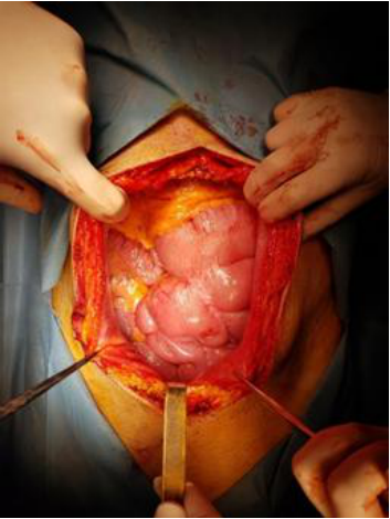
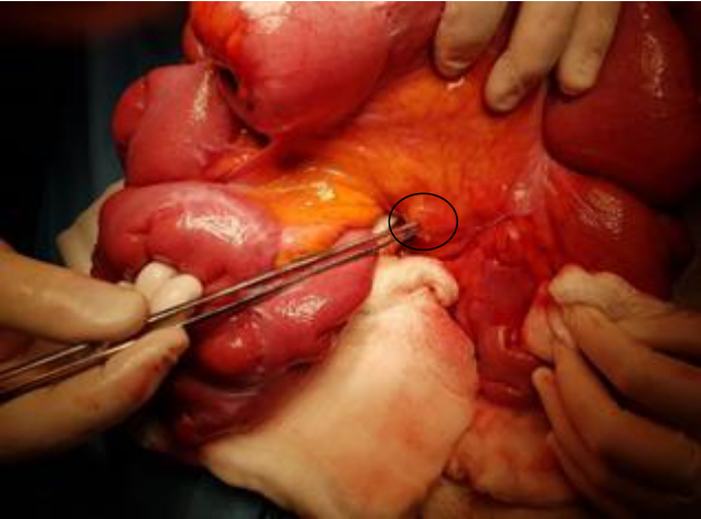
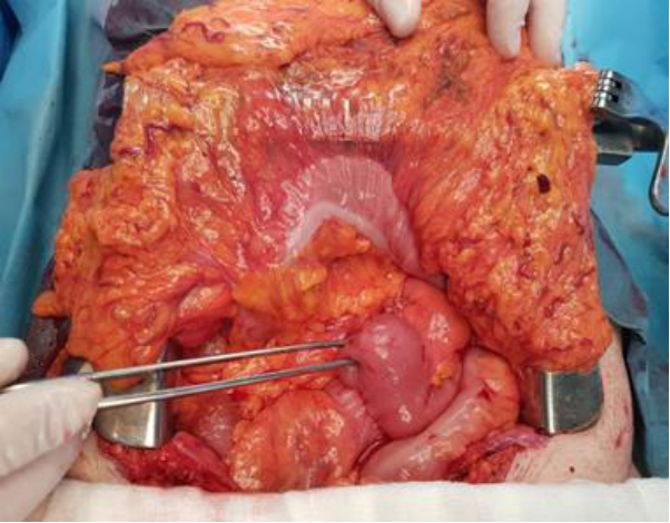
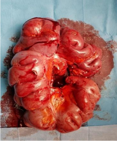
The patient underwent emergency surgery, consisting of exploratory laparotomy. Intraoperatively, an intense adhesive process (Figure 1), multiple whitish jejunal tumor masses up to 3 cm in size, with two mesenteric tumoral masses measuring 3 cm and 5 cm in diameter with adherence to neighboring loops and incarcerated left inguinal hernia (Figure 2) were found, without ascites and no peritoneal or liver nodule. Extensive enterectomy with side-to-side entero-enteric anastomosis and non-mesh hernia repair were performed (Figures 3, 4). There were no signs of ischemia in the bowel loop contained within the herniated sac, hence, there was no need for resection. The non-mesh surgical technique was preferred in order to prevent the risk of infection associated with the mesh.
During hospitalization, the patient had a favorable evolution and was discharged on the 7th postoperative day.
Histopathologic result confirmed the diagnosis of neuroendocrine tumor involving multiple levels of the small intestine with lymph node metastases (3/10) (TNM: pT4 N2 Mx).
The immunohistochemical examination revealed that CDX2 (EPR2764Y) exhibited a uniform positive reaction with both intensity and distribution in the tumor cells, indicating consistent expression throughout the sample. Similarly, CHROMO (LK2H10) showed a uniform positive reaction with intensity, while Synaptophysin (SP11) demonstrated a uniform positive reaction with diffuse intensity in the tumor cells, suggesting widespread expression. Furthermore, Ki67 (30-9) displayed an intense nuclear reaction in 1% of the tumor cells, indicating a low proliferative index within the examined sample. These findings collectively support the diagnosis and provide insights into the tumor’s characteristics.
| CDX2 (EPR2764Y): Uniform positive reaction with intensity and distribution in tumor cells |
| CHROMO (LK2H10): Uniform positive reaction with intensity in tumor cells |
| Synaptophysin (SP11): Uniform positive reaction with intensity, diffuse in tumor cells |
| Ki67 (30-9): Intense nuclear reaction in 1% of tumor cells |
Immunophenotypic data correlated with the morphological cells in the examined fragment are compatible with a neuroendocrine tumor of reduced histological grade (G1).
The patient did not receive chemotherapy, somatostatin analogues, or radiotherapy. He underwent routine follow-ups and regular imaging investigations. At the five-year evaluation, no tumor recurrences or new pathological findings were detected.
Primary tumors in the jejunum or ileum are frequently very small, requiring meticulous palpation of the small intestine from the ligament of Treitz to the ileocecal valve. In 25% to 44% of patients, there are multifocal primary tumors, many of which are subcentimeter in size and can only be detected through careful digital palpation [24]. The use of laparoscopy in these cases is controversial, as it may not allow the detection of such small tumors and does not enable the comprehensive palpation of the entire small intestine that is essential for identifying multifocal disease. Therefore, the limitations of laparoscopy must be taken into account
when determining the surgical strategy for these patients. The literature contains limited studies on the laparoscopic resection of small bowel neuroendocrine tumors (SBNETs). Figueiredo et al. [25] reported successful laparoscopic resections in 12 patients, while Reissman et al. [26] documented similar success in 20 patients. Wang et al. [27] described successful laparoscopic or minimally invasive resections of ileal neuroendocrine tumors (NETs) in six patients who presented with NETs of unknown primary origin. In their study, the authors highlighted the critical role of palpation during minimally invasive surgery to identify the small, often multifocal primary tumors. To facilitate this, they employed a hand-assisted laparoscopic device or a soft tissue wound retractor to exteriorize the jejunum and ileum. This technique allows for comprehensive palpation, resection of the primary tumors, dissection of mesenteric lymph nodes and associated fibrosis, and subsequent intestinal anastomosis.
Current literature regarding the optimal management of neuroendocrine tumors recommends achieving an R0 resection to increase survival rates. However, an R0 resection is feasible in only 20% of cases due to the typically advanced stage at diagnosis [28]. It is important to highlight that the patient presented as an emergency case with symptoms caused by intestinal obstruction due to an incarcerated inguinal hernia, and not from the intestinal tumors, which were discovered incidentally. Therefore, it cannot be claimed that there was an intent for radical treatment, even though an R0 resection was achieved.
Adjuvant treatment options for neuroendocrine tumors may include chemotherapy, somatostatin analogues, and radiotherapy. However, following curative surgery for small intestinal neuroendocrine tumors, there is no established benefit for adjuvant systemic therapy. Somatostatin analogues are recommended for their antiproliferative properties as a first-line treatment for slowly progressive, well-differentiated metastatic small intestinal neuroendocrine tumors. Additionally, they are used for antisecretory purposes to manage persistent secreting syndromes or refractory diarrhea by inhibiting gastrointestinal secretions [29-31].
The overall survival rate appears favorable when compared to other malignancies. However, achieving reliable survival data is challenging due to variations in terminology, inconsistencies in coding practices, conflicting results from smaller cohort studies, and recent advancements in treatment modalities [32].
Several studies have indicated that the long-term recurrence rate for small intestinal neuroendocrine tumors (SI-NETs) following curative R0 resection, in the absence of distant metastases, is approximately 50%. This high recurrence rate underscores the need for vigilant postoperative surveillance and ongoing management strategies to monitor for disease recurrence. Factors contributing to this recurrence include residual microscopic disease and the inherent biological behavior of SI-NETs. Consequently, patients require comprehensive follow-up protocols, including regular imaging and biochemical assessments, to promptly identify and address any recurrence or progression of the disease [33].
The case highlights the importance of considering gastrointestinal neuroendocrine tumors in the differential diagnosis of nonspecific abdominal pain.
Advancements in diagnostic and treatment methods have significantly improved the outlook for many patients with NET, but challenges remain, especially for advanced or poorly differentiated cases.
Although neuroendocrine tumors are rare, their increasing incidence and clinical complexity underline the need for a multidisciplinary approach for effective diagnosis and treatment. Progress in the genetic and molecular understanding of these tumors has opened new avenues for targeted therapies, offering improved hopes for patients.
The authors declare no conflict of interest. No artificial intelligence automatically generated text was inserted in this manuscript, and no image was previously published in another journal or is under consideration for publication elsewhere. This research received no external funding.
MT, BI, JC, HS performed the surgical procedure. MT contributed to the manuscript’s conceptualization, methodology, investigation, discussion, review, editing, and supervision. BI contributed to the conceptualization, investigation, data curation, review, and writing of the manuscript. JC contributed to the manuscript’s conceptualization, investigation, review, and editing. HS contributed to the editing and review of the manuscript. All authors have read and agreed to the published version of the manuscript.
The study was conducted under the Declaration of Helsinki. The research was conducted under ethical guidelines and regulations, ensuring compliance with all necessary protocols.
Written informed consent has been obtained from the patient to publish this paper.