
Ovarian cancer is a heterogeneous disease that is the eighth most common cancer in women and the leading cause of gynecologicrelated cancer death [1-3].
In relation to the postoperative histopathological/immunohistochemical diagnosis for each operated patient, the contribution of Computer Tomography was evaluated for the preoperative characterization of ovarian tumors and assessment of the postinterventional evolution through monitoring at 6 and 12 months, respectively [4-6].
The Computer Tomography (CT) examination is an imaging method that uses ionizing radiation, but demonstrates good sensitivity and specificity, thanks to advancements in multidetector technology. Computed tomography using new multidetector technologies (MDCT) achieves a better characterization of adnexal masses, with a sensitivity of at least 90% and a specificity of 80%. This type of imaging is recommended for preoperative staging of ovarian cancer [4]. This is especially recommended for the preoperative staging of ovarian tumors [7,8,9]. The CT examination is usually used in addition to an inconclusive abdominopelvic ultrasound for the differential diagnosis of pelvic pain syndrome [8]. Although the accuracy of CT is generally lower than that of magnetic resonance imaging (MRI) or transvaginal ultrasound for assessing ovarian pathology, its multiplanar reconstructions enable effective evaluation of the extent of ovarian lesions and identification of coexisting pathologies [4,10,11].
The ovaries are visualized near the iliac vascular axes as oval structures with low densities due to the presence of peripheral follicles. Ovarian ligaments may appear as dense linear structures, especially when free intraperitoneal fluid outlines them. The corpus luteum is thick-walled and shows peripheral contrast uptake [5,7,10,11].
This study aimed to evaluate the performance of diagnostic imaging concerning postoperative histopathological results and the features that may be useful for the classification of tumors. The diagnostic imaging introduction should briefly place the study in a broad context and highlight its importance. It should define the purpose of the work and its significance.
This retrospective study investigated a group of patients with ovarian tumors (n = 63), operated in the Gynecology Department of the Central University Emergency Military Hospital, who underwent preoperative CT examinations. The studied group included patients with benign ovarian tumors, histopathologically diagnosed postoperatively (n=30), and with malignant ovarian tumors (n = 33); 3 patients with malignant ovarian tumors were considered borderline.
The main imaging characteristics that were considered suspicious for malignancy were the following: solid components; endo/exocystic vegetations; wall with irregular thickness and intramural nodules; irregular intratumoral septa with thicknesses over 3mm; contrast uptake of the solid components (intratumoral, parietal or septal) with enhancement curve type II/III appearance (early contrast uptake of lower intensity/equal to the myometrium, persistent in the plateau, respectively intense early contrast uptake, more pronounced than the myometrial and dynamic “wash-out”); dimensions (>3 cm); secondary lesions involving the peritoneal lining, mesenteric or omental fat; ascites; pelvic extension; locoregional nodal disease; distant metastases (extraperitoneal) [5,8].
Fisher’s test and Mann Mann-Whitney (Wilcoxon) test were used to determine the significance between groups. The correlation between age at diagnosis and CA125 values was performed using Pearson’s correlation coefficient.
The Sensitivity [TPR =TP/(TP+FN)], False negative ratio [FNR=FN/(TP+FN)]; False positive ratio [FPR=FP/(FP+TN)], Specificity [TNR =TN/(FP+TN)], Predictability of positive values [Precision PPV=TP/(TP+FP)]; Omission rate [Omission rate = FN/(FN+TN)] and Accuracy [Accuracy = (TP+TN)/Total] were estimated. For this estimation, the values for True positive (TP); True negative (TN); False positive (FP) and False negative (FN) were used.
This retrospective study analyzed 63 patients with ovarian tumors who were preoperatively CT investigated. Nine patients with malignant tumors and eight with benign tumors had bilateral tumors. Histopathological diagnosis established the presence of epithelial (n = 40), germ cell (n = 17), or stromal and sex cord tumors (n = 6). Epithelial tumors are more common in patients with malignant tumors compared to benign tumors. (31 vs 9, p<0.0001). Multilocular tumors were identified in 29 patients with malignant tumors and 4 patients with benign tumors (p<0.001). Endo or exocytic vegetations were identified in 29 of the patients with malignant or borderline tumors, but were not present in those with benign tumors.
Of the patients analyzed, 23 resided in rural areas (9 with benign tumors) and 36 (21 with benign tumors) in urban areas. In this sample, the distribution of malignant and benign tumors was not associated with place of residence (p>0.05). The age at diagnosis of patients from urban and rural areas was similar (44.58±13.72 vs 50.44±10.50, p>0.05).
The median age of these patients was 46 years (23-72 years old). The age of patients with benign tumors (average: 41.40±12.17 years; 23-67 years old) was significantly lower (p=0.0007) than that of those with malignant tumors (average: 51.88±10.61; 35-72 years old). The age at diagnosis in the subplot of women with tumors over 4 cm was lower in subjects with benign tumors when compared with carriers of malignant tumors (40.12±12.88 vs 51.88±10.61 vs p=0.003). The age of patients with malignant tumors, stratified according to the presence or absence of metastases, was similar (52.44±10.77 vs 51.35±10.76, p>0.05).
CA125 was elevated more frequently found to be elevated in carriers of benign tumors (15 patients with benign tumors, all 3 patients with borderline tumors, in 5 patients with malignant tumors, p<0.05). The values of this parameter were not significantly correlated with the age of the patients in the entire group of subjects (r = 0.25) or the groups of patients with benign tumors (r = 0.16) or malignant tumors (r = -0.06).
The ROMA score had increased values in all patients with malignant tumors and 53.33% of patients with benign tumors. The size of the tumors over 4 cm was recorded in all 33 malignant and borderline tumors and 17 of the benign lesions. Analysis of the intratumoral solid component. A solid intratumoral component was detected in 52 of the studied ovarian tumors (30 malignant tumors and 22 of benign tumors, p<0.05). The presence of irregular intratumoral septa with a thickness of over 3mm was found in 28 malignant and borderline tumors and only in 4 benign tumors (p<0.0001).
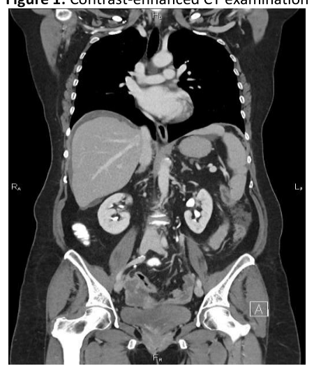
Contrast uptake analysis of the solid/parietal/septal tumor components. Of the benign tumors, 80% showed contrast uptake (80% with a progressive loading curve type I), and in the remaining 20%, contrast uptake was absent. Bilateral ovarian serous cystadenocarcinoma, with extensive peritoneal disease (especially at the level of the great omentum – “omental cake”) and a small amount of ascites near the liver, spleen and in the pouch of Douglas. Ascites were detected in 22 of 30 malignant tumors, in none of the borderline tumors, and in one case of a benign tumor (p<0.0001).
Analysis of locoregional lymph node disease. Several 32 tumors from the 63 cases presented with enlarged locoregional lymph nodes. Of these, most were malignant and borderline tumors (78% of this group), confirmed histopathologically with positive lymph node disease. Out of the total of benign tumors, only 20% of benign tumors had locoregional ganglionic hypertrophies, confirmed histopathologically as reactive lymph nodes (inflammatory). These differences between malignant and benign tumors are statistically significant (p<0.0001).
Analysis of cases with macroscopic peritoneal disease. Macroscopic peritoneal disease was highlighted only in malignant tumors, 78% of malignant tumors presented this aspect. The age of women with or without macroscopic peritoneal disease was similar (51.80±10.89 vs 52.13±10.40, p>0.05).
Analysis of pelvic parietal extension/to the neighboring organs. Most of the malignant tumors (70%) and 2 of the borderline ones showed pelvic parietal extension/to the neighboring organs. Among the benign tumors, only one presented this aspect, which represents 3%.
Analysis of cases with distant metastasis. This category included cases in which distant (non-peritoneal) metastases occurred. Among the malignant tumors, 53% showed distant metastases, while benign or borderline tumors did not show such metastases.
Evaluation of the sensitivity and specificity of CT examination. From the sample of 63 cases investigated by CT examination (the groups of patients evaluated by ultrasound and CT or ultrasound, CT, and MRI), the number of cases identified as positive for malignant tumors was 36, in relation to the post-interventional histopathological examination that established the diagnosis of malignancy in 33 of the analyzed cases.
There were 27 cases evaluated by CT with a benign appearance, and the post-interventional histopathological examination established the diagnosis of benignity in 30 of them.
The confusion matrix for assessing the sensitivity and specificity of CT evaluation of malignant ovarian tumors showed a sensitivity of 91%, with a specificity of 80% and an accuracy of 85%.
| Results | Final Positive | Final Negative | |
|---|---|---|---|
| CT positive | TP=30 | FP=6 | Predictability of positive values, =0.83 |
| CT negative | FN=3 | TN=24 | Omission rate =0.11 |
| Sensitivity = 0.91 | False positive ratio =0.2 | Accuracy=0.85 | |
| False negative ratio=0.09 | Specificity =0.80 |
CT evaluation was particularly beneficial in cases of large tumors with distant extension, allowing for the assessment of secondary lesions in other organs and the presence of associated comorbidities, in order to optimize therapeutic management.
From the group of 63 cases investigated by CT examination, the number of cases identified as positive for malignant tumors was 36, compared to the post-interventional histopathological examination, which confirmed the diagnosis of malignancy in 33 of the analyzed cases.
The method remains useful in assessing nodal extension and distant metastatic disease and allows a sensitivity of 90%, a specificity of 89%, a positive predictive value of 78%, and a negative predictive value of 95%, with an overall accuracy in diagnosing malignancy of about 89% [2,7,10].
This retrospective study analyzed 63 patients with ovarian tumors who were preoperatively CT investigated. Histopathological analysis reveals that epithelial tumors, multilocular tumors, and cystic vegetations were more common in malignant tumors. These differences highlight the heterogeneity and differences between ovarian tumors.
The median age of patients investigated in this study was 46 years (23-72 years old). This result is concordant with data previously published [15], the majority of women diagnosed with ovarian cancer are in the perimenopausal state.
The size of the tumors over 4 cm was recorded in all 33 malignant and borderline tumors and 17 of the benign lesions. The large sizes found in patients diagnosed with malignant tumors may be due to the non-specific symptoms at the onset and the late presentation at the gynecological examination, which determines a long time until diagnosis, ovarian tumors being most frequently discovered incidentally during a routine examination.
A solid intratumoral component was detected in 52 of the studied ovarian tumors (30 malignant tumors and 22 benign tumors, p<0.05). The presence of a solid intratumoral component with enhancement curve type II/III is correlated with a higher risk of malignancy, as the Ovarian-Adnexal Reporting and Data System shows [16].
The presence of irregular intratumoral septa with a thickness of over 3mm was significantly more frequent in malignant and borderline tumors than in benign tumors (p<0.0001). The irregular septa with a thickness of over 3mm in any ovarian tumor seen on imaging studies should raise suspicion of malignancy, requiring further investigations.
Ascites was detected only in 22 of 30 malignant tumors. In most cases, ascites is present in ovarian cancer because neoplastic cells in the peritoneal lining affect the physiological drainage of peritoneal fluid [17]. Very rarely, ascites can be associated with a benign ovarian tumor, as we found in our patient who had Demos-Meigs syndrome [18].
Most of the malignant tumors (70%), 2 of the borderline ones, and one benign tumor showed pelvic parietal extension/to the neighboring organs. Parietal extension/invasion to the neighboring organs is an imaging feature of malignancy until proven otherwise. Sometimes, in imaging studies, it can be misinterpreted as malignancy in cases of chronic infections or previous surgeries [4].
The ROMA score had increased values in all patients with malignant tumors and 53.33% of patients with benign tumors. In some studies, high ROMA scores were correlated with advanced ovarian cancer before surgery [19,20].
Different imagistic methods can be used to differentiate between physiologic, benign, and malignant ovarian conditions in women [21,22]. Computer Tomography has increased value, especially for the preoperative staging of ovarian cancer, considering the increased spatial resolution of the method, the increased accuracy in the detection and characterization of masses through the new multi-detector technologies (MDCT) implemented, and the reduced radiation doses due to low-dose protocols optimization [6,23].
Computed tomography (CT) is the imaging modality of choice for staging ovarian cancer. Multidetector computed tomography (MDCT) provides clinically relevant information about these tumors (e.g., size of the primary tumor, size and location of peritoneal implants, and lymph nodes). These data can be used to help evaluate surgical options. The overall accuracy of MDCT in diagnosing malignant adnexal masses is as high as 89%. MDCT signs predictive of malignancy include the presence of papillary projections in a cystic lesion, necrosis in a solid mass, and peritoneal metastases. CT is frequently used to detect ovarian carcinoma and to assess response to therapy. CT has limitations in detecting small peritoneal metastases. CT can detect tumors larger than 1 cm with a sensitivity of 85– 93% and a specificity of 91–96% [24].
Computed Tomography has significant value in the management of ovarian cancer patients, especially in the preoperative staging of ovarian cancer, given its high spatial resolution and improved accuracy in detecting and characterizing ovarian tumor masses and metastatic lesions. This is achieved through newly implemented multi-detector tomography (MDCT) technologies and significantly reduced radiation doses using optimized low-dose protocols. The method remains useful in assessing lymph node extension and distant metastatic lesions, offering high sensitivity and specificity, as demonstrated in this article.
The authors declare no conflict of interest. No artificial intelligence automatically generated text was inserted in this manuscript, and no image was previously published in another journal or is under consideration for publication elsewhere. This research received no external funding.
Conceptualization, N.I.A. and N.M.F.; methodology N.N, S.N.M.E.D, A.F.C, A.D.S and D.L.M; software, N.F. and S.N.M.E.D.; validation, N.I.A. ,C.I.M, A.D.S, A.F.C. and N.M.F.; formal analysis, N.M.F.; investigation N.I.A.; resources A.D.S, S.N.M.E.D.; data curation N.F.; writing—original draft preparation, N.I.A.; writing—review and editing, N.I.A, N.M.F, D.L.M; visualization, N.I.A.; supervision, N.M.F; project administration, N.I.A. All authors have read and agreed to the published version of the manuscript.
The present study was approved by the hospital’s ethics committee Nr. 601/ 24.05.2023 and is performed under the ethical standards laid down by the Declaration of Helsinki.
Informed consent was obtained from all subjects involved in the study.