
Non-fermentative Gram-Negative bacilli (NFGNB) represent a diverse group of aerobic, non-spore-forming bacteria that either do not use glucose as an energy source or metabolize it oxidatively. They account for approximately 20% of all Gram-Negative bacilli (GNB) [1]. These bacteria are typically found as saprophytes in natural environments like soil and water, and they also live as commensals in humans and other animals.
While they are often isolated incidentally, their role as pathogens has become more prominent due to the widespread use of antibiotics and the increasing number of patients with weakened immune systems.
Non-fermentative bacteria (NFs) are increasingly recognized as causes of opportunistic and sometimes severe infections, as well as nosocomial infections [2]. These bacteria are frequently isolated from patients with severe underlying conditions who have undergone extensive use of broad-spectrum antibiotics, prolonged surgical procedures, extended hospital stays, insufficient mechanical ventilation or tracheotomy, genitourinary procedures, or who are burn victims or low-birth-weight infants. Infections caused by these bacteria are commonly observed in vulnerable age groups, including neonates, children, and the elderly [3]. They are also commonly found in cases of septicemia, meningitis, pneumonia, urinary tract infections, and surgical wound infections [4].
NFGNB can cause a variety of infections and account for approximately 15% of all Gram-negative bacilli isolated from clinical samples [5]. These infections are increasingly problematic in hospital environments, especially among immunocompromised patients [6]. Among the opportunistic pathogens affecting immunocompromised hosts, whether due to disease or treatment, Pseudomonas aeruginosa is especially significant.
Other important species include Acinetobacter baumannii, Pseudomonas fluorescens, Pseudomonas stutzeri, Stenotrophomonas maltophilia, Pseudomonas putida, and Burkholderia cepacia [7]. Rates of colonization by NFGNB increase in hospitalized patients, especially those who have been in the hospital for extended periods or have received broad-spectrum antimicrobial therapy or chemotherapy. These non-fermenters frequently cause nosocomial blood-stream infections, particularly in debilitated and immunocompromised individuals, and are often multidrug-resistant. According to data from the Surveillance and Control of Pathogens of Epidemiological Importance (SCOPE) study, approximately one-fourth of Gram-negative bacteremia were attributed to NFGNB [8].
Treating infections caused by these pathogens is challenging due to their multidrug resistance (MDR) and the rapid emergence of high-level MDR to various antibiotic classes, including β-lactams, aminoglycosides, and fluoroquinolones. This resistance complicates both treatment and infection control efforts [9]. Pseudomonas aeruginosa has a high potential for developing resistance to most antibiotics, as evidenced by its genome containing the largest resistance island with over 50 resistance genes. The mechanisms behind this antibiotic resistance include the production of enzymes that degrade or inactivate antibiotics, outer membrane proteins that expel antibiotics, and mutations that alter the antibiotic targets [10].
Production of antibiotic-degrading enzymes, such as extended-spectrum beta-lactamases, AmpC cephalosporinases, carbapenemases, and aminoglycoside-modifying enzymes, has been reported among non-fermentative Gram-negative bacilli [11]. NFGNBs exhibit resistance to many antibiotics and are known to produce extended-spectrum beta-lactamases (ESBLs) and metallobeta-lactamases (MBLs) [12]. Non-fermentative Gram-negative bacteria have developed resistance to many commonly used antibiotics, including cephalosporins and carbapenems. This resistance complicates treatment, extends hospital stays, increases mortality rates, and drives up healthcare costs [13].
The aim of our study was to determine the antibacterial susceptibility patterns of non-fermentative Gram-negative bacilli isolated from clinical samples.
A prospective study was carried out at Mahatma Gandhi Medical College and Hospital (MGMCH), focusing on patients exhibiting signs and symptoms of non-fermenter infections. The study period was conducted from June 2019 to May 2020. All NFGNB isolates were obtained from different clinical samples received in the clinical microbiology laboratory of Mahatma Gandhi Hospital (MGH), Sitapura, Jaipur, Rajasthan, included in the study.
Non-fermentative Gram-negative bacilli isolated from endotracheal (ET), pus swab, sputum, blood, urine, pus, and other body fluids, etc.
All Gram-negative bacilli except non-fermentative Gram-negative bacilli from different clinical samples.
Various types of samples were collected, including urine, blood, pus, discharge from the skin and soft tissue sites, sputum, ET, and other fluids such as Cerebrospinal fluid (CSF), Ascitic fluid, and Pleural fluid. Additionally, miscellaneous swabs (ear, throat, vaginal, wound, etc.), tissue samples, and central line tips were gathered. These samples were received in the clinical microbiology laboratory from both inpatients and outpatients at the hospital. The samples were collected using universal precautions and sterile techniques and were transported to the laboratory promptly under optimal conditions. Detailed patient histories were recorded, including age, sex, any use of indwelling medical devices, and duration of hospital or ICU stays.
After collection, each sample container was carefully labeled with the patient’s name, ID number, and other relevant details. The specimens were promptly transported to the laboratory, typically within 1 hour of collection, and processed as quickly as possible. If processing was delayed, the samples were stored at 4°C until they could be analyzed.
Routine microscopy was performed on all samples. Gram staining was carried out for all samples except urine. Wet microscopy was used to detect bacteria and pus cells in urine samples. Each clinical sample was inoculated onto Blood agar and MacConkey agar plates, which were incubated at 37°C for 18-24 hours. Colony characteristics were then observed. Organisms that produced pale or colorless colonies on MacConkey agar and appeared as gram-negative bacilli on gram staining were identified as NFGNB. Further identification was conducted using the VITEK-2 compact system [14]. All NFGNB isolates underwent antimicrobial susceptibility testing using the Kirby-Bauer method according to CLSI guidelines 2020 [15].
In our study, out of 10707 clinical samples, 879 NFGNB isolates were obtained, with a prevalence of 8.20%. Also, out of the 879 NFGNB isolates, 415 (47.21%) were Pseudomonas aeruginosa, followed by Acinetobacter baumannii 380 (43.23%), Burkholderia cepacia 29 (3.30%), Burkholderia pseudomallei 17 (1.93%), Stenotrophomonas maltophilia 16 (1.82%), Acinetobacter lwoffii 7 (0.80%), Pseudomonas putida 6 (0.68%), Acinetobacter coffee 2 (0.23%), Pseudomonas fluorescens 2 (0.23%), Pseudomonas stutzeri 2 (0.23%), A. junii 1 (0.11%), A. haemolyticus 1 (0.11%) and Pseudomonas luteola 1 (0.11%) as shown in Table 1. Out of the total 879 isolates of NFGNB, the majority of the specimens were from ET (30.60%), followed by pus swabs (19.11%), blood (16.15%), and urine (10.01%), as shown in Table 2. Out of the total positive NFGNB cases, the maximum number was from inpatients (IPD) 775 (88.17%), followed by outpatients (OPD) 104 (11.83%). Out of the total 879 NFGNB isolates, the majority of isolates were the males in the age group 61-70, i.e.121 (13.76%), followed by the age group 41-50, i.e.113 (12.85%), as shown in Table 3 and Figure 1.
| NFGNB spp. | Total No. | Percentage |
|---|---|---|
| Pseudomonas aeruginosa | 415 | 47.21% |
| Acinetobacter baumannii | 380 | 43.23% |
| Burkholderia cepacian | 29 | 3.30% |
| Burkholderia pseudomallei | 17 | 1.93% |
| Stenotrophomonas maltophilia | 16 | 1.82% |
| Acinetobacter lwoffii | 7 | 0.80% |
| Pseudomonas putida | 6 | 0.68% |
| Acinetobacter coffee | 2 | 0.23% |
| Pseudomonas fluorescens | 2 | 0.23% |
| Pseudomonas stutzeri | 2 | 0.23% |
| Acinetobacter junii | 1 | 0.11% |
| Acinetobacter haemolyticus | 1 | 0.11% |
| Pseudomonas luteola | 1 | 0.11% |
| TOTAL | 879 | 100% |
| Sl. No. | Sample | No. of cases | Percentage |
|---|---|---|---|
| 1 | ET | 269 | 30.60% |
| 2 | PUS SWAB | 168 | 19.11% |
| 3 | BLOOD | 142 | 16.15% |
| 4 | URINE | 88 | 10.01% |
| 5 | SPUTUM | 87 | 9.90% |
| 6 | CSF | 44 | 5.01% |
| 7 | PUS | 19 | 2.16% |
| 8 | DRAIN | 16 | 1.82% |
| 9 | PLEURAL FLUID | 11 | 1.25% |
| 10 | BAL | 11 | 1.25% |
| 11 | TIPS | 6 | 0.68% |
| 12 | TISSUE | 5 | 0.57% |
| 13 | ASCITIC FLUID | 4 | 0.46% |
| 14 | OTHER BODY FLUID | 3 | 0.34% |
| 15 | SYNOVIAL FLUID | 2 | 0.23% |
| 16 | SEMEN | 2 | 0.23% |
| 17 | BILE | 2 | 0.23% |
| TOTAL | 879 | 100% |
ET: Endotracheal secretion, CSF: Cerebrospinal fluid, BAL: Bronchoalveolar lavage, TIPS: Transjugular Intrahepatic Portosystemic Shunt
| Distribution of OPD/IPD | Number of Isolates | Percentage |
|---|---|---|
| OPD | 104 | 11.83% |
| IPD | 775 | 88.17% |
| Total | 879 | 100% |
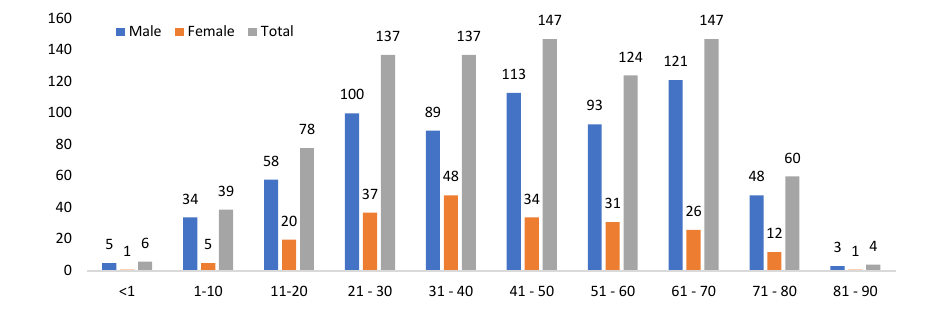
Table 4 shows the distribution of various bacterial isolates from different clinical samples. In this study, P. aeruginosa was most frequently isolated from pus swabs (13.42%), followed by endotracheal samples (8.19%), sputum (7.17%), urine (6.94%), and blood (4.44%). Ac. baumannii was predominantly isolated from ET samples (21.4%), followed by blood (7.85%), pus swabs (4.44%), CSF (3.53%), sputum (2.05%), and urine (1.82%). B. cepacia was most commonly found in blood samples (1.48%), with fewer instances in pus swabs (0.68%), sputum (0.34%), ET samples (0.23%), and other body fluids (0.23%) as shown in Table 4.
| SAMPLE | P. AERUGINOSA | A. BAUMANNII | B. CEPACIA | |||
|---|---|---|---|---|---|---|
| N | % | N | % | N | % | |
| ET (n=269) | 72 | 8.19 | 190 | 21.6 | 2 | 0.23 |
| PUS SWAB (n=168) | 118 | 13.42 | 39 | 4.44 | 6 | 0.68 |
| BLOOD (n=142) | 39 | 4.44 | 69 | 7.85 | 13 | 1.48 |
| URINE (n=88) | 61 | 6.94 | 16 | 1.82 | 1 | 0.11 |
| SPUTUM (n=87) | 63 | 7.17 | 18 | 2.05 | 3 | 0.34 |
| CSF (n=44) | 12 | 1.37 | 31 | 3.53 | 0 | 0 |
| PUS (n=19) | 14 | 1.59 | 2 | 0.23 | 1 | 0.11 |
| DRAIN (n=16) | 12 | 1.37 | 3 | 0.34 | 0 | 0 |
| PLEURAL FLUID (n=11) | 5 | 0.57 | 4 | 0.46 | 0 | 0 |
| BAL (n=11) | 5 | 0.57 | 5 | 0.57 | 0 | 0 |
| TIP (n=6) | 5 | 0.57 | 0 | 0 | 1 | 0.11 |
| TISSUE (n=5) | 3 | 0.34 | 2 | 0.23 | 0 | 0 |
| ASCITIC FLUID (n=4) | 2 | 0.23 | 1 | 0.11 | 0 | 0 |
| OTHER BODY FLUID (n=3) | 0 | 0 | 0 | 0 | 2 | 0.23 |
| SYNOVIAL FLUID (n=2) | 0 | 0 | 0 | 0 | 0 | 0 |
| SEMEN (n=2) | 2 | 0.23 | 0 | 0 | 0 | 0 |
| BILE (n=2) | 2 | 0.23 | 0 | 0 | 0 | 0 |
| TOTAL | 415 | 47.21 | 380 | 43.23 | 29 | 3.3 |
As shown in Table 5, the present findings showed that the resistance ratio of various antibiotics was higher than that of sensitivity against NFGNB. The antimicrobial susceptibility testing conducted on all 879 non-fermenting Gram-negative bacteria revealed the following results: Cefoperazone/Sulbactam showed a sensitivity rate of 37.54%, making it the most effective among the tested agents.
This was followed by Meropenem at 35.26% & Ceftazidime at 34.47%. On the other hand, Ticarcillin/Clavulanic Acid exhibited the highest resistance rate at 90.78%. This was closely followed by Ciprofloxacin with 73.49% resistance, Levofloxacin at 72.81%, and Piperacillin/Tazobactam at 70.19%, as detailed in Table 5.
| S. No. | Antibiotics | Sensitive | Resistance | ||
|---|---|---|---|---|---|
| No. of cases | % | No. of cases | % | ||
| 1 | Ticarcillin/Clavulanic Acid | 81 | 9.21% | 798 | 90.78% |
| 2 | Piperacillin/Tazobactam | 262 | 29.80% | 617 | 70.19% |
| 3 | Ceftazidime | 303 | 34.47% | 576 | 65.52% |
| 4 | Cefoperazone/Sulbactam | 330 | 37.54% | 549 | 62.45% |
| 5 | Cefepime | 288 | 32.76% | 591 | 67.23% |
| 6 | Imipenem | 285 | 32.42% | 594 | 67.57% |
| 7 | Meropenem | 310 | 35.26% | 569 | 64.73% |
| 8 | Ciprofloxacin | 233 | 26.50% | 646 | 73.49% |
| 9 | Levofloxacin | 239 | 27.18% | 640 | 72.81% |
Among the 879 NFGNB isolates obtained, P. aeruginosa showed high sensitivity to several antibiotics. Cefepime was the most effective, with 258 out of 415 isolates (62.16%) demonstrating sensitivity. This was followed by Imipenem with 254 isolates (61.20%) and Ceftazidime with 250 isolates (60.24%). In contrast, P. aeruginosa exhibited high resistance rates to Ticarcillin/Clavulanic Acid, affecting 349 isolates (84.09%). The bacteria also showed considerable resistance to Levofloxacin (226 isolates or 54.45%) and Ciprofloxacin (208 isolates or 50.12%).
Out of the 380 total A. baumannii isolates, the highest sensitivity was observed for Cefoperazone/Sulbactam, with 70 isolates (18.42%) showing susceptibility. In contrast, A. baumannii exhibited very high resistance rates to Ticarcillin/Clavulanic Acid (366 isolates, 96.82%), and Ciprofloxacin (363 isolates, 96.03%). For B. cepacia isolates, which numbered 29 in total, the highest sensitivity was to Meropenem, with 19 isolates (65.51%) showing susceptibility. This was followed by Ceftazidime with 14 isolates (48.27%) and Levofloxacin with 11 isolates (37.93%). All 29 B. cepacia isolates were resistant to Ticarcillin/Clavulanic Acid, Piperacillin/Tazobactam, Cefepime, and Imipenem, as shown in Table 6.
| ANTIBIOTIC | P. aeruginosa | A. BAUMANNII | B. CEPACIA | ||||
|---|---|---|---|---|---|---|---|
| (n=415) | (n=380) | (n=29) | |||||
| N | % | N | % | N | % | ||
| Ticarcillin/Clavulanic Acid | R | 349 | 84.09% | 366 | 96.82% | 29 | 100.00% |
| S | 66 | 15.90% | 14 | 3.68 % | 0 | 0.00% | |
| Piperacillin/Tazobactam | R | 185 | 44.57% | 357 | 94.44% | 29 | 100.00% |
| S | 230 | 55.42% | 23 | 6.05% | 0 | 0.00% | |
| Ceftazidime | R | 165 | 39.75% | 363 | 96.03% | 15 | 51.72% |
| S | 250 | 60.24% | 17 | 4.47% | 14 | 48.27% | |
| Cefoperazone/Sulbactam | R | 169 | 40.72% | 310 | 82.01% | 28 | 96.55% |
| S | 246 | 59.27% | 70 | 18.42% | 1 | 3.44% | |
| Cefepime | R | 157 | 37.83% | 358 | 94.70% | 29 | 100.00% |
| S | 258 | 62.16% | 22 | 5.78% | 0 | 0.00% | |
| Imipenem | R | 161 | 38.79% | 358 | 94.70% | 29 | 100.00% |
| S | 254 | 61.20% | 22 | 5.78% | 0 | 0.00% | |
| Meropenem | R | 167 | 40.24% | 360 | 95.23% | 10 | 34.48% |
| S | 248 | 59.75% | 20 | 5.3% | 19 | 65.51% | |
| Ciprofloxacin | R | 208 | 50.12% | 363 | 96.03% | 28 | 96.55% |
| S | 207 | 49.87% | 17 | 4.47% | 1 | 3.44% | |
| Levofloxacin | R | 226 | 54.45% | 359 | 94.97% | 18 | 62.06% |
| S | 189 | 45.54% | 21 | 5.5% | 11 | 37.93% | |
Resistance and Sensitivity patterns of various antibiotics from IPD & OPD Patients are shown in Table 7. Out of a total positive 879 NFGNB isolates, a maximum number of patients were from IPD 775 (88.17%), followed by OPD 104 (11.83%). The antimicrobial susceptibility pattern varied in both IPD & OPD patients, as results shown in Table 7.
The present results showed that IPD patients manifest high sensitivity to several antibiotics. Cefoperazone/Sulbactam showed a sensitivity rate (33.80%,) making it the most effective among the tested agents, followed by Meropenem (31.48%), and Ceftazidime (30.70%). On the other hand, Ticarcillin/Clavulanic Acid exhibited the highest resistance rate at 92.12%. This was similarly followed by Ciprofloxacin with 76.25% resistance, Levofloxacin (74.70%) & Piperacillin/Tazobactam (74.19%). Among the 104 OPD patients showed high sensitivity to Cefoperazone/Sulbactam with a sensitivity rate of 65.38%, followed by Meropenem (63.46%), Cefepime (63.46%), Ceftazidime (62.5%) against the tested agents and high resistance rates also observed against Ticarcillin/Clavulanic Acid (80.76%) followed by Levofloxacin (58.65%), Ciprofloxacin (52.88%).
| S. No. | Antibiotics | IPD=775 | OPD=104 | ||||||
|---|---|---|---|---|---|---|---|---|---|
| Sensitive | Resistance | Sensitive | Resistance | ||||||
| No. of cases | % | No. of cases | % | No. of cases | % | No. of cases | % | ||
| 1 | Ticarcillin/Clavulanic Acid | 61 | 7.87 % | 714 | 92.12% | 20 | 19.23% | 84 | 80.76% |
| 2 | Piperacillin/Tazobactam | 200 | 25.80% | 575 | 74.19% | 62 | 59.61% | 42 | 40.38% |
| 3 | Ceftazidime | 238 | 30.70% | 537 | 69.29% | 65 | 62.5% | 39 | 37.5% |
| 4 | Cefoperazone/Sulbactam | 262 | 33.80% | 513 | 66.19% | 68 | 65.38% | 36 | 34.61% |
| 5 | Cefepime | 222 | 28.64% | 553 | 71.35% | 66 | 63.46% | 38 | 36.53% |
| 6 | Imipenem | 223 | 28.77% | 552 | 71.22% | 62 | 59.61% | 42 | 40.38% |
| 7 | Meropenem | 244 | 31.48% | 531 | 68.51% | 66 | 63.46% | 38 | 36.53% |
| 8 | Ciprofloxacin | 184 | 23.74% | 591 | 76.25% | 49 | 47.11% | 55 | 52.88% |
| 9 | Levofloxacin | 196 | 25.29% | 579 | 74.70% | 43 | 41.34% | 61 | 58.65% |
In the current study, a total number of 10707 specimens were received in the microbiology laboratory for culture. Out of these, 879 specimens yielded positive isolates for non-fermenting Gram-negative bacteria (NFGNB). The distribution of these isolates was as follows: Among them, P. aeruginosa, 415 (47.21%) was the most commonly isolated, followed by Ac. baumannii 380 (43.23%), B. cepacia 29 (3.30%), B. pseudomallei 17 (1.93%), S. maltophilia 16 (1.82%), Ac. lwoffii 7 (0.80%), P. putida 6 (0.68%), Ac. coffee 2 (0.23%), P. fluorescens 2 (0.23%), P. stutzeri 2 (0.23%), Ac. junii 1 (0.11%), Ac. haemolyticus 1 (0.11%) and P. luteola 1 (0.11%).
The current study’s findings align with those reported by Simgamsetty et al., who isolated 260 NFGNB strains. Of these, 151 (58%) were identified as P. aeruginosa, 94 (36.1%) as Ac. baumannii, 1.92% as B. cepacia, 1.15% each as Ac. lwoffii and P. stutzeri, and 0.38% each as Achromonas xylosoxidans and Sphingomonas paucimobilis [16]. Similarly, other studies by Maniyan et al., Prashant et al., and Mahajan [17-19] reported comparable findings. In the present study, out of a total of 879 NFGNB isolates, the majority were from endotracheal (ET) specimens (30.60%), followed by pus swabs, blood, and urine samples.
The present study’s results are similar to those reported by Wadhwa et al., who found that the majority of clinical specimens were from endotracheal (ET) samples (67.44%), followed by tracheal secretions (15.11%), sputum (9.30%), and bronchoalveolar lavage (BAL) (8.72%).
These findings are also consistent with studies conducted by Patel et al. [20,21]. In the present study, of the total 879 positive NFGNB isolates, 775 (88.17%) were from IPD, while 104 (11.83%) were from OPD. The present study is consistent with the findings of Nazir et al., who reported that among 120 NFGNB strains, 91 (75.8%) were isolated from IPD and 29 (24.16%) from OPD. These results align with those of Rashid et al. [22,23]. In the present study, the majority of isolates were from males in the age group of 61-70 years, accounting for 121 (13.76%) of the cases, followed by the age group of 41-50 years, with 113 (12.85%) of the isolates.
The present study’s findings are similar to those reported by Mahajan et al., who observed that the majority of patients (35.13%) were in the age group of 40-60 years. Additionally, out of 342 NFGNB isolates, 59.79% were from males and 40.20% were from females. In the study conducted by Nazir et al., males were more commonly affected than females in the age group under 10 years (34.1%), followed by those over 60 years (32.5%) [19,22]. In the present study, P. aeruginosa was most commonly isolated from pus swabs, followed by endotracheal (ET) samples, sputum, urine, and blood. In the study conducted by Rashid et al., 10.5% of P. aeruginosa isolates were obtained from wound swab samples.
A similar study by Sarkar et al. reported that P. aeruginosa was most commonly isolated from pus (58%), followed by urine, stool, and other specimens. This finding is consistent with studies conducted by Mahajan and Madkey [23,24,19,25]. In the present study, Ac. baumannii was most commonly isolated from endotracheal (ET) samples (21.39%), followed by pus swabs, cerebrospinal fluid (CSF),
sputum, and urine. These findings are similar to those reported by Madkey et al., who isolated 58.82% of Ac. baumannii from endotracheal (ET) samples, followed by pus, urine, blood, and sputum. This is consistent with other studies conducted by Mahajan et al. and Maniyan et al. [17,19,25]. In the present study, B. cepacia was isolated from blood (1.48%), followed by pus swabs, sputum, and endotracheal (ET) samples. These findings are consistent with reports from other studies, which indicated that 5.00% of B. cepacia isolates were obtained from blood, followed by sputum. Similar results were found in studies conducted by Maniyan, Madkey, and Kalidas [17,25,26].
The extensive use of antibiotics in the hospital environment has led to the selection of strains with resistance to multiple antibiotics. Increased antibiotic resistance of NFGNB associated with increased morbidity and mortality makes the infections they produce a major public health problem. In the present study, resistance to various antibiotics against NFGNB was noted. This finding was similar to Buzila [27], who also reported Acinetobacter spp. presented over 70% resistance to the majority of antibiotics. Three pan-drugresistant P. aeruginosa strains were identified. The rate of colistin resistance was 2.91% for P. aeruginosa and 3.33% for Acinetobacter spp. Mechanical ventilation and hospitalization in intensive care units are known risk factors for contracting infections with microorganisms that have become resistant to multiple antibiotics. Increased antibiotic resistance of the strains isolated from tracheobronchial aspirate suggests that using such medical devices (endotracheal intubation tubes and urinary catheters) promotes infections with MDR strains. In the present study, among the 879 NFGNB isolates, the highest sensitivity was observed with Cefoperazone/Sulbactam (37.54%), followed by Meropenem (35.26%), Ceftazidime (34.47%). On the other hand, Ticarcillin/Clavulanic Acid exhibited the highest resistance rate at 90.78%. This was closely followed by Ciprofloxacin with 73.49% resistance, Levofloxacin at 72.81%, and Piperacillin/Tazobactam at 70.19%. This is similar to the findings of Bhargava et al. and Sarkar et al., who reported that Cefoperazone/Sulbactam was the most effective antibiotic against NFGNB [28,29].
In the present study, out of a total of 879 NFGNB isolates, P. aeruginosa showed high sensitivity to Cefepime, with 258 isolates (62.16%) being sensitive, followed by Imipenem with 254 isolates (61.20%), and Ceftazidime with 250 isolates (60.24%). This is similar to the study by Malini et al., which found sensitivity to Cefepime in 69 (38.33%) of isolates, followed by Imipenem in 156 (86.67%), and Ceftazidime in 61 (33.39%) of isolates. These findings coincide with those of earlier studies, which also reported that P. aeruginosa is highly sensitive to Cefepime, followed by Imipenem [30]. In the present study, out of 380 Ac. baumannii isolates, the highest sensitivity was observed to Cefoperazone/Sulbactam with 70 isolates (18.42%), In the present study, out of 29 B. cepacia isolates, the highest sensitivity was observed to Meropenem (19 isolates, 65.51%), followed by Ceftazidime (14 isolates, 48.27%), and Levofloxacin (11 isolates, 37.93%). These results are consistent with the findings of Sharma et al., Simgamsetty et al., and Kalidas et al. [31,16,26]. So, to prevent the spread of infections, it is essential to implement strict infection control measures. Clinicians should adhere to established hospital hygiene protocols and guidelines.
The study of Pseudomonas aeruginosa and Acinetobacter baumannii is crucial for improving patient outcomes in healthcare settings, especially in high-risk environments like ICUs and burn units. By advancing our understanding of their resistance mechanisms, persistence in the hospital environment, and diagnostic challenges, we can develop more effective strategies to manage infections and reduce the spread of antimicrobial resistance. Research in this area is pivotal for both the clinical management of infections and the developing of new antimicrobial therapies.
The study on Pseudomonas aeruginosa and Acinetobacter baumannii is subject to several limitations that may affect the accuracy. Addressing these limitations through broader geographic sampling, more detailed resistance profiling, better control of confounding factors, and more sophisticated study designs can enhance our understanding of these pathogens and improve strategies for managing infections in healthcare settings.
Pseudomonas aeruginosa and Acinetobacter baumannii are among the most frequently reported pathogens worldwide, posing significant challenges in healthcare settings. These organisms have become notable for their resistance to multiple antibiotics, making them particularly difficult to control and treat. They are especially problematic in patients admitted to burn units, intensive care units, and wards where central intravenous catheters and respiratory devices are used. These settings are prime environments for these hardy pathogens due to the increased risk of infection associated with invasive procedures and compromised patient conditions. Nonfermenters are significant pathogens responsible for both hospital- and community-acquired infections. NFGNBs are increasingly recognized as major contributors to various nosocomial infections. In our study, P. aeruginosa and Ac. baumannii were identified as the most common NFGNB isolates.
Accurate species-level identification of NFGNB, coupled with regular monitoring of their susceptibility patterns, is crucial for effective infection management. The high rates of isolation and antimicrobial resistance observed are concerning for healthcare professionals. These organisms can persist in hospital environments, underscoring the need for rigorous housekeeping practices, thorough equipment decontamination, and adherence to strict sterilization protocols. Continued research is essential to enhance our understanding of the evolving antimicrobial resistance patterns of these pathogens.
The authors declare no conflict of interest. No artificial intelligence automatically generated text was inserted in this manuscript, and no image was previously published in another journal or is under consideration of being published elsewhere.
None.
Author SK contributed to the design of experiments, review, discussion, and editing of the manuscript. Author VPM contributed to the manuscript review. Author RS contributed to editing and reviewing the manuscript.
This study was approved by the Institutional Ethics Committee, dated 26/09/2019, MGMC&H/IEC/JPR/2019.