
The brain structures involved in cardiac autonomic activity regarding the modulation of the force of contraction, heart rate, and stress response are the anterior cingulate cortex, the parabrachial nucleus, the hypothalamus (the paraventricular nucleus, the dorsomedial nucleus, the lateral hypothalamic area), insula (especially its anterior portion), periaqueductal gray matter, and some parts of the medulla (ventrolateral rostral medulla, nucleus ambiguus, nucleus of the solitary tract) [1].
Right insular lesions lead to increased parasympathetic activity to the detriment of sympathetic activity, and the opposite occurs in left insular lesions [2]. Thus, it can be stated that there is a lateralization of vegetative influences at the cerebral level. This lateralization hypothesis applies to the intrinsic control of the cardiac vegetative system [3].
In a study of approximately 865,000 patients, it was observed that the incidence of acute myocardial infarction (MI) in patients with acute stroke reaches 1.6% (approximately 80% being non-STEMI and 20% STEMI). Advanced age is the strongest predictor of MI in stroke patients, along with ischemic heart disease, chronic kidney disease (CKD), and atrial fibrillation (AF) [4,5]. In the trial RANTTAS [6], it has been observed that in 6% of acute ischemic strokes, cardiac complications may occur, such as angina pectoris, acute MI, and cardiac ischemia. Cardiac monitoring is crucial after a stroke. Prolonged cardiac monitoring is a weak link in stroke care in Romania overall – evaluation past the first 24-48 hours is a rarity [7]. Acute MI after stroke
may have the following etiopathogenic mechanisms: concomitant coronary artery occlusion or exacerbation of a pre-existing coronary artery disease secondary to excessive and abnormal catecholamine discharge in the acute phase of stroke [4,8,9]. Sympathetic hyperactivity in the acute phase of stroke leads to excessive stimulation of beta receptors, with the opening of Ca<sup>2+</sup> channels with the intracellular flow of Ca<sup>2+</sup> , contractile myocardial dysfunction, and ATP depletion, leading to mitochondrial dysfunction, and finally, cell death, translated into the paraclinical image of ECG changes and increased troponin (Tn) values [10].
In patients with acute ischemic stroke, myofibrillar degeneration, myocytolysis or the appearance of contraction band necrosis may occur, the death of myocardial
cells occurring in a state of exaggerated contraction, followed by the rapid appearance of calcifications and infiltration of mononuclear cells. In contrast, in acute MI, the important occurrence of necrosis contraction bands is not identified, and the process of necrosis of – myocardial cells takes hours days, occurs in a state of relaxation, and calcifications are delayed. Thus, neurogenic myocytolysis involves the subendocardium with a high arrhythmogenic potential, different from the coagulative necrosis encountered in acute MI [11]. Scheiz et al. [12] proposed a method of classifying Tn growth in patients with acute stroke into two categories: patients with an acute Tn increase and patients with a chronic Tn increase. In those with an acute troponin (Tn) increase, after excluding acute MI, it was observed that, most likely, this increase is the consequence of neurogenic lesions.
We conducted a prospective study on 110 patients hospitalized in the Neurovascular Emergency Unit who presented with acute ischemic stroke (symptom onset taking place a maximum of 24 hours before admission). We excluded patients with:
The study’s objective was to highlight the correlation between enzymatic changes (their dynamic during the first five days after stroke onset) and insular involvement/insular lateralization, stroke etiology, severity of acute ischemic stroke (using the NIHSS score at admission and at seven days), and in-hospital mortality.
Before inclusion in the study, patients were informed about the methodology and the clinical, paraclinical, imaging, and other explorations performed. Each patient (or caregiver if the patient has severe language disorder, impaired understanding, cognitive impairment, or impaired alertness) has signed informed consent, respecting the conditions of medical ethics. The approval of the Scientific Research Ethics Board of the University of Medicine and Pharmacy “Carol Davila” Bucharest, Romania, was obtained for this study.
The etiological diagnosis of stroke was performed using the TOAST classification [13] after paraclinical tests (brain CT / MRI, Doppler ultrasound, standard ECG, 24-hour Holter monitoring, 5-day continuous ECG monitoring, and transthoracic echocardiogram).
The clinical severity of ischemic stroke was calculated using the NIHSS (National Institutes of Health Stroke Scale) by a neurologist certified to use the NIHSS scale. According to the NIHSS scale, stroke patients were divided into 3 categories:
For both Tn and creatine-kinase MB (CK-MB) values, the Kolmogorov-Smirnov normality test (which determines to what extent the data have a normal/Gaussian distribution) was applied, which invalidated normality, which is why the Student t-test could not be applied. We resorted to a non-parametric test, Mann-Whitney (MW), which demonstrated the absence of a significant difference between troponin/CK-MB values on any 2 days (of the 5 days studied). The Mann-Whitney test was used to analyze the correlation
between stroke subtype, insular location/insular lateralization, stroke severity, hospital mortality, and enzymatic elevations (Tn, TnT, CK, CK-MB). The chi-squared test was applied to investigate the possible correlation between increased values of myocardial lesion indicators (choosing those with a higher cardiac specificity: TnT and CK-MB) and acute ischemic stroke.
The mean age of the patients was 67.2+/-12.93 years, and the male-to-female ratio was 64.55%:35.55%. The prevalence of vascular risk factors and associated pathologies was the following – high blood pressure (HBP) 73.64%, obesity 31.82%, diabetes 23.64%, dyslipidemia 46.36%, active smoking 50%. Chronic alcohol usage was recorded in 27.27% of patients, stroke history in 21.82%, transient ischemic attack (TIA) in 1.82%, heart failure (HF) NYHA class 1 or 2)= in 5.45%, past MI in 8.18%, dementia in 10%, peripheral arteriopathy in 3.64%, chronic obstructive pulmonary disease in 1.82%, CKD (stage 1 or 2) in 0.91% of patients. Death during admission occurred in 14 of 110 patients (12.72%).
The etiological classification of stroke was performed based on the TOAST system criteria (Trial of Org 10172 in Acute Stroke Treatment) [13] in cardioembolic (29.09%), atherosclerosis of large vessels (50.90%) [including a small proportion atheroembolic (6.36%)], lacunar (3.64%), other causes (internal carotid artery dissection 1.81%) and cryptogenic (14.54%).
Regarding the topographic location of stroke, out of the total of 110 patients included in the study, 63 patients (42.73%) had a stroke located in the insula. Of the 63 patients with insular localization, 23 patients (36.5%) had right insular localization and 40 patients (63.5%) had left insular localization.
The growth dynamic of cardiac enzymes was analyzed. There was no sustained increase in cardiac enzymes during the 5 days of followup. In all calculations concerning cardiac enzymes, the 3 patients who were found to have had acute MI after stroke onset were excluded.
values above the upper limit (>0.02ng/ml). A similar division of patients was performed according to the CK-MB value: patients with normal 110 patients, 60.86% had at least one elevated TnT value (in at least one of the 5 days of follow-up) (Figure 1), and 36.52% had at least one increased CK-MB value (in at least one of the 5 days of follow-up) (Figure 2).
For each patient, we took into consideration the highest value obtained in the 5 days of follow-up. The relatively high percentage of patients with elevated TnT is within the limits demonstrated in the literature, in a review [14] on increased Tn in patients with acute stroke (ischemic and hemorrhagic), this percentage difference is pending on various factors, such as the chosen day of reference, the number of patients and the arbitrary baseline. When applying the chi-squared test to analyze the correlation of TnT value above the normal limit of every 5 days with insular localization, on day 4 and 5, the TnT value correlation with insular involvement was statistically significant (Table 1). No statistically significant correlation with insular lateralization was observed. The increase in Tn (less specific for cardiac distress than TnT) did not correlate with insular involvement nor with left-right insular lateralization.
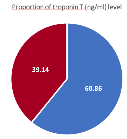
<!– Start of picture text –> Proportion of troponin T (ng/ml) level<br>39.14<br>60.86<br><!– End of picture text –>
TnT>0.02
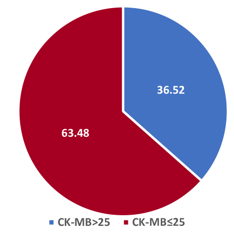
<!– Start of picture text –> Proportion of CK-MB (UI/L) level<br>36.52<br>63.48<br>CK-MB>25 CK-<br><!– End of picture text –>
| Parameter | Insular | Non-insular | P-value |
|---|---|---|---|
| TnT day 4, [median (Q1-Q3)] | 0.029(0.01-0.029) | 0.01(0.01-0.029) | 0,007 (Mann-Whitney) |
| TnT day 5 [median (Q1-Q3)] | 0.029(0.01-0.029) | 0.01(0.01-0.029) | 0,02 (Mann-Whitney) |
On average, CK-MB was significantly higher in patients with insular localization on each follow-up day (day 1 to day 5) (Table 2), but this was not statistically significant depending on insular lateralization. There was also no statistically significant correlation between insular location or insular lateralization and the CK value.
| Parameters | Insular | Non-insular | P-value |
|---|---|---|---|
| CK-MB day 1, [median (Q1-Q3)] | 17 (14-23) | 14 (12-16) | 0.006 (Mann-Whitney) |
| CK-MB day 2, [median (Q1-Q3)] | 17 (13,21.5) | 13 (11-15.25) | 0,0004 (Mann-Whitney) |
| CK-MB day 3, [median (Q1-Q3)] | 16.25 (13.25,20.75) | 14 (11.25-17.75) | 0,03 (Mann-Whitney) |
| CK-MB day 4, [median (Q1-Q3)] | 19 (13.75-26.25) | 13 (12-16) | 0.0002 (Mann-Whitney) |
| CK-MB day 5, [median (Q1-Q3)] | 16 (13-18.75) | 13 (10.25-15) | 0,003 (Mann-Whitney) |
Regarding stroke etiology, a statistical correlation between stroke subtype and an increase in CK-MB or TnT during any of the 5 days of follow-up has not been demonstrated (neither by the Mann-Whitney test nor by the Chi-Square test).
When analyzing the correlation between stroke severity and cardiac enzyme elevations, the mean CK-MB on days 2, 3, 4, and 5 was troke (NIHSS<16). CK (day 3 and 4) and Tn (day 4) were, on average, statistically significantly higher in patients with severe stroke than in patients with moderate to mild stroke (Table 3). TnT, much more sensitive for myocardial injury, did not record statistically significantly higher values in patients with severe stroke.
| Parameters | NIHSS<16 (moderate stroke) | stroke) | P-value |
|---|---|---|---|
| Tn day4, [median(Q1-Q3)] | 0.2(0.2-0.2) | 0.2(0.2-0.26) | 0.003 (Mann-Whitney) |
| CK day3, [median(Q1-Q3)] | 91(51.9-139) | 132(82-254) | 0.001 (Mann-Whitney) |
| CK day4(median(Q1-Q3)) | 100(59-147) | 115.5(71.75-296) | 0.004 (Mann-Whitney) |
| CK-MB day2, [median(Q1-Q3)] | 13(12-16) | 17(13-20.75) | 0.002 (Mann-Whitney) |
| CK-MB day3, [median(Q1-Q3)] | 14(12-18) | 17(13-21) | 0.002 (Mann-Whitney) |
| CK-MB day4, [median(Q1-Q3)] | 15(12-19.5) | 18.5(13.75-26.25) | 0.002 (Mann-Whitney) |
| CK-MB day 5, [median (Q1-Q3)] | 13 (11-16) | 15.5 (13-18.25) | 0.002 (Mann-Whitney) |
The correlation between enzyme increases and in-hospital mortality was analyzed. For Tn, a statistically significant correlation was observed between its increase on days 3, 4, 5 and hospital mortality. TnT on day 4 was, on average, significantly higher in patients who died during hospitalization than in those who survived. The increase in CK and CK-MB on days 4 and 5 correlated significantly with death during hospitalization (Table 4).
| Parameters | Death | No death | P-value |
|---|---|---|---|
| Tn day 3 [median (Q1-Q3)] | 0.378 (0.2-1.129) | 0.2 (0.2-0.2) | 0,01 (MW) |
| Tn day 4 [median (Q1-Q3)] | 0.506 (0.31-1.33) | 0.2 (0.2-0.2) | 0.002 (MW) |
| Tn day 5 [median (Q1-Q3)] | 0.426 (0.26-0.81) | 0.2 (0.2-0.2) | 0,009 (MW) |
| TnT day 4 [median (Q1-Q3)] | 0.049 (0.02-0.2) | 0.01 (0.01-0.03) | 0,008 (MW) |
| CK day 4, median (Q1, Q3) | 326 (180.75-1621) | 101 (61-177) | 0,02 (MW) |
| CK day 5 [median (Q1-Q3)] | 15 (188-4468) | 288 (55.5-160) | 0,01 (MW) |
| CK-MB day 4 [median (Q1-Q3)] | 38 (21.5-55.75) | 16 (12-20) | 0.006 (MW) |
| CK-MB day 5 [median] (Q1-Q3)] | 29 (19-63) | 14 (12-17) | 0,01 (MW) |
MW=Mann-Whitney
With regard to the growth dynamics of cardiac enzymes, a time-based increase was not observed as in other studies [15]. No statistical correlation has been demonstrated (either by the Mann-Whitney test or by the chi-square test) between the stroke subtype and CKMB or TnT elevation on any of the 5 follow-up days.
The increase in myocardial injury indicators has been interpreted differently in existing studies, with some authors considering it a marker of stroke severity and not an independent factor of poor prognosis [16] and others see it as an independent predictor of disability or mortality [17,18]. Apart from the current study, the literature has revealed only one study [19] that previously targeted the timed growth (days 1, 2, 3, 4, 5) of cardiac enzymes in stroke patients; other studies chose a variety of values of cardiac enzymes.
The possible pathophysiological mechanisms of increasing TnT and CK-MB in patients with acute stroke are multiple, but none of the studies existing so far in the literature could establish the exact cause. There is evidence of neurocardiogenic influences on myocytolysis in the acute phase of stroke, but there is great controversy regarding the cardiogenic increase in CK-MB. This increase may be possible in the case of a stroke location with a high potential for heart damage. Supporting the theory of a cardiac CK-MB increase secondary to neurogenic influences, in our study, 36.52% of patients had values above the normal limit of CK-MB, and the value of CK-MB (and TnT) correlated with insular involvement.
The current study offers different results than other literature references [20] – which concluded that TnT, unlike CK-MB, does not increase in patients with acute ischemic stroke. Also, the current study demonstrates contradicting conclusions to Barber et al. [21], which showed that in patients with acute ischemic stroke and elevated TnI, this increase did not correlate with insular location, one explanation being that in the cited study, only one TnI value was measured upon admission, whereas in the current study, statistically significant results were obtained on day 4 and 5 from stroke onset. Other examples from the literature [22] demonstrated a correlation between increased TnI and right hemisphere ischemia, but no insular involvement was specified, and no differences in insular lateralization were observed. One explanation could be that this increase is not related to other brain locations except insular ones, although further studies on other brain locations will need to be conducted to prove this claim. An argument against the theory mentioned above would be that the results obtained are controversial when it comes to the correlation of TnT alone with cardiac function, which makes it possible that CK-MB increase is not of cardiac origin and demonstrates that TnT is a much more reliable parameter to highlight the cardiac impact of chronic ischemia.
The statistical significance of elevated TnT and CK-MB values at days 4 and 5 with insular location may actually be due to a large ischemic stroke, and the increased TnT may reflect the systemic severity of stroke that may worsen the neurological status. A possible pathophysiological mechanism would be that in the case of large ischemic strokes, the growth curve of cerebral edema is maximum on days 4-5-6 from the onset of cerebral ischemia. Cerebral edema during these days may cause an exacerbation of sympathetic activity; this explanation is supported in a previous study [15], which revealed that CK-MB values are higher on day 5 of stroke onset. Obviously, this hypothesis will require further research in the future, by measuring catecholamines and enzymes simultaneously. Increased CK-MB value may originate elsewhere (muscle damage due to venous punctures, fluid restriction, and negative caloric
intake) [20]. In support of this argument comes the association of growth and CK.
In fact, it is well known that TnT is much more sensitive for assessing cardiac ischemia, either acute or chronic. The authors are of a similar opinion to the study conducted by Jensen et al. [18] that the increase in TnT is rather the consequence of HF. Another plausible explanation for the increase in TnT could be the existence of unstable atheroma plaques in the coronary arteries that could cause recurrent myocardial ischemia. It is probably necessary to dose other types of Tn (hsTnI) to correctly evaluate cardiac myocytolysis in patients with acute ischemic stroke and to redefine the concept of neurogenic cardiac ischemia.
Although this study cannot accurately state the pathophysiology of the increase in cardiac enzymes, we demonstrated that a higher value is associated either with stroke severity or with a higher risk of death in the short term, this growth being a marker for a more severe prognosis of a large stroke rather than an independent cause of mortality.
and CK-MB was related to stroke severity, which indicates that in well-selected cases cardiac enzyme dosing may be useful for stratifying the risk of cardiac complications. It cannot be stated that the dosing of TnT and CK-MB should be performed routinely in all patients with acute stroke, being an expensive method and not providing additional net benefit .
Cardiac complications are common in patients with acute ischemic stroke, some of which may be potentiated by autonomic hyperactivity. Careful monitoring of patients at high risk of heart complications may change the short-term prognosis.
The authors declare no conflict of interest. This research received no external funding.
Conceptualization and design, MMM, DD, ST; data analysis, MMM, DD; preparing the manuscript, MMM, IIE, ICS, MIG, OV, ST, DD, CV, CAS; writing, review and editing, MMM, DD.; visualization, MMM, DD; supervision, MMM. All authors have read and agreed to the published version of the manuscript.
The current manuscript does not contain previously published materials or self-generated AI text.
The study was conducted in accordance with the Declaration of Helsinki, and approved by the Ethics Committee of “CAROL DAVILA” UNIVERSITY OF MEDICINE AND PHARMACY (protocol code PO-35-F-03 no. 83 and date of approval: 29.02.2016).
Written informed consent has been obtained from the patient(s) to publish this paper.