
Parkinson’s disease (PD) is a neurodegenerative disorder that affects quality of life (QoL) through a range of motor and non-motor symptoms [1]. It is usually associated with motor symptoms [2] such as tremor, bradykinesia, and postural instability [3,4]. These motor dysfunctions result from the progressive loss of dopaminergic neurons in the substantia nigra (SN) [5–7], leading to reduced dopamine levels in the striatum. Beyond dopaminergic neuronal loss, oxidative stress and mitochondrial dysfunction are increasingly recognized as important contributors to the complex neurodegenerative processes underlying PD [8]. Evidence indicates that PD patients may also exhibit non-motor symptoms: psychiatric, cognitive, autonomic, and other, which tend to become more pronounced as the disease progresses [9].
Deep brain stimulation (DBS) is a neurosurgical procedure known for alleviating motor symptoms and improving QoL in many PD patients [10]. It involves implanting
electrodes into specific brain regions, usually the subthalamic nucleus (STN) or globus pallidus internus (GPi) [1,11]. Despite its numerous benefits, DBS has several limitations and challenges. Being a symptomatic treatment [7], it does not halt neurodegeneration or cure PD. Outcomes vary among patients [11] and some symptoms, such as speech, postural instability and gait, may be resistant to DBS or even worsen after surgery [12].
The aim of this narrative review is to provide a comprehensive synthesis of current knowledge on DBS in PD patients, summarizing the current state of the procedure, including its mechanisms, clinical outcomes, predictive factors, selection criteria, risks, and future directions.
The research was conducted using PubMed, Scopus, and Web of Science, using the following search formula: (“DBS” OR “deep brain stimulation”) AND (“PD” OR “Parkinson’s Disease”) AND “outcome”. Titles and abstracts were screened for relevance. We selected articles published in English within the last 5 years to ensure recent and accessible data. Eligible studies included adult and elderly PD patients who received DBS and reported clinical outcomes or predictive factors. We excluded case reports, ongoing trials, and studies based solely on animal models. A total of 129 articles were included in the final analysis and organized thematically according to clinical domain.
Before the widespread use of levodopa in the late 1960s, PD was primarily considered a surgical disease, treated by making lesions in the thalamus (thalamotomy) or globus pallidus (pallidotomy) to improve symptoms [7]. After the introduction of levodopa, these surgeries became less common until the 1980s, when pallidotomy regained attention for its benefits. In 1987, Benabid and Pollak offered a reversible alternative to lesions by introducing thalamic DBS [7,13]. This expanded to the GPi (1994) and STN (1993–95), with STN DBS uniquely reducing medication needs [7]. Following a multicenter trial in 2001, STN and GPi DBS gained FDA approval and became the global standard of care [7].
The DBS procedure involves two steps. First, electrodes are implanted into specific brain targets and connected to an Implantable Pulse Generator (IPG), a small device, typically placed under the skin of the chest during the same surgery or in a separate procedure [1,12]. The IPG modulates abnormal neural activity by generating continuous, high-frequency electrical pulses to the targeted brain regions [1,14,15]. Unlike older surgical methods involving permanent brain lesions, such as pallidotomy [7], DBS is reversible and adjustable through modification of stimulation [7,16].
The exact mechanisms of action of DBS are still not fully understood; however, it is believed to work by modulating complex neural networks and reestablishing more normal movement patterns [17–19]. In PD, specific brain regions, particularly the STN and GPi, become overactive, leading to an imbalance in brain circuitry that contributes to motor symptoms like rigidity and akinesia [7]. DBS is thought to “silence” or disrupt this pathological, excessive neuronal activity, thereby restoring better control over movement [7,13,20]. This modulation involves direct effects on local cell bodies, perturbation of axons (nerve fibers), and interactions with oscillatory brain rhythms [20–22]. For example, in the STN, excessive beta-band oscillatory activity (12–30 Hz) within the cortico-basal ganglia network is considered a well-established pathophysiological biomarker associated with motor impairment in PD, particularly bradykinesia and rigidity [21,23]. Elevated beta synchronization is thought to reflect an “anti-kinetic” state that interferes with normal motor processing and movement initiation. Conventional high-frequency DBS suppresses pathological beta activity and reduces abnormal neuronal synchrony, which correlates with clinical motor improvement [20,21,23]. The relevance of beta oscillations has become even more important with the development of adaptive DBS (aDBS), in which beta power recorded from implanted electrodes is used as a real-time feedback biomarker to dynamically adjust stimulation delivery according to the patient’s clinical state (20). Compared with conventional continuous stimulation, aDBS may reduce unnecessary stimulation, improve energy efficiency, and potentially decrease stimulation-related adverse effects while maintaining motor benefit.
Traditionally, DBS surgery is performed while the patient is awake under local anesthesia because it allows surgeons to test the stimulation during the operation and monitor side effects [24–26]. However, advances in imaging and intraoperative MRI have supported the growing adoption of asleep surgery under general anesthesia. Microelectrode recording (MER) remains reliable in this setting and is crucial for accurate placement [27]. Combining MER with deep learning may help predict clinical improvement [28]. Many patients prefer asleep surgery because it is perceived as more tolerable than awake surgery and, on average, 26 minutes shorter than awake surgery [29].
Multiple studies have reported no significant differences in motor outcomes between the two approaches [24,27,30]. In 2021, Holewijn et al. [29] conducted a randomized clinical trial which showed no significant difference in the improvement of off-medication Movement Disorders Society Unified Parkinson’s Disease Rating Scale (MDS-UPDRS) Motor Examination scores between groups (awake: −27.3 points or 52% improvement; asleep: −25.3 points or 50% improvement). The subscores for tremor, rigidity, bradykinesia, and gait and balance were also similar [29]. This study also showed no significant differences in cognition, mood, and behavior. Izzo et al. [27] reported in a 2024 meta-analysis that complication rates were similar between awake and asleep surgery. Later, in 2025, Roldán et al. [31] observed a low complication rate for asleep surgery of 1 event among 89 patients. Holewijn et al. [29] noted one case of symptomatic intracranial hemorrhage after awake surgery and four after asleep surgery, but classified this as a “random finding” with no statistical differences in incidence between the two approaches.
The key stimulation parameters used in DBS are frequency, pulse width, and amplitude/voltage [32–37]. Frequency represents the delivery rate of the electrical pulses. It is usually 130 Hz, but can range from 125 Hz to 180 Hz [7,20,27,38–40]. High-frequency stimulation is commonly used to suppress pathological network activity and improve motor symptoms in PD [41]. In parallel, oscillatory activity in the gamma range has attracted growing interest as a neurophysiological biomarker. Excessive narrow-band gamma activity, particularly around 60–90 Hz, has been associated with levodopa-induced dyskinesia and may reflect hyperkinetic motor states [24,42–44]. Recent studies suggest that gamma oscillations could help guide adaptive DBS strategies aimed at balancing motor improvement while minimizing dyskinesias. Gamma-related modulation has also been linked to cognitive and speech effects. Increased gamma stimulation may contribute to declines in verbal fluency, unlike theta frequencies (4–10 Hz), which show episodic improvement in verbal fluency but less robust motor benefits [45]. Despite this, combined theta-gamma frequencies improved verbal fluency without affecting motor benefits, with improvements observed in both non-episodic (p = 0.038) and episodic (p = 0.030) verbal fluency scores after one month of STN-DBS [46]. For specific symptoms such as gait disturbances or dyskinesia, low-frequency stimulation is sometimes used [24,42–44]. Low-frequency paradigms have also been explored for memory modulation during NREM sleep and for speech improvement [40,41].
Pulse width, the duration of each electrical pulse, is usually 60 μs, but can range from 40 μs to 90 μs [7,21,27,38–40,47]. Short pulse widths have been shown to improve speech intelligibility and reduce dyskinesia and pyramidal side effects [24,40].
Amplitude, the strength of the electrical current, varies between 1 and 4 mA or 1.5 to 4.0 V, adjusted based on clinical response and side effects [7,21,27,38–40,47]. Monopolar stimulation (IPG as anode, optimal contact as cathode) is common, but bipolar or double monopolar configurations are also used [7,25,39,48,49].
Recent advances in DBS technology include the development of directional leads, which allow current steering toward specific neural regions while avoiding adjacent structures associated with adverse effects [24,40]. Unlike conventional ring-shaped electrodes that deliver stimulation circumferentially, directional segmented contacts provide greater spatial selectivity and may enlarge the therapeutic window by improving motor benefit at lower stimulation thresholds [24]. Directional stimulation is increasingly used in contemporary DBS practice because it can reduce stimulation-induced side effects, including dysarthria, capsular effects, and sensory disturbances, particularly when electrode placement is suboptimal or located near eloquent pathways [24,40]. This approach also facilitates more individualized programming strategies and may improve overall tolerability and energy efficiency.
Precise DBS electrode placement is crucial for maximizing motor benefit and minimizing side effects [36,50,51]. Suboptimal placement occurs in up to 17% of cases, leading to reduced efficacy and higher side effect rates [50]. In one study, the electrode reached the planned target within 2 mm in only 42.9% of cases, meaning that more than half of the surgeries required depth or track adjustments [27]. Lead location also plays a critical role. One study pointed out that anterior sensorimotor STN placement improved voice outcomes, whereas posterior contacts worsened them [38].
The subthalamic nucleus (STN) and globus pallidus internus (GPi) are the most well-known and established targets for DBS (Figure 1) [13,26,35,52]. Their extensive benefits and differences in outcomes have created a vast debate regarding their selection [7,12,52]. Understanding their strengths and differences will help personalize the treatment based on the patient’s needs.
The STN is the most frequently targeted site for DBS in the treatment of PD [5,53,54]. It is neuronally dense, highly vascularized [55], measuring approximately 6–8 mm anterior-posterior, 9–11 mm mediolateral, and 3–5 mm dorsoventral [24]. Between the cerebral peduncle and the zona incerta [55], its location is referenced using the anterior commissure–posterior commissure (AC-PC) line coordinates [53]. The dorsolateral STN is believed to be a sweet spot [33,56,57] due to slower motor progression [33] and improvement of gait after DBS [57]. Stimulating this target helps alleviate a range of motor symptoms, such as tremor, akinesia, and rigidity [7,44,58], while also significantly reducing levodopa dosage, thereby avoiding drug-induced motor complications [59,60].
GPi
The GPi is another commonly targeted structure for DBS in PD. It is larger than STN, measuring approximately 10-12 mm anteriorposterior, 15-18 mm mediolateral, and 4-6 mm dorsoventral [24]. It is a primary output nucleus of the basal ganglia [7], and it is separated from the globus pallidus externus (GPe) by the internal medullary lamina [24]. GPi-DBS is particularly effective for controlling dyskinesias [7,8,25,60,61]. It offers greater flexibility in medication adjustments, even if it leads to less overall drug reduction compared to STN-DBS [8,25,61]. Moreover, GPi is associated with fewer neuropsychiatric issues [26] and less harsh on cognition than STN [12]. Bucur and Papagno’s meta-analysis [4] suggests that GPi-DBS may cause fewer side effects, including cognitive impairment, than STN-DBS. Supporting this point, França et al. [12] and Murcia Carretero et al. [26], who cite Boogers and Fasano, believe that GPi should be considered the main target for frail older patients with cognitive and neuropsychiatric problems. In contrast, Tröster et al. [62] suggest caution when interpreting meta-analyses comparing the cognitive effects of the two targets, as subject selection bias is likely to occur. Due to GPi being perceived as safer for cognitive effects, patients with poor cognition may be assigned to GPi-DBS, resulting in different sample characteristics –demographic and disease traits– between the two groups pre-surgery [62]. They believe that STN and GPi may be more similar, with GPi only having slightly milder effects on verbal fluency and processing speed [62].
While STN and GPi are the primary targets, research is exploring alternative regions to address DBS-resistant symptoms, particularly axial symptoms and cognitive impairment.
The ventral intermediate nucleus (VIM) of the thalamus was the first modern DBS target and is mainly used for treating tremors in PD and essential tremor when medications do not work. VIM DBS reliably reduces tremors over the long term, but it does not help much with other movement problems like slowness or balance issues. Side effects, especially when both sides are stimulated, can include speech difficulties, imbalance, reduced effectiveness over time, and, rarely, problems with coordination. For these reasons, VIM DBS is usually chosen for people with tremor-dominant Parkinson’s disease, rather than those with more widespread movement symptoms [7].
Caudal Zona Incerta (cZi), below the thalamus, has shown excellent effects on both Parkinsonian and essential tremor, possibly with fewer side effects than VIM DBS [7,16,24].
Substantia Nigra Pars Reticulata (SNr), ventrolateral to the STN, has shown a favorable response for axial motor symptoms, including gait impairment and postural instability [16]. Combined STN and SNr stimulation has shown significant improvement in freezing of gait in a pilot trial, though not consistently for other axial symptoms [12,16,42]. This combination might also improve intonation and maximum phonation time [63].
The Pedunculopontine Nucleus (PPN), part of the brainstem locomotor area, has been investigated for therapy-resistant gait and balance disturbances [16,24]. It was suggested that PPN stimulation improves gait freezing and falls, but results remain conflicting [7,24]. Having undefined boundaries, precise targeting is challenging [24].
Nucleus Basalis of Meynert (NBM) provides the primary cholinergic input to the cortex, and it atrophies in PD dementia [7,64]. It’s been hypothesized that NBM may slow cognitive decline, but no significant cognitive outcomes have been reported [7,12].
Advanced PD patients mostly undergo bilateral, symmetric lead placement surgery, due to often showing motor symptoms on both sides of the body [43]. However, studies have also analyzed the unilateral effects. When the affected site is targeted, the first lead is often the most beneficial [65,66], potentially postponing or canceling the second surgery for the other side [66]. The left-side stimulation may contribute to slower gait velocity than the right-side [67], but better outcomes of rightward visually-guided saccades [47]. Bilateral stimulation also improves these saccades, but doesn’t always outperform left, suggesting that unilateral stimulation may be enough in some cases [47].
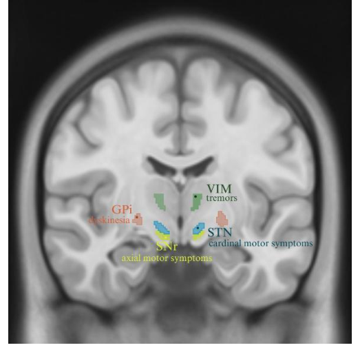
Coronal section (MNI ICBM152 [68,69] template) showing key DBS targets and their principal clinical domains. VIM (green, Morel [70] atlas), GPi (red), STN (blue), and SNr (yellow; CIT168 atlas) are depicted with their predominant therapeutic effects: tremor control (VIM), dyskinesia reduction (GPi), amelioration of cardinal motor symptoms (STN), and modulation of axial motor function (SNr). Given the heterogeneity of Parkinson’s disease manifestations and DBS outcomes, contemporary DBS management increasingly relies on personalized approaches integrating patient characteristics, target selection, programming strategies, and expected clinical outcomes (Figure 2).
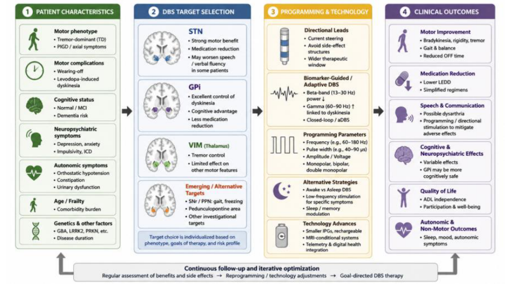
Schematic overview of individualized DBS management integrating patient characteristics, target selection, programming strategies, emerging technologies, and expected clinical outcomes. The framework highlights the interaction between motor and non-motor features, DBS target choice, adaptive programming approaches, and longitudinal optimization of therapy. Abbreviations: aDBS, adaptive deep brain stimulation; DBS, deep brain stimulation; GPi, globus pallidus internus; LEDD, levodopa equivalent daily dose; MCI, mild cognitive impairment; PD, Parkinson’s disease; PIGD, postural instability and gait difficulty; PPN, pedunculopontine nucleus; SNr, substantia nigra pars reticulata; STN, subthalamic nucleus; TD, tremordominant; VIM, ventral intermediate nucleus.
Choosing the right patients for DBS in Parkinson’s Disease is key to getting the best results and reducing risks [24,71]. Since outcomes differ from person to person, careful selection helps avoid poor results, extra costs, or complications.
In 1999, the Core Assessment Program for Surgical Interventional Therapies in Parkinson’s Disease (CAPSIT-PD) was published, becoming the first set of selection criteria that specialists relied on [14,71]. However, the current knowledge on PD evolution and genotyping led to the opinion that CAPSIT-PD is often restrictive and outdated [14,71]. Barbosa et al. [71] reported that CAPSIT-PD initially limited selection eligibility to 1.6% of PD patients, while flexible criteria limited it to 4.5%.
Patients must have a confirmed diagnosis of idiopathic PD based on criteria such as the UK Parkinson’s Disease Society Brain Bank criteria [72–74]. In diagnostically uncertain cases, functional neuroimaging may provide complementary information, with brain [¹⁸F]FDG PET/CT demonstrating characteristic metabolic patterns that can support the differential diagnosis of PD and atypical parkinsonian syndromes (75). Atypical Parkinsonism does not benefit from DBS [12,14,71]. DBS is mainly indicated in advanced PD when medication can’t control disabling motor complications, such as wearing-off, levodopa-induced dyskinesias, or painful dystonia [7,12,22,26,29].
Levodopa responsiveness is supposed to improve by at least 30% in the Unified Parkinson’s Disease Rating Scale Part III (UPDRS-III) during a levodopa challenge test (LCT) [24,36,53,55,73,76,77]. Improvement exceeding 50% may indicate a greater likelihood of benefit [24,76,78]. To exclude atypical Parkinsonism, the disease duration should be at least four years [14,71,77]. Patients must be able to attend regular follow-up appointments for device programming and ongoing care [71,79].
The European Academy of Neurology/Movement Disorder Society (EAN-MDS) 2022 Guideline suggests DBS for patients with “early fluctuations” (less than three years of onset) who meet other criteria [71]. Early-stage surgeries may offer greater long-term medication reduction [80]. Younger age at the time of DBS surgery is generally associated with longer-lasting and more robust motor outcomes and a better cognitive status [7,62,81]. While traditionally, older patients (e.g., over 69-70 years) might have been considered less suitable due to less improvement and more side effects, recent data suggest DBS can be effective and safe in elderly patients, though they may face higher risks of surgical complications [81–83].
Genetic factors were not part of the original CAPSIT-PD criteria. The increased accessibility to genetic testing made these factors considered when deciding on DBS surgery and as possible predictors of outcomes [42].
Glucocerebrosidase (GBA) mutations are a frequent genetic risk factor for PD [84]. GBA carriers are often younger at disease onset and surgery, with stronger levodopa responsiveness and earlier motor complications [85]. Motor outcomes after DBS are generally similar between GBA and non-GBA patients, with sustained improvements in motor symptoms, dyskinesias, fluctuations, and reduced medication burden [85]. However, cognitive outcomes remain the main concern. GBA-PD is linked to faster cognitive decline, and several long-term studies suggest accelerated deterioration after DBS, with higher dementia rates at 5 years (25% vs. 11% in non-GBA PD) [85]. Still, most GBA carriers continue to benefit motorically, and DBS remains a valid option [85,86].
Leucine-Rich Repeat Kinase 2 (LRRK2) is another mutation that can be present in PD. LRRK2 mutation carriers generally show excellent levodopa responsiveness, and their good outcomes from DBS are often long-lasting [7,13]. The G2019S mutation has demonstrated an excellent response to STN-DBS in over 85% of subjects, unlike the R1441G variant, which has shown more unsatisfactory outcomes [24].
Patients with parkin (PRKN) and PINK1 gene mutations often respond very well to dopamine [7]. PRKN-associated PD may present with foot dystonia and depression [71]. Barbosa et al. [71] reported good and sustained motor outcomes (46–84% improvement), with no significant decline in cognitive function generally reported. Other gene variants, like SNCA RS356220, may also be associated with positive QoL outcomes [87].
As mentioned in the “Selection criteria” section, an improvement greater than 50% in LCT may predict meaningful results from DBS [24,76,78]. However, the LCT’s ability to accurately predict individual patient improvement is debated, and its long-term predictive value may be limited, especially for non-motor or axial symptoms [24,71].
The optimal contact orientation is linked to strong evoked cortical responses leading to smoother movement. This leads to the idea that the magnitude is correlated with motor benefits, making the DBS-evoked cortical responses a possible physiological biomarker [88].
Gray Matter Volume (GMV) has potential as a predictive factor, yet its results are debatable for now. Some authors believe that GMV may influence DBS outcomes. For example, Koivu et al. [58] and Yim et al. [19] noted that lower GMV or higher GMV in certain regions may predict medication reduction and greater UPDRS-III scores. Wang et al. [64] reported that less motor improvement is linked to low GMV in the motor cortex and thalamus and to declined cognition linked to low volume in the hippocampus. However, Peter et al. [89] point out that GMV is not predictive of axial symptom improvement. The discrepancies highlight the need for larger, more diverse cohorts to validate GMV as a predictive factor.
The investigation of gender differences in DBS outcomes is important for personalized medicine, but it can also give us insight into women’s participation in trials [90]. Kübler et al. [90] pointed out that women are underrepresented in trials, with 30% of participants being women. This percentage is also present in another cohort, with 21 females out of the 70 participants (30%) [91]. A higher percentage of women, with 17 out of 43 (39.5%) was present in Somma et al. [92], a retrospective study, and in Mori et al. [93] with 28 out of 57 (49%), the first study that analyzes gender outcomes of PD in Japan. While both sexes showed similar motor
improvements, only women showed improvements in cognition and only men in depression and impulsivity scores [90]. In the Japanese cohort, women had greater short-term improvements, with 66.3% improvement in women vs. 53.9% in men (p = 0.028), when no medication was active [93]. However, these differences disappeared five years post-DBS [93].
The preoperative motor and non-motor symptoms may influence the outcome of DBS. Sauerbier et al. [94] reported that severe impulsive and compulsive behaviors (ICB) and lower doses of dopamine agonists before DBS lead to favorable ICB results after STNDBS. On the other hand, severe attention or memory deficits pre-DBS are linked to worse ICB after the surgery [94]. Preexisting cognition problems might also influence the amount of benefits after DBS. Patients with mild cognitive impairment have lower motor improvements than those with normal cognition off medication (−41.4% vs −52.2%, p = 0.013) [95]. However, mild cognitive impairment isn’t linked to dementia development after the procedure [96,97].
Voruz et al. [98] analyzed how motor symptom asymmetry may predict the non-motor outcomes following DBS. The results show that patients with right-sided motor symptoms have great QoL improvements and preserve their cognition, unlike left-sided ones, who have modest QoL improvements and are vulnerable to cognitive decline [98].
Comorbidities complicate DBS. Patients with additional health issues have higher risks of complications and readmissions, though DBS usually results in lower readmission rates than other neurosurgical procedures [71]. Managing patient expectations is also important. Unrealistic hopes for a cure or major improvement can lead to disappointment. Clinicians should discuss the main symptoms patients want to address [24], as perceived symptom severity often shapes expectations [99].
Multiple studies have consistently demonstrated the efficacy of DBS in improving motor symptoms [100]. A meta-analysis reported that the UPDRS-III scores improved by 50.5% after STN-DBS and by 29.8% after GPi-DBS [52]. The cardinal motor symptoms (tremor, rigidity and bradykinesia) have shown significant improvements [101]. One study reported that tremors improved by a mean of 74%, rigidity by 57%, and bradykinesia by 49% [12]. These motor benefits are sustained as noted by De Noordhout et al. [102], after a 12year comparative study of STN-DBS and best medical therapy in PD patients.
While DBS is associated with good motor outcomes, its effects on axial functions such as speech may be less consistent or even adverse. Postural abnormalities may improve after DBS [103], with one study having 67.8% of Camptocormia and 72.2% of Pisa patients with PD exhibiting positive responses after DBS [104]. Another study noted better scores of axial symptoms such as neck rigidity (p < 0.0001), freezing of gait (p = 0.0031) and postural stability (p = 0.0220), except speech, which did not improve [91]. In patients predisposed to dystonia, STN-DBS may have an additive boost, one study reporting that 74.4% of post-DBS on-dystonia cases were new-onset [105]. Despite better scores on objective scales, some PD patients under DBS perceived speech tasks as more effortful than hand movements, especially in dual treatment states [106]. This highlights the fact that clinical improvement is not always the same as subjective improvement, making patient-perceived effort useful when assessing DBS effectiveness.
Based on motor subtype [(tremor-dominant (TD), akinetic-rigid, postural instability/gait difficulty (PIGD)], DBS outcomes may differ [25,61,71]. After STN-DBS, TD patients often exhibit better short-term motor outcomes [71], but fewer non-motor benefits than PIGD patients [73]. PIGD patients may also demonstrate a greater decline in temporal asymmetry and dyscoordination of walking than TD patients [107].
Speech outcomes are heterogeneous. Castillo-Triana et al. [108] found no consistent group-level changes, though individual variability was high. However, another study noted that 15% of patients developed dysarthria at 1 year after STN-DBS [109]. GPi-DBS also worsened speech, potentially inducing several types of dysarthria or stuttering [110].
Olfaction, a non-motor symptom affecting over 70% of PD patients and impacting quality of life, appears to be negatively influenced by STN-DBS. A prospective pilot study, utilizing the objective University of Pennsylvania Smell Identification Test (UPSIT), observed a statistically significant worsening of olfaction following bilateral STN-DBS. The mean UPSIT scores declined from 20.0 ± 6.6 (severe hyposmia) before surgery to 17.5 ± 5.7 (total hyposmia) after three months (p = 0.03). This objective decline contrasts with some earlier subjective reports, suggesting a potential direct effect of DBS or the natural progression of the disease, rather than changes in mood, as anxiety and depression scores remained stable [111].
Conversely, gastrointestinal symptoms, particularly constipation, which affects up to 70% of PD patients, showed objective improvements after STN-DBS. In the pilot study, 63% (5 of 8) of patients with baseline constipation, as defined by Rome III criteria, experienced resolution of the condition three months post-DBS (p = 0.03). Significant improvements were noted in several objective measures: the number of complete spontaneous bowel movements (CSBM) per week increased from 2.2 ± 1.9 to 4.7 ± 2.4 (p = 0.04), and the proportion of bowel movements associated with straining decreased from 28% ± 12% to 7% ± 4% (p = 0.09). Patient satisfaction with constipation treatment improved from 44 ± 27 mm to 64 ± 25 mm on a visual analog scale (p = 0.02). These benefits
were not associated with changes in laxative use, dopaminergic medication dosage, or mood, suggesting a direct effect of DBS, possibly through improved motor function and rectoanal coordination, or modulation of autonomic centers [111].
Despite PD being associated with weight loss, one study found that after DBS patients gained weight, which was positively linked to survival (R2 = 0.14, p < 0.001) [112]. The patients who had bilateral STN-DBS showed the most weight gain compared to bilateral GPiDBS (3.03 ± 0.45% vs 1.89 ± 0.31%, p < 0.01) [112].
Sleep disturbances are among the most debilitating non-motor symptoms in PD, heavily influencing quality of life. A prospective, controlled, 36-month observational study compared bilateral STN-DBS with standard-of-care medical therapy (MED) in propensity score-matched cohorts (n=40 per group). STN-DBS led to significantly better overall Parkinson’s Disease Sleep Scale-1 (PDSS) scores (p < 0.001) at 36-month follow-up, indicating substantial improvement in sleep quality. Specific domains demonstrating significant improvement in the STN-DBS group included overall quality of night’s sleep, sleep onset and maintenance insomnia, nocturnal restlessness, and nocturnal motor symptoms. In contrast, the MED group experienced a significant worsening in nocturia and nocturnal motor symptoms. STN-DBS was observed to stabilize nocturia and sleep refreshment, while these aspects worsened in the MED group (P = 0.027 and P = 0.032, respectively). These sleep improvements were significantly correlated with enhanced quality of life (rs = –0.40, p < 0.001) and motor symptom amelioration (rs = –0.35, p = 0.002), and remarkably, were independent of changes in depression, anxiety, or dopaminergic medication requirements. Potential mechanisms for improved sleep include enhanced motor function, modulation of brain regions adjacent to the STN, like the pedunculopontine nucleus and improved sensory processing [72].
The influence of DBS on the diverse spectrum of neuropsychiatric symptoms associated with PD is equally significant, albeit more complex and variable, profoundly affecting patients’ overall quality of life. These non-motor disturbances, including mood disorders, apathy, impulsivity, and changes in personality and perception, can manifest either as improvements or, in some cases, as exacerbations or de novo occurrences following DBS [4,74,113,114].
A comprehensive meta-analysis of long-term (1–3 years) neuropsychological outcomes indicated a mild, statistically significant improvement in anxiety and depression scores for STN-DBS patients, with a Hedges’ g of 0.34 (p = 0.02) for STN stimulation. Similarly, GPi-DBS showed a small, significant improvement in depression, with a standardized mean difference (SMD) of 0.11 (p = 0.03). This meta-analysis further noted an anxiety improvement with an SMD of 0.30 (p = 0.01) [4].
In contrast, other studies reported no significant long-term changes in anxiety and depression levels after STN-DBS when compared to standard medical therapy [72,111].
Changes in depression and anxiety scores are predictive of QoL after STN-DBS. However, apathy and depression were more frequent in the DBS group after 12 years compared to the drug-treated group [102].
Apathy, characterized by a loss of motivation and reduced initiative, represents a frequently reported and often challenging neuropsychiatric symptom in PD that can significantly impact QoL [114–116]. The effects of STN-DBS on apathy are notably controversial. A meta-analysis of 33 articles involving 1,286 patients revealed that apathy scores were significantly higher after STNDBS compared to both the pre-operative state (Hedges’ g = 0.34, 95% CI = 0.19–0.48, p < 0.001) and control groups receiving only medication (Hedges’ g = 0.36, 95% CI = 0.03–0.65; p = 0.004) [115]. This increase in apathy appeared independent of the reduction in dopaminergic medication, and was even greater if patients with baseline apathy were excluded (Hedges’ g = 0.79, P < 0.001) [115]. Factors such as inaccurate electrode placement within the STN, particularly in associative or limbic areas, rather than the motor portion, have been implicated in inducing or exacerbating apathy [116]. This study suggests a correlation between apathy and more dorsolateral stimulation of the left STN [116]. Apathy scores (AES) showed a slight, non-significant decrease after STN-DBS (54.00 ± 10.30 to 52.69 ± 8.88) at 6 months post-DBS, attributed to a conservative and gradual reduction of dopaminergic drugs [116]. In contrast to STN-DBS, GPi-DBS has been associated with no significant changes in apathy scores postoperatively, possibly due to less pronounced reductions in LEDD [116].
ICB are important considerations given their prevalence in PD and their association with dopaminergic therapy [94]. STN-DBS can lead to complex and variable effects on ICB. While ICB often improves post-DBS, particularly due to the typical reduction in dopaminergic medication, de novo ICB can also emerge [74,94]. 6 months post-DBS, patients with preoperative ICB showed significant improvement in scores on the Questionnaire for Impulsive-Compulsive Disorders in PD-Rating Scale (QUIP-RS). However, there was considerable interindividual variability, with 27.3% experiencing worsening and 29.1% showing improvement [94]. Another study investigating personality traits noted that STN-DBS might have effects on impulsivity, especially a decline in persistence. This was observed even with lowered medication doses, suggesting an effect of DBS beyond pharmacological changes [117]. Impulsivity, as evaluated by scales like the Barratt Impulsiveness Scale 11 (BIS11) and Behavioral Inhibition System/Behavioral Activation System (BIS/BAS), has also been significantly associated with postoperative quality of life at different follow-up points after STN-DBS [114].
Personality and self-perception can be affected following DBS, although early reports raising concerns about unwanted identity or personality changes were often based on retrospective data or case reports with methodological limitations. A recent prospective,
patient-centered cohort study specifically investigated changes in characteristics that patients with PD themselves identified as personally meaningful and feared losing. This study found that, at a group level, DBS did not result in negative changes in these valued personal characteristics. Instead, participants often reported greater (more positive) manifestations of these traits 6- and 12-months post-DBS. While negative changes were observed in 4 out of 49 patients, the majority reported no change or increased manifestation across domains such as prosociality, cognition, self-regulation, and positive emotionality. These changes were observed to be unrelated to improvements in motor scores, and behaviors related to positive emotions were among the characteristics patients most feared losing [113].
Regarding perceptual problems and hallucinations, a long-term follow-up study reported that hallucinations were present in 58% of PD patients after at least ten years of DBS treatment [4]. However, a more recent prospective, observational multicenter study indicated improved perceptual problems and hallucinations specifically in patients with the PIGD motor subtype, while outcomes remained stable in tremor-dominant patients after 6 months of DBS [73]. This improvement was partly attributed to the postoperative reduction of dopaminergic medication [73].
DBS has presented benefits in QoL in multiple studies. For example, one study noted that 80.8% of the cohort reported “much or very much improvement” in Health-Related Quality of Life (HRQoL) [118]. Tandra et al. reported an improvement in the Parkinson’s Disease Questionnaire (PDQ-39) by 49% [18]. These results are sustained even after 5 years post-DBS, unlike the patients just on medication who exhibit QoL deterioration [119]. Combining scalp nerve block with intercostal nerve block improves the quality of recovery after DBS [120].
Medication usage is also decreasing. One study shows a 47% reduction in the LEDD at one year post-surgery [121]. Furthermore, another study noted that 10 years after the STN-DBS, mean LEDD was 600 mg in DBS patients vs. 1316 mg in controls (p < 0.001), sustaining the initial benefits [122]. When implemented in earlier stages of PD, DBS has been shown to lower medication costs by a factor of 2.4 over a five-year period compared to standard medical therapy alone [123].
Degirmenci et al. [17] analyzed STN-DBS from the caregivers’ perspective via telephone interviews. Caregivers noted improvements in both motor symptoms (tremor 58.1%, dyskinesia 59.6%, gait/ataxia 59.6%) and non-motor symptoms (depression 46.8%, sleep 43.6%, pain 41.9%). Notably, 80.6% of patients showed better daily life functioning post-DBS, reducing caregivers’ burden [17].
As with any surgical procedure, DBS is associated with a spectrum of potential complications and limitations that must be comprehensively addressed during the preoperative informed consent process. These encompass acute neurological disturbances, such as delirium, to diverse surgical and hardware-related problems, as well as other specific postoperative concerns [30,124].
A notable neurocognitive adverse event observed is postoperative delirium (POD), which manifests as an acute disruption of attention and awareness, coupled with cognitive alterations. Its fluctuating symptoms often include hallucinations, confusion, and disorientation. While incidence ranges from 5.8% to 42.6%, it is often insufficiently studied in the context of DBS. Different diagnostic criteria and detection methods may contribute to this variation. A prospective study focusing on POD showed that in 21 out of 57 (36.8%) of PD patients after the procedure. These cases generally presented with mild severity. The typical onset was rapid, approximately 1.7 ± 2.0 days after surgery, and lasted for an average of 7.5 ± 6.4 days, even up to 21 days in some cases. The clinical significance of POD is underscored because it is linked to worse prognoses, elevated mortality, and accelerated PD progression [124].
Beyond postoperative delirium, individuals undergoing DBS are vulnerable to various surgical and hardware-related complications. An investigation involving 800 PD patients who received STN-DBS reported that 8.4% encountered intracranial complications resulting in neurological deficits; of these, 7.4% were temporary, while 1.0% led to lasting impairment. The most critical acute complication observed was ICH, manifesting symptomatically in 2.8% (22 patients) of this patient group. Out of these, 0.6% (5 patients) sustained permanent neurological deficits, encompassing conditions like hemiparesis, severe cognitive impairment, hemiplegia, persistent diplopia, dysarthria, and balance disorders. The other 16 patients experienced transient deficits, such as aphasia, dysarthria, central facial palsy, frontal lobe syndrome, delirium, and cognitive decline, which typically resolved within six months to a year. Although previous research has suggested various risk factors for ICH, including the quantity of MER trajectories, age, anesthesia technique, hypertension, and sex, this extensive retrospective study did not establish a statistically significant impact for these factors across the entire cohort. However, a higher risk was noted for patients who did not undergo MER compared to those with a single MER trajectory. Strategies to mitigate ICH include precise preoperative trajectory planning to bypass blood vessels, positioning the trajectory entry over a sulcus, and applying fibrin glue in the burr hole to minimize brain shift [30].
The necessity for revision surgeries constitutes a notable limitation, with 18.4% of patients in a particular cohort undergoing an additional surgical procedure after their initial DBS system implantation. Notably, this rate significantly diminished over time, falling from 27.3% among the first 300 cases to 13.0% in the following 500 cases (p < 0.001), highlighting an evident learning curve and improvements in surgical methodologies. Frequent causes for revision included infection (3.4% overall), which also saw a reduction
from 5.3% to 2.2% over the study period and often mandated complete hardware extraction, antibiotic therapy, and later reimplantation. Lead migration also represented a reason for revision, impacting 2.1% of patients. However, the prevalence of lead migration was substantially lowered using lead anchoring devices (1.1% of electrodes) when contrasted with previous miniplate fixation techniques (6.3% of electrodes; OR: 0.15, 95% CI: 0.05–0.32, p < 0.001). Other triggers for revision included skin erosion, hardware malfunctions, and pain, with pain emerging as the most common reason in this study [30].
Delayed-onset edema localized around the DBS leads was noted in 1.6% (13 patients). This condition typically manifests with temporary symptoms such as cognitive decline, dysarthria, apathy, and confusion, with only a single patient experiencing persistent mild cognitive impairment. This form of edema was more commonly observed with segmented leads (3.1% of 390 patients with segmented leads) when contrasted with traditional ring electrodes, though enhanced awareness and reporting in more recent years might contribute to this perceived difference. The overall incidence of delirium documented within this cohort was 4.3%, a figure recognized as being lower than in some other studies, potentially attributable to underreporting in a retrospective study design without standardized delirium assessment. Fatalities directly associated with DBS electrode implantation were infrequent, with one instance of cardiac arrest following IPG replacement and another death due to a hip fracture, neither of which was deemed directly caused by the electrode placement itself [30].
It is crucial to acknowledge the inherent limitations of the studies themselves. The retrospective design of some analyses, particularly the extensive cohort study, inherently carries a risk of incomplete data or the underreporting of milder, transient complications. The lengthy observational periods, spanning over two decades in the retrospective study of surgical complications, introduce heterogeneity resulting from ongoing advancements in surgical techniques, imaging, and hardware. This makes direct comparisons across the entire cohort challenging [30]. Moreover, the comparatively small sample size in certain prospective studies, such as the one investigating POD severity (n=57), restricts the statistical power to identify all significant predictors, especially when numerous potential factors are explored. Although the predictive models developed for POD severity showed performance better than random chance, they might not yet offer clinically meaningful predictive capabilities, underscoring the necessity for validation through larger, international multicenter trials. Finally, distinguishing acute delirium symptoms from pre-existing PD symptoms, such as fluctuating hallucinations or attention deficits, can present a diagnostic difficulty [124].
The increasing integration of digital technologies, artificial intelligence, and wearable devices into neurological practice may further facilitate objective and longitudinal monitoring of patients [125]. Machine learning (ML) is paving the way for personalized care, being used in various stages such as patient selection, outcome prediction, and parameter management [20]. To predict clinical outcomes, Chang et al. [126] used a DLRad model, which combines multi-instance learning (MIL) and radiomics features. It achieved the highest discriminative ability with an Area Under the Curve (AUC) of 0.871 in the test cohort, higher than radiomics (AUC 0.825) and MIL models (AUC 0.846) alone. When age was added to the combined model, a decrease in AUC 0.858 appeared. This suggests that age didn’t add predictive value, underlining the need for careful feature selection [126].
In another study, ML found predictors of QoL after DBS. It was found that the PDQ-39 scores and upper beta band activity (>20 Hz) in the left STN can influence QoL. Lower initial QoL burden predicted worsening, while higher beta activity was linked to improvement [127].
Deep learning combined with microelectrode recording (MER) was used to predict motor improvements after bilateral STN-DBS. The learning model presented 80.21% maximal accuracy and an AUC of 0.88, proving deep learning’s capabilities to predict clinical improvements [28].
Vision-based machine learning models were also tried for analyzing changes in gait in PD patients. While the model couldn’t predict gait scores with accuracy, it could detect weak but real improvements in walking after medication and DBS use. For now, they are hard to apply universally, due to differences in gait and camera angles. Large, diverse datasets are needed to improve the current state of this technique [128].
The future is adaptive. Despite being a great option for PD patients, conventional DBS (cDBS) lacks the ability to adjust to the patient’s changing needs [129]. Adaptive DBS (aDBS) aims to overcome the limitations of cDBS by using real-time detection of brain signals to automatically adjust stimulation parameters in response to patients’ needs [129]. A study showed that aDBS is effective, providing more energy when needed and less when it’s not, works well with medication, and is safe [129]. Above that, ML is explored as a way to identify electrophysiological biomarkers for aDBS [20].
Robot-Assisted DBS (RAS-DBS) can be a reliable and safe approach for PD patients. It offers greater accuracy than traditional methods, with a pooled vector error of 1.09 mm versus 1.11 to 3.70 mm. However, RAS-DBS shows similar improvements in motor symptoms and reduction of medication, compared to the standard procedure [130].
DBS is an effective therapy for PD, offering sustained symptom benefits, medication reduction, and better QoL. However, outcomes may be heterogeneous: while tremor and rigidity show sustained improvements, axial symptoms like speech remain challenging to treat. Future advances in adaptive stimulation and machine learning promise more personalized and precise DBS, expanding its therapeutic reach while minimizing risks.
Conflicts of interest and sources of funding
The authors declare no conflict of interest. This research received no external funding.
Acknowledgments
This article was written as part of the “Tech-Review Summer School: academic writing summer camp for young researchers” organized by the Center of Innovation and e- Health, Carol Davila University of Medicine and Pharmacy in Bucharest.
Authors’ contribution
Conceptualization, G.-S.D., M.-C.M., A.-R.T. and O.A; methodology, G.-S.D., M.-C.M. and A.-R.T.; validation, A.-R.T., O.A., O.M.S., M.M.M.; formal analysis, G.-S.D. and M.C.M.; investigation, G.-S.D., M.-C.M. and A.-R.T.; resources, O.A., O.M.S and M.M.M.; data curation, G.-S.D., M.-C.M. and A.-R.T.; writing—original draft preparation, G.-S.D. and M.-C.M.; writing—review and editing, A.-R.T., O.A., O.M.S. and M.M.M; visualization, G.-S.D., M.-C.M. and A.-R.T.; supervision, A.-R.T. O.M.S and O.A.; project administration, A.-R.T. O.A, and M.M.M. All authors have read and agreed to the published version of the manuscript.
Ethics approval and consent to participate
Not applicable.
Patient consent for publication
Not applicable.