
Primary brain and other central nervous system (CNS) tumors have an average annual age-adjusted incidence of 26.05 per 100,000 population in the United States, with glioblastoma accounting for 52.2% of malignant tumors and meningioma for 42.6% of all tumors [1]. The five-year relative survival after a malignant CNS tumor remains only 34.8% in the latest report [1], almost unchanged from the 35.7% reported for the 2016-2020 period [2], and malignant primary brain tumors are among the most difficult cancers to treat [3]. Brain metastases add considerably to this burden, developing in an estimated 20% of patients with cancer [4]. The 2021 World Health Organization classification of CNS tumors has made molecular markers central to diagnosis [5], and genomic profiling has revealed recurrent alterations, such as epidermal growth factor receptor rearrangements in glioblastoma [6]. Some of these markers are directly relevant to neuropsychiatric care, since gliomas carrying a mutation in isocitrate dehydrogenase (IDH) present with seizures in 59-74% of patients, compared with 18-34% of IDH-wild-type gliomas, probably because the oncometabolite D-2-hydroxyglutarate, which is structurally similar to glutamate, increases neuronal excitability [7]. Seizure burden, in turn, determines exposure to antiseizure medications and to their psychiatric adverse effects (as highlighted further in Section 5). Meningiomas, in contrast, are mostly benign and often curable by gross total resection [8].
Behind these figures lies a clinical reality that is less often discussed, i.e., many patients with brain tumors first come into contact with the health system because of changes in mood, behavior, or cognition, and psychiatric symptoms are occasionally the only manifestation of the tumor [9]. Once the diagnosis is established, depression, anxiety, distress, and cognitive impairment accompany patients through surgery, adjuvant therapy, and survivorship. In this Perspective, we argue that psychiatry should not be an occasional consultant in neuro-oncology but a structural component of care. This view is argued based on the published evidence and on our experience in a liaison psychiatry service working alongside neurosurgery. The focus of this article is on adults, because pediatric neuro-oncology involves different tumor types, developmental considerations, and assessment tools that deserve separate discussion.
Both major classifications of psychiatric disorders (Diagnostic and Statistical Manual of Mental Disorders, Fifth Edition, Text Revision— DSM-5-TR, and International Classification of Diseases, Eleventh Revision—ICD-11) [10,11] recognize that a brain tumor can directly cause a mental illness or at least a corresponding syndrome, but they frame this possibility differently (Table 1). The DSM-5-TR places categories such as depressive disorder, bipolar and related disorder, anxiety disorder, obsessive-compulsive and related disorder, psychotic disorder, catatonia, and personality change due to another medical condition within the corresponding diagnostic chapters [10]. Such a diagnosis requires evidence from the history, physical examination, or laboratory findings that the disturbance is the direct pathophysiological consequence of the medical condition, that it is not better explained by another mental disorder, including an adjustment disorder in which the medical illness is the stressor, and that it does not occur exclusively during delirium [10]. The ICD-11 groups the equivalent conditions in a single block of secondary mental or behavioral syndromes associated with disorders or diseases classified elsewhere (6E60-6E6Z), which includes secondary psychotic, mood, anxiety, obsessive-compulsive or related, dissociative, impulse control, neurocognitive, and catatonia syndromes as well as secondary personality change [11]. ICD-11 speaks of syndromes rather than disorders, which highlights that the psychiatric picture is a manifestation of the underlying disease; it asks for the secondary code to be used in addition to the code for that disease, and it states explicitly that the symptoms must not be a psychologically mediated response to a severe medical condition, such as depressive symptoms in reaction to a life-threatening diagnosis [11].
Applying these criteria to patients with brain tumors is less straightforward than the definitions suggest, because the tumor, its treatment, the psychological impact of the diagnosis, and premorbid vulnerability usually act together, and their relative weight changes over the course of the disease. Both classifications also assign syndromes induced by medications, such as corticosteroids or levetiracetam, to separate substance- or medication-induced categories rather than to the secondary categories [10,11], so a single patient may legitimately move between diagnostic labels as treatment evolves. The two cases presented in Section 3 illustrate the complexity of distinguishing organic from psychological factors in assessing psychiatric syndromes with onset in neuro-oncology cases. The depressive syndromes at presentation were attributable to the tumors on clinical grounds, yet the persistence of core depressive symptoms after successful resection raises the possibility of residual network injury, of a primary depressive disorder unmasked by the illness, or of a reaction to the diagnosis and surgery. Rather than forcing a binary choice between primary and secondary disorders, we therefore propose that clinicians formulate each case across four etiological domains (Table 2). This formulation is not equivalent to a new diagnostic category but proposes a practical framework that links the diagnostic label to management, because each active domain points to a different intervention, and it should be revisited at each phase of care.
| Feature | DSM-5-TR | ICD-11 |
|---|---|---|
| Terminology | Mental disorder due to another medical condition | Secondary mental or behavioral syndrome associated with disorders or diseases classified elsewhere |
| Place in the classification | Distributed across the corresponding diagnostic chapters (e.g., depressive, psychotic, and anxiety disorders) | Grouped in a single block (6E60-6E6Z) within the chapter on mental, behavioral or neurodevelopmental disorders |
| Core requirement | Evidence from history, physical examination, or laboratory findings that the disturbance is the direct pathophysiological consequence of the medical condition | Syndrome judged to be a direct pathophysiological consequence of the health condition, based on history, examination, or laboratory findings |
| Exclusions | Not better explained by another mental disorder, including adjustment disorder with the illness as stressor, and not occurring exclusively during delirium | Not accounted for by delirium or another mental disorder, and not a psychologically mediated response to a severe medical condition |
| Categories relevant to brain tumors | Depressive disorder (with depressive features, with major depressive-like episode, or with mixed features), bipolar and related, anxiety, obsessive-compulsive and related, and psychotic disorders, catatonia, personality change (including apathetic and disinhibited types), and neurocognitive disorder due to another medical condition | Secondary psychotic (6E61), mood (6E62, with depressive, manic, or mixed symptoms), anxiety (6E63), obsessive-compulsive or related (6E64), dissociative (6E65), impulse control (6E66), and neurocognitive (6E67) syndromes, secondary personality change (6E68), and secondary catatonia syndrome (6E69) |
| Coding | The medical condition is coded first, followed by the mental disorder | The secondary code is used in addition to the code for the underlying disease |
| Medication effects | Coded as substance/medication-induced disorders | Coded among substance-induced disorders rather than as secondary syndromes |
| Domain | Clinical clues | Diagnostic correlate | Management implications |
|---|---|---|---|
| Direct tumor effect | Onset or worsening linked to tumor growth, edema, or progression, late or atypical onset, neurological or cognitive signs, apathy or personality change, improvement after resection or edema control | Disorder due to another medical condition (DSM-5-TR) or secondary syndrome (ICD-11) | Tumor-directed and antiedema treatment, psychotropic drugs as adjuncts, vigilance for progression |
| Treatment-related and endocrine | Onset after starting or increasing corticosteroids, antiseizure drugs, or dopamine agonists; symptoms during steroid taper; late fatigue and low mood after cranial irradiation; abnormal pituitary function | Substance/medication-induced disorder, or secondary syndrome due to an endocrine disorder | Dose reduction or switching, endocrine testing and hormone replacement, joint review with neuro-oncology |
| Psychological reaction | Content centered on diagnosis, prognosis, disability, or loss, onset after disclosure or a treatment milestone, preserved emotional reactivity | Adjustment disorder, or a primary disorder if full criteria are met | Psycho-oncological support, psychotherapy, involvement of caregivers |
| Pre-existing vulnerability | Previous episodes, family history, persistence after the other domains have been addressed | Primary mental disorder (e.g., major depressive disorder) co-occurring with the tumor | Standard evidence-based treatment adapted for seizure risk, drug interactions, and cognition |
Neurobehavioral presentations of brain tumors have long been recognized in clinical settings and in medical literature as challenging aspects. In a series of eight patients, frontal tumors presented with abulia, personality change, or depression, whereas temporolimbic tumors presented with hallucinations, mania, panic attacks, or amnesia, and after treatment the syndromes abated or resolved in seven patients [12]. In another series, eight patients with brain lesions, including four meningiomas and two high-grade gliomas, were diagnosed and treated for a significant period as having Alzheimer’s disease, schizoaffective disorder, schizophrenia, or depression before neurological signs led to neuroimaging [13]. Meningiomas are particularly prone to such delays because of their slow growth. In a retrospective hospital sample, 15 of 72 patients (21%) with meningioma presented with psychiatric symptoms in the absence of neurological symptoms, most commonly affective disorders [14]. An individual patient data analysis of published meningioma cases confirmed a wide range of presentations, from mood disorders to psychosis and obsessive-compulsive symptoms [15].
| Feature | Examples |
|---|---|
| Onset or course | First psychiatric episode in later life (usually after about 50 years of age), particularly when combined with any other feature in this table, subacute, progressive, or fluctuating course |
| Atypical presentation | Personality change, apathy without sadness, abulia, or disinhibition, anorexia without body-image concerns, new-onset psychosis or mania without a personal or family history |
| Treatment response | Poor or paradoxical response to adequate psychotropic treatment, prolonged confusion or new neurological signs after electroconvulsive therapy |
| Cognitive signs | Memory impairment, disorientation, inattention, or language disturbance (e.g., receptive dysphasia misread as thought disorder) |
| Neurological signs | Headache, vomiting, papilledema, or other signs of raised intracranial pressure, seizures, focal deficits, urinary incontinence, visual disturbances, anosmia |
| Endocrine context | Features suggesting a sellar lesion, hyperprolactinemia that is very high, persistent, or not temporally linked to antipsychotic treatment |
| Systemic context | History of cancer (risk of brain metastases, and of paraneoplastic or immune-mediated encephalitis when imaging is unrevealing), treatment with immune checkpoint inhibitors |
Anatomical localization is, however, a weak guide to establish correspondence between the neurological problems and the psychiatric ones. A meta-analysis of published case reports found a statistically significant association only between anorexia and hypothalamic tumors [20]. Lesion network mapping offers a more modern explanation for this inconsistency, since in 461 patients with focal lesions of different etiologies, lesions associated with depression were spread across many regions but mapped onto a common functional circuit centered on the left dorsolateral prefrontal cortex [21]. Thinking in terms of networks rather than lobes may help explain why tumors located far from the prefrontal cortex can still present with depression. Clinicians should therefore rely less on predicting tumor site from symptoms and more on recognizing features that make a primary psychiatric disorder unlikely. These features, summarized in Table 3, include new-onset symptoms in later life, atypical presentations, poor response to standard treatment, and any neurological or cognitive sign [9,12,15]. Late onset is a weak criterion on its own, because depression in later life is common and only rarely caused by a tumor, so the threshold for imaging should fall mainly when late onset coincides with at least one other feature in Table 3 [9]. The question of routine imaging is not settled, but a meta-analysis of first-episode psychosis found a clinically relevant magnetic resonance imaging (MRI) abnormality in 5.9% of patients, meaning that one such finding was detected for every 18 patients scanned [16]. Imaging may also reveal lesions whose relevance to the psychiatric picture is uncertain, such as the multinodular and vacuolating neuronal tumor, a rare, benign neuronal tumor that is frequently asymptomatic and most often discovered incidentally, and that, when it causes no symptoms, is often managed by observation with serial MRI rather than by resection [22]. Finding such a lesion does not by itself establish that the psychiatric syndrome is secondary to it, a conclusion that still requires the evidence of a direct pathophysiological link discussed in Section 2.
In patients with known systemic cancer, a new psychiatric syndrome with unrevealing or nonspecific imaging should also raise the possibility of autoimmune encephalitis. The clinical criteria for possible autoimmune encephalitis require a subacute onset, over less than three months, of working memory deficits, altered mental status, or psychiatric symptoms, together with at least one supportive finding, such as new focal neurological signs, unexplained seizures, cerebrospinal fluid pleocytosis, or suggestive MRI changes, once reasonable alternative causes have been excluded [17]. The updated criteria for paraneoplastic neurological syndromes classify limbic encephalitis as a high-risk phenotype and grade antibodies according to their association with cancer [23]. Because immune checkpoint inhibitors, now widely used in metastatic cancers, can also trigger immune-mediated neurological syndromes, a careful oncological treatment history is part of the differential diagnosis [23].
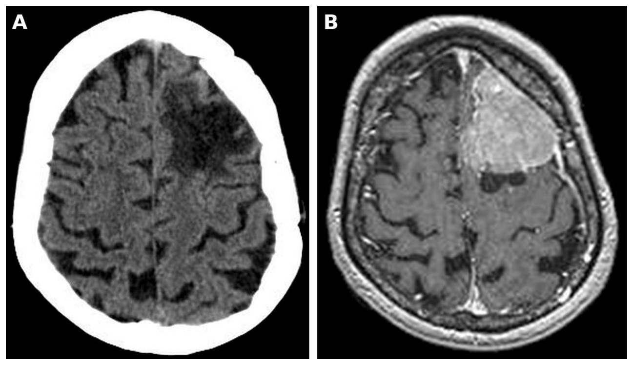
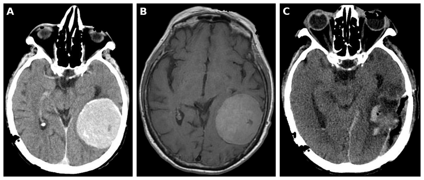
Two cases from our service are further presented to illustrate these points. Both patients were referred by their general practitioners for psychiatric evaluation of depressive symptoms. The first, a 60-year-old patient with obesity and diabetes, presented with memory disturbances, a depressive syndrome, behavioral changes, and non-fluent, effortful speech, and neuroimaging showed a left frontal parasagittal, extra-axial, contrast-enhancing lesion with perilesional edema (Figure 1). The second, a 67-year-old patient, presented with depressive symptoms and, on closer examination, signs of intracranial hypertension and a receptive dysphasia, which neuroimaging explained as a left temporo-parietal convexity meningioma compressing Wernicke’s area (Figure 2). A receptive language deficit can easily be misread as confusion, inattention, or disorganized thinking in a patient labeled as depressed. Both diagnoses were reached through a team-based approach with successive investigations. After surgery, which in the second case was a gross total resection, most neurological and cognitive symptoms remitted significantly. However, core depressive symptoms persisted at follow-up in both patients and required continued psychotropic treatment. These observations are consistent with population data showing that depression often does not resolve after surgery, especially in patients with a history of depression or residual disability [24], and they show that surgical success does not end the need for psychiatric care. Table 4 applies the four-domain formulation proposed in Section 2 to both cases and shows that the direct tumor effect, which dominated the initial presentation, could not fully account for the depressive symptoms that persisted after resection, so residual network injury and the remaining domains had to be reassessed at follow-up.
| Domain | Case 1 | Case 2 |
|---|---|---|
| Direct tumor effect | Left frontal parasagittal, extra-axial, contrast-enhancing lesion with perilesional edema. The depressive syndrome was accompanied by memory disturbances, behavioral changes, and non-fluent, effortful speech, and apathy was a plausible contributor given the frontal location. Most neurological and cognitive symptoms remitted after surgery. | Left temporo-parietal convexity meningioma compressing Wernicke’s area. The depressive symptoms were accompanied by signs of intracranial hypertension and receptive dysphasia. Most neurological and cognitive symptoms remitted after gross total resection. |
| Treatment-related and endocrine | Not available in the source documents | Not available in the source documents |
| Psychological reaction | Depressive cognitions of helplessness and incurability | Receptive dysphasia made self-report unreliable before surgery, which favored observer-rated assessment of mood. |
| Pre-existing vulnerability | No previous psychiatric episodes, and no family history of psychiatric illnesses. Obesity and diabetes as medical comorbidities. | No previous psychiatric episodes, and no family history of psychiatric illnesses. |
| Formulation at follow-up | Core depressive symptoms persisted after surgery and required continued psychotropic treatment, so residual network injury, a reaction to the diagnosis and surgery, and a primary depressive disorder unmasked by the illness remained the hypotheses to be weighed. | Core depressive symptoms persisted despite gross total resection and required continued psychotropic treatment, with the same three hypotheses to be weighed. |
Apathy deserves separate attention, because it is one of the most frequent ways in which a frontal tumor is mistaken for depression. The 2018 international consensus criteria define apathy as a quantitative reduction of goal-directed activity, compared with the patient’s previous level of functioning, that persists for at least four weeks, affects at least two of three dimensions (behavior and cognition, emotion, and social interaction), and causes identifiable functional impairment [25]. Unlike depression, apathy is not driven by sadness, guilt, hopelessness, or negative cognitions, and patients often show little concern about their own passivity, which is why informant ratings are more informative than self-report. The Apathy Evaluation Scale, available in self-, informant-, and clinician-rated versions, can quantify the syndrome and follow its course [26]. In the DSM-5-TR paradigm, a persistent apathetic presentation caused by the tumor corresponds to personality change due to another medical condition, apathetic type, and in ICD-11 to secondary personality change [10,11]. The distinction has direct therapeutic consequences, since escalating antidepressant doses is unlikely to help when the core problem is loss of motivation rather than low mood, whereas structured activity, caregiver education, and cognitive rehabilitation are more rational options. Psychostimulants may be considered in selected cases, although the evidence in this population is limited to uncontrolled data and a negative trial of modafinil for fatigue, mood, and cognition [27,28]. In Case 1, the parasagittal frontal location made apathy a plausible contributor to the depressive presentation, and this possibility should be assessed explicitly in similar patients.
Depression is the best-studied psychiatric complication of brain tumors. A meta-analysis of 37 studies estimated a pooled prevalence of depression or depressive symptoms of 21.7% [29]. Estimates depend heavily on how depression is measured: in a systematic review of glioma studies, the median frequency was 15% by clinical interview but 39% with the Beck Depression Inventory [30]. In the Glioma Outcomes Project, physicians reported depression in 15% of patients early after surgery for high-grade glioma, rising to 22% at 3 and 6 months, whereas 93% of patients reported depressive symptoms, and antidepressants were given to only 60% of those whom physicians recognized as depressed [31]. In a population-based cohort, 35% of patients had depression before surgery, and lower functional status was associated with higher depression scores at 3 months and 1 year [24]. Anxiety is similarly common and frequently coexists with depression, with female sex, lower tumor grade, lower education, and a psychiatric history among its predictors [32]. Shortly after craniotomy, 48.4% of patients report clinically relevant distress, which cannot be predicted from tumor stage or sociodemographic data [33].
These conditions matter beyond quality of life, since they can have devastating consequences at multiple levels of mental health and daily functionality. Depression is consistently associated with reduced physical function, cognitive impairment, and poorer quality of life [30], and it may shorten survival. Preoperative depression was independently associated with reduced survival after resection of malignant astrocytoma [34], and a meta-analysis found a borderline increased risk of death among depressed patients with highgrade glioma [35]. In glioblastoma, depression may also reduce treatment adherence [36]. Suicide risk deserves explicit attention, since the standardized mortality ratio for suicide among patients with cancer is 4.44 compared with the general population [37]. In a population-based analysis of 87,785 patients with brain cancer, the risk of suicide in the first year after diagnosis was three times higher than expected (observed-to-expected ratio 3.05, 95% CI 2.04-4.37), rising to 5.04 in patients older than 64 years, and most of those who died by suicide were men with glioblastoma [38]. Suicide risk assessment therefore belongs to the first months after diagnosis, when patients are also undergoing surgery and adjuvant treatment. Tumors of the sellar region raise specific issues, since depression scores improved after surgery for pituitary adenoma in one cohort [24], but patients cured of Cushing’s disease show persistent psychopathology and maladaptive personality traits years after remission, suggesting lasting effects of glucocorticoid excess on the brain [39].
Screening is feasible in this population, and should be applied thoroughly whenever possible. In adults with glioma, the depression subscale of the Hospital Anxiety and Depression Scale at a threshold of 8 or more and the Patient Health Questionnaire-9 at 10 or more performed well, whereas the Distress Thermometer was inferior, although positive screens still require clinical assessment [40]. However, self-report instruments become unreliable when receptive or expressive aphasia is present, as in our second case, and observer-rated tools developed for stroke offer a practical alternative. The Stroke Aphasic Depression Questionnaire is completed by a caregiver or nurse on the basis of observable behaviors and also exists in shorter and hospital versions [41], whereas the Aphasic Depression Rating Scale is rated by clinical staff and was validated in patients with aphasia in the subacute phase of stroke [42]. Neither instrument has been validated in patients with brain tumors, but both are preferable to omitting mood assessment altogether.
Postoperative delirium is another frequent and underrecognized complication. A meta-analysis of patients undergoing intracranial surgery estimated an incidence of 19%, ranging from 12% to 26% according to clinical features and assessment method [43], and a more recent meta-analysis found delirium in 17% of hospitalized patients with brain tumors [44]. After craniotomy for brain metastases, delirium was associated with postoperative hematoma and with poorer survival [45]. Hypoactive delirium is easily mistaken for depression or apathy, and fluctuating attention can be misread as a new psychiatric disorder, so routine screening with validated instruments, such as the Confusion Assessment Method [46], should be part of perioperative care. An episode of delirium should also prompt closer cognitive follow-up, because delirium and later cognitive decline are distinct but related problems.
A distinctive feature of psychiatric care in neuro-oncology is that the treatments themselves can cause or worsen psychiatric symptoms. Corticosteroids, usually dexamethasone, remain essential for controlling peritumoral edema [47,48], yet severe psychiatric reactions occur in nearly 6% of patients receiving systemic corticosteroids and mild-to-moderate reactions in about 28%, with shortterm therapy most often inducing euphoria or hypomania, long-term therapy tending to cause depression, and delirium or psychosis occurring at higher doses [49]. Steroid-related insomnia, agitation, or mood changes should therefore be considered before a new psychiatric diagnosis is made, and the lowest effective dose should be the goal. The taper deserves equal attention, because steroid withdrawal and secondary adrenal insufficiency can produce fatigue, anorexia, and low mood that closely resemble depression [48].
Seizures are common, affecting 60-75% of patients with low-grade glioma, 20-50% of those with meningioma, and 20-35% of those with brain metastases [50]. Current guidelines, updating the 2000 practice parameter [51], advise against anticonvulsant prophylaxis in patients who have never had a seizure and suggest levetiracetam over older agents when treatment is needed [52]. Levetiracetam, however, has its own psychiatric burden, as 14.1% of patients with epilepsy develop a psychiatric symptom or disorder within 2 years of starting it, and a history of depression or anxiety substantially increases this risk [53]. When irritability, aggression, or depressive symptoms emerge, switching the antiseizure drug is often more effective than adding a psychotropic, and in a retrospective series of patients switched overnight from levetiracetam to brivaracetam, levetiracetam-related neuropsychological adverse events improved in 76% [54]. Valproate has mood-stabilizing properties and may therefore be attractive in patients with comorbid bipolar disorder, but in the European Organization for Research and Treatment of Cancer and National Cancer Institute of Canada (EORTC/NCIC) trial it was associated with more grade 3-4 thrombocytopenia and leukopenia during temozolomide-based chemoradiotherapy, although the same post hoc analysis also suggested longer survival in patients taking valproate alone [55]. This survival signal was not confirmed in a pooled analysis of four prospective trials in newly diagnosed glioblastoma, which found no survival benefit for either valproate or levetiracetam and concluded that these drugs should be used for seizure control rather than to improve survival [56]. Enzyme-inducing antiseizure drugs, in turn, interact with chemotherapeutic agents and should be avoided where possible [50,57].
Endocrine pathways add a further and often overlooked source of psychiatric morbidity. The hypothalamic-pituitary axis is highly radiosensitive, and radiation-induced hypopituitarism develops insidiously over the years after cranial irradiation, is usually irreversible and progressive, and affects growth hormone secretion first, followed by gonadotropin, corticotropin, and thyrotropin deficiencies [58]. Its manifestations, including fatigue, low mood, reduced libido, and cognitive slowing, overlap so extensively with depression that long-term survivors presenting with new depressive symptoms should have pituitary function assessed before an antidepressant is started. Sellar tumors carry their own risks. Prolactinomas are treated with dopamine agonists, which can induce impulse control disorders such as pathological gambling, compulsive shopping, and hypersexuality, and men with dopamine agonisttreated prolactinomas were almost ten times more likely to develop such disorders than men with non-functioning adenomas [59]. Conversely, psychiatrists frequently encounter hyperprolactinemia caused by antipsychotics, and endocrine guidelines recommend pituitary imaging when the drug cannot safely be stopped or replaced and the onset of hyperprolactinemia does not coincide with the start of treatment [19].
Oncological treatments have improved survival, which increases the need for long-term psychiatric care. Radiotherapy plus temozolomide extended median survival in glioblastoma from 12.1 to 14.6 months [60], tumor-treating fields added further benefit [61], and vorasidenib more than doubled progression-free survival in grade 2 IDH-mutant glioma [62]. European guidelines now define multimodal standards for diffuse gliomas [63] and meningiomas, including watch-and-scan strategies for many asymptomatic meningiomas [64]. Cognitive deficits, however, arise from the tumor itself, from surgery, from antiseizure and steroid treatment, and from radiotherapy [65]. In brain metastases, memantine given during whole-brain radiotherapy did not significantly reduce the decline in delayed recall at 24 weeks, the primary endpoint (p = 0.059), probably because only 149 patients remained analyzable, but it prolonged the time to cognitive failure (hazard ratio 0.78, 95% CI 0.62-0.99) and lowered the probability of cognitive failure at 24 weeks from 64.9% to 53.8% [66]. Hippocampal avoidance plus memantine reduced the risk of cognitive failure further compared with whole-brain radiotherapy plus memantine (adjusted hazard ratio 0.74, 95% CI 0.58-0.95) [67]. These advances show that neurotoxicity can be modified, and psychiatrists should be involved when treatment options with different cognitive risks are weighed.
The most intriguing finding in this field is the absence of controlled trials of antidepressants. The latest Cochrane review found no eligible study of any pharmacological treatment of depression in people with a primary brain tumor [68]. In practice, clinicians extrapolate from other populations and choose agents on the basis of tolerability, seizure risk, and drug interactions, and selective serotonin reuptake inhibitors (SSRIs) are generally favored [9]. Pharmacological pathways shared by depression and glioblastoma, and possible interference of some antidepressants with tumor treatment, add a further layer of uncertainty [36]. These choices should be made jointly with the neuro-oncology team.
Several practical points follow from this approach. Sertraline, citalopram, and escitalopram have a low potential for pharmacokinetic interactions and are reasonable first choices, whereas bupropion, clomipramine, maprotiline, and amoxapine lower the seizure threshold in a dose-dependent manner and are best avoided in patients with tumor-related epilepsy [69]. SSRIs increase the risk of bleeding, with a pooled odds ratio of 1.41 for severe bleeding in a meta-analysis of 42 observational studies [70], which matters in the perioperative period and during temozolomide treatment, when thrombocytopenia is common, so platelet counts should be checked, and mirtazapine may be preferred when bleeding risk is high. All antidepressants except mianserin were associated with hyponatremia in a large register-based study, with the strongest association for citalopram [71], and the risk is compounded by carbamazepine or oxcarbazepine and by the sodium disturbances that frequently follow intracranial surgery, so sodium should be monitored after an antidepressant is started. Mirtazapine can also relieve insomnia, poor appetite, and nausea, which are common during chemoradiotherapy. When a hypnotic is still required, for example for corticosteroid-related insomnia, it should be prescribed at the lowest effective dose and for a short period, because Z-drug hypnotics such as zolpidem, as well as some benzodiazepines, antidepressants, and antipsychotics, can trigger sleep-related eating disorder, a parasomnia that may involve hazardous behaviors during nocturnal eating episodes [72]. For corticosteroid-induced mania or psychosis, dose reduction is the first step, with short-term low-dose antipsychotic treatment when symptoms are severe [49]. When antipsychotics are needed for psychosis due to the tumor, the lowest effective dose should be used with attention to sedation, QTc prolongation, and seizure threshold, and clozapine, which carries a dose-dependent seizure risk, should be reserved for exceptional cases [69]. Cardiac safety also deserves attention, because brain lesions can affect the heart through autonomic pathways, as suggested by the frequent elevation of cardiac enzymes in acute ischemic stroke, particularly when the insula is involved [73], so a baseline electrocardiogram is advisable before an antipsychotic is started.
Electroconvulsive therapy (ECT) poses a particular dilemma. For decades, an intracranial mass was considered an absolute contraindication, a view based largely on early reports of neurological deterioration [74]. A review of 75 patients who received ECT in the presence of a brain tumor over 80 years found that, among the more recently reported cases, meningiomas were the most common lesions and adverse effects occurred in 15%, and it concluded that ECT can be applied safely in patients with small, benign, or otherwise clinically insignificant lesions [18]. Large lesions, multiple metastases, peritumoral edema, mass effect, and raised intracranial pressure nevertheless remain major risk factors, because seizures transiently increase cerebral blood flow and intracranial pressure, so the decision should be taken jointly with neurosurgery and anesthesia, with consideration of premedication such as corticosteroids [18]. Two further implications are relevant for psychiatrists. Neuroimaging is advisable before ECT in patients whose depression has atypical features or late onset, and unexpectedly prolonged confusion or new neurological signs after ECT should prompt imaging rather than a mere change in stimulation parameters.
Evidence for other drugs is also limited. Methylphenidate improved cognition, mood, and function in an early uncontrolled study [27], but modafinil did not outperform placebo for fatigue, mood, or cognition in a randomized crossover trial [28]. Donepezil did not improve the overall cognitive composite score in irradiated survivors but modestly improved memory and motor speed, especially in more impaired patients [75]. Psychological interventions face similar challenges: an internet-based guided self-help program based on problem-solving therapy did not reduce depressive symptoms in glioma patients, although it may have reduced fatigue, and the trial was underpowered [76]. For caregivers, a Cochrane review found only eight small trials and could not draw reliable conclusions [77].
Cognitive rehabilitation has stronger support. In a randomized controlled trial of 140 adults with mostly low-grade glioma in remission, a program of six weekly two-hour sessions combining computer-based attention retraining with compensatory strategy training improved attention and verbal memory at six months and reduced mental fatigue compared with usual care [78]. Because cognitive complaints are often intertwined with low mood, apathy, and fatigue, cognitive rehabilitation should be coordinated with psychiatric treatment rather than offered in parallel.
Cognition deserves a central place in outcome assessment. Verbal memory performance independently predicted survival in recurrent malignant glioma [79], and cognitive deterioration preceded radiographic progression by about 6 weeks [80]. A change in cognition or behavior during follow-up may therefore be an early sign of tumor progression rather than a new psychiatric disorder. Repeated cognitive screening and clear distinction between delirium and progressive cognitive decline are core tasks of the liaison psychiatrist. In advanced disease, palliative care guidelines identify disorders of behavior and mood, fatigue, and caregiver needs as areas with scarce evidence, and recommend an early palliative approach [81].
Our experience and the evidence reviewed above support a phase-based model in which psychiatric tasks are defined in advance for each stage of the disease (Table 5). In neuro-oncology, the key milestones are disclosure of the diagnosis, surgery, adjuvant treatment, and the transition to survivorship or palliative care. The core team should include psychiatrists, psychologists, neurologists, neurosurgeons, oncologists, endocrinologists, physical therapists, speech and language therapists, social workers, and specialized nurses, each with clear roles in managing psychiatric and organic symptoms in the short and long term. In the prediagnostic phase, the priority is recognition, which means that psychiatrists and general practitioners should know the red flags in Table 3 and have rapid access to neuroimaging [9,16]. In the perioperative phase, baseline assessment of mood, apathy, cognition, suicide risk, decision-making capacity, and delirium risk allows postoperative changes to be interpreted correctly [31,33,43,82]. During adjuvant therapy, review of corticosteroid and antiseizure regimens is as important as psychotropic prescribing [49,53]. In survivorship and palliative care, the focus shifts to persistent depression and apathy, endocrine late effects, cognitive rehabilitation, caregiver support, and advance care planning [58,77,78,81].
| Phase | Psychiatric tasks | Key considerations |
|---|---|---|
| Prediagnostic | Recognize red flags, request neuroimaging (including before ECT in atypical or late-onset depression), avoid premature psychiatric labeling | Close communication with general practitioners and neurology, consider autoimmune encephalitis in patients with known cancer |
| Perioperative | Baseline assessment of mood, anxiety, apathy, cognition, suicide risk, and decision-making capacity, delirium screening with validated instruments | Distress is high after craniotomy and is not predicted by tumor stage; observer-rated tools are needed in aphasia |
| Adjuvant treatment | Monitor corticosteroid- and antiseizure drug-related psychiatric effects, treat depression and anxiety, check drug interactions, sodium, and platelet counts | Lowest effective steroid dose and careful taper, preference for non-enzyme-inducing antiseizure drugs, switching rather than adding when levetiracetam causes behavioral effects |
| Follow-up and survivorship | Repeated screening of mood, apathy, and cognition, psychotherapy, cognitive rehabilitation, endocrine review after cranial irradiation, return-to-work support | New cognitive or behavioral change may signal tumor progression |
| Palliative and end of life | Management of delirium, agitation, and mood, caregiver support, reassessment of capacity and advance care planning | Early integration of palliative care |
Such a model does not require new specialized units. It requires routine screening with validated instruments [40], shared protocols for steroid and antiseizure drug review, and a liaison psychiatrist who follows the patient across departments rather than being called only in a crisis. Our two cases illustrate the value of continuity: without follow-up visits after successful surgery, the persistent depression of both patients would probably have gone untreated. The psychometric evaluation of ClinFIT COVID-19 illustrates standardized measurement of functioning across care settings, although use in neuro-oncology would require adaptation and validation [83].
Decision-making capacity runs through every phase of this model. Soon after diagnosis, patients with malignant glioma performed worse than healthy controls on the understanding, appreciation, and reasoning standards of a structured capacity instrument, and these impairments were explained mainly by deficits in short-term verbal memory, which led the authors to recommend repeated assessment of capacity throughout the disease [82]. Capacity is decision-specific and may fluctuate with edema, seizures, delirium, or steroid effects, and aphasia can make a capable patient appear incapable unless communication is supported with simplified language, written or pictorial aids, and the help of a speech and language therapist. The liaison psychiatrist is well placed to assess capacity for consent to surgery, adjuvant treatment, and trial participation, and to encourage advance care planning while capacity is preserved, so that surrogate decision-makers can later act on the patient’s known wishes.
Several gaps should be addressed. First, randomized trials of antidepressants and psychotherapies in patients with brain tumors are needed, ideally stratified by tumor type, grade, and phase of treatment [68]. Second, screening thresholds and diagnostic criteria for depression should be validated in patients with aphasia, cognitive impairment, or fatigue, in whom self-report instruments may be unreliable, and observer-rated tools developed for stroke, such as the Stroke Aphasic Depression Questionnaire and the Aphasic Depression Rating Scale, should be tested in neuro-oncology [30,40-42]. Third, prospective studies should clarify whether treating depression improves adherence, functional outcome, and survival, a hypothesis already raised by the Glioma Outcomes Project [31,35]. Fourth, psychiatric and cognitive outcomes, including apathy and decision-making capacity, should be included as endpoints in neuro-oncology trials, alongside survival and imaging response [67,80]. Fifth, the four-domain formulation proposed here should be tested for inter-rater reliability and for its ability to predict treatment response. Nonlinear dynamics and fractal metrics explored in simulation-based post-COVID respiratory rehabilitation offer another methodological direction, whose usefulness in neurooncology would require investigation [84]. Finally, the biological links between tumors, inflammation, glucocorticoid exposure, and mood merit translational research, and lesion network mapping applied to tumor cohorts could clarify why lesions in different locations produce similar psychiatric syndromes [21,36,39].
This Perspective has several limitations. First of all, it is not a systematic review, and the literature was selected to support a clinical argument rather than identified through a predefined search strategy with formal appraisal of study quality, so relevant evidence may have been missed. The two cases come from a single liaison psychiatry service and were chosen because they illustrate diagnostic pitfalls, which means that they cannot establish how often tumors present as depression or how often depression persists after surgery. Much of the evidence on psychiatric comorbidity and treatment derives from patients with glioma, and its applicability to meningioma, brain metastases, and sellar tumors is uncertain. Several recommendations, including the use of observer-rated mood scales in aphasia and of structured capacity assessment, are extrapolated from stroke, dementia, or other populations, and the fourdomain formulation and the phase-based model are proposals that have not yet been validated. Finally, our discussion is limited to adults and is shaped by the organization of care in our own setting, which may limit its transferability to health systems with different resources.
Psychiatric symptoms can be the first sign of a brain tumor and remain a common, prognostically relevant, and undertreated problem after diagnosis. Psychiatrists should keep a low threshold for neuroimaging when psychiatric symptoms are atypical, start late in life, or are accompanied by neurological, cognitive, or endocrine signs, and they should distinguish apathy from depression, because the two call for different treatments. Classifying a syndrome as secondary to the tumor is only the first step, and a formulation that also weighs treatment-related and endocrine effects, psychological reactions, and pre-existing vulnerability offers a more useful guide to management, particularly when symptoms persist after successful surgery. After diagnosis, depression, anxiety, distress, delirium, suicide risk, and cognitive impairment should be screened for and treated as part of oncological care, with attention to the psychiatric effects of corticosteroids, antiseizure drugs, dopamine agonists, and cranial irradiation, to the specific risks of psychotropic drugs and electroconvulsive therapy in this population, and to the patient’s capacity to take part in decisions. Until controlled evidence becomes available, a multidisciplinary, phase-based model with regular psychiatric follow-up offers the most reasonable way to protect patients’ quality of life before, during, and after surgery.
Conflicts of interest and sources of funding
The authors declare no conflicts of interest.
The authors declared that generative AI (Claude Opus 5.5, Anthropic) was used for grammar and style revisions of this text. The authors take full responsibility for the final manuscript.
Authors’ contribution
Conceptualization, C.N., B.M.P., O.V., C.A.C., and A.G.M.; methodology, C.N., B.M.P., O.V., C.A.C., and A.G.M.; literature search and data curation, C.N., C.A.C., and A.G.M.; formal analysis, C.N., B.M.P., O.V., C.A.C., and A.G.M.; visualization, O.V.; writing—original draft preparation, C.N., B.M.P., O.V., C.A.C., and A.G.M.; writing—review and editing, C.N., B.M.P., O.V., C.A.C., and A.G.M.; supervision, B.M.P. and O.V.; project administration, A.G.M. All authors have read and agreed to the published version of the manuscript.
Ethics approval and consent to participate
The study was conducted in accordance with the Declaration of Helsinki and approved by the Local Ethics Committee of the “Dr. Carol Davila” University Emergency Central Military Hospital, Bucharest, Romania (approval no. 631, 14 September 2023).
Patient consent for publication
Written informed consent was obtained from both patients for the publication of their clinical data and anonymized images.