
Coronary CT angiography (CCTA) has become established as the first-line non-invasive test for patients with suspected chronic coronary syndrome. This position is supported by a Class I recommendation in both the 2019 and the 2024 European Society of Cardiology guidelines [1, 2]. One essential reason for its clinical usefulness is its high ability to rule out disease. When the test is negative, obstructive coronary disease can be safely excluded. This spares the patient an unnecessary diagnostic cardiac catheterization [3, 4]. Evidence from randomized controlled trials — among them SCOT-HEART, PROMISE and DISCHARGE — also suggests that an approach centered on anatomical assessment is not inferior to a strategy based on functional testing in predicting and subsequently preventing adverse cardiovascular events [5, 6, 7].
CCTA has high diagnostic performance, but it also has a widely known anatomical limitation: it frequently overestimates the severity of some stenoses. Part of this comes from the plaque itself. Non-calcified plaque with positive remodeling, low-attenuation areas, and irregular calcification are graded as more severe on computed tomography than on invasive angiography [8, 9].
Beyond these intrinsic differences, technical factors add to the impression of narrowing. Partial volume averaging, the blooming effect produced by dense microcalcifications, and vessel motion all contribute to additional overestimation of luminal stenosis [10]. As a result, a substantial proportion of the patients referred for catheterization solely on the basis of a CCTA report turn out, in the end, to have nonobstructive disease. This means these patients took on the risks, the contrast exposure, the radiation exposure, and the costs of an invasive procedure without obtaining any therapeutic benefit.
Solid real-world data describing how the two methods correlate remain limited in Romania. An unselected outpatient registry from Cluj-Napoca, for example, reported the frequency of significant lesions on CCTA, but it did not then follow the patients up to obtain invasive confirmation [11]. To our knowledge, no Romanian study has specifically examined the situation in which CCTA describes a severe stenosis in the presence of a low Agatston score. This gap matters because, when the coronary calcium burden is low, assessment of the vessel lumen relies largely on the morphological features of non-calcified plaque. Under these conditions, CCTA is most exposed to the risk of overestimation. Establishing how often such cases are ultimately confirmed is therefore an essential condition for calibrating local referral thresholds.
We carried out a study on a patient cohort from two clinical centers (in Sibiu, Romania). It included 124 patients who underwent coronary CT angiography (CCTA) followed later by invasive coronary angiography. An institutional registry from the same centers was analyzed in a past study in order to characterize referral patterns by age and sex, using a proportionally stratified subsample of 397 examinations [12]. The cohort analyzed here comes from the same registry, but it is designed to answer a different question — invasive confirmation, rather than the characterization of referral patterns. The objectives of the study were: (1) to assess how well the Agatston score can identify obstructive coronary artery disease; (2) to identify the factors that, independently of one another, are associated with the presence of coronary obstruction; and (3) to assess how well the CCTA findings match the other available data in patients who had a low coronary calcium level but in whom CCTA showed a stenosis.
The study was observational, retrospective, and bicentric. The source population comprised 2,742 consecutive CCTA examinations performed at the Sibiu Pediatric Clinical Hospital between March 2019 and January 2025. This is the public hospital that provides part of the outpatient cardiovascular computed tomography services for Sibiu County. The same patient registry was also used in an earlier study, which analyzed age- and sex-related patterns of referral to CCTA. That study analyzed a subsample of 397 examinations, selected by proportional stratification [12].
Although the two studies are based on the same population, they had different objectives and different selection criteria. The earlier study used a proportionally selected sample, whereas the present study included patients according to whether they subsequently underwent catheterization. The two studies also examined different outcomes. From the source population, we identified the patients who subsequently underwent invasive coronary angiography (ICA) at the Sibiu County Emergency Clinical Hospital. We then included 124 patients for whom both the CCTA and the ICA examinations were available and analyzable. These accounted for 4.5% of the source population (124 of 2,742 examinations).
Referral for ICA was driven by symptoms, by the results of non-invasive risk assessment, or by the identification of an abnormal finding on CCTA. This way of selecting patients may introduce verification bias, because ICA was not performed in the catheterization laboratory of the SCJU for every patient in the initial population who had an indication for invasive investigation. No weighting or statistical adjustment method was applied for the patients who were not referred for catheterization or who underwent this investigation in another center. For inclusion in the study, the CCTA examination had to be clear enough for interpretation and had to allow calculation of the Agatston score [13]. The patient also had to have subsequently undergone a diagnostic ICA, with no revascularization procedure performed between the two examinations. Patients were excluded if severe motion artifacts prevented calculation of the calcium score, if catheterization was performed solely for valvular assessment, or if the available medical data were incomplete. Clinical data were extracted from the electronic medical records using a standardized chart review procedure [14, 15]. The following information was collected for each patient: age, sex, coronary dominance, Agatston calcium score, stenosis severity for each coronary vessel, the presence of previously implanted stents, the existence of diffuse coronary disease, and whether a revascularization procedure was subsequently performed. Stenosis severity in invasive coronary angiography (ICA) was taken from the report of the interventional cardiologist who performed the procedure. The data were extracted by a single researcher, using the same criteria for all patients. Setting these criteria before the results were analyzed reduced the risk that certain data would later be selected according to the results obtained. The data were not independently verified by a second researcher and, consequently, agreement between two raters was not assessed.
The primary outcome of the study, termed CAD_70, was defined as the presence of a stenosis of at least 50% in the left main coronary artery or of at least 70% in one of the main coronary vessels. These included the left anterior descending artery, the circumflex artery and the right coronary artery. Dominant side branches were also included: the diagonal branches, the obtuse marginal branches and the ramus intermedius. Coronary fistulae and myocardial bridges without systolic compression were not considered obstructive coronary artery disease, because these findings are not caused by atherosclerosis. The threshold used in this study is the one used in the studies that compared coronary CT angiography with invasive coronary angiography, without the use of functional testing. This threshold should not be confused with the criteria used by the 2024 European Society of Cardiology guidelines to indicate revascularization. In the guidelines, the revascularization decision is based on functional criteria and requires at least one of the following values: fractional flow reserve ≤0.80, instantaneous wave-free ratio ≤0.89, or quantitative flow ratio ≤0.80. These measurements are used in particular for intermediate lesions and, in routine practice, for stenoses visually estimated at 40–90%, with the exception of stenoses in the left main coronary artery [2].
The secondary outcome, termed CAD_50, was defined as the presence of a stenosis of at least 50% in any epicardial vessel. Thirteen cases had initially been coded ambiguously because they had intermediate lesions. All these cases were re-adjudicated deterministically according to the rule above. Three met the primary threshold, and ten did not. Consistent application of the rule changed other cases as well, in both directions. Two further cases were reclassified as non-obstructive during the final check of the database: one with a 60% diagonal lesion and all major vessels below 30%, whose angiographic conclusion was itself negative, and another for which the only finding above the threshold was an arterio-cavitary fistula, not an atherosclerotic stenosis.
The subgroup with discordant findings (n = 33) was defined on the basis of the reason for referral. It included the patients referred for catheterization because the CCTA report described a severe stenosis even though their calcium score was low or disproportionate to the severity of the stenosis described. To check whether the results depend on the calcium score threshold chosen, sensitivity analyses were planned using two thresholds: CAC <400 AU and CAC <100 AU.
Continuous variables were checked for normality with the Shapiro–Wilk test and are reported as mean with standard deviation or as median with interquartile range. Categorical variables are summarized as frequencies, expressed as absolute numbers alongside the corresponding percentages. When comparing groups, we used the Student t test for suitable continuous variables, the Mann–Whitney U test when a non-parametric approach was needed, and the chi-square test for categorical comparisons, according to the requirements of each analysis. To assess the ability of the Agatston score to distinguish patients by CAD_70 and CAD_50, we applied ROC (receiver operating characteristic) curve analysis. Within this approach, we reported the area under the curve together with 95% confidence intervals obtained by bootstrapping, and we selected the optimal operating point using Youden’s J statistic. We then calculated sensitivity, specificity, predictive values and likelihood ratios across the standard Agatston score intervals. Multivariable logistic regression was used to assess whether log10(CAC+1), age measured per decade, and male sex were each independently associated with CAD_70. Goodness of fit was reported using the McFadden pseudo-R² and was further assessed by a likelihood ratio test comparing the full model with the null model. Statistical significance was established using two-sided p-values, with values below 0.05 considered significant.
The study was carried out with the approval of four separate institutional bodies. Ethics approval no. 31 / 24.04.2026 was formally issued by the Ethics Council of the Sibiu Military Emergency Clinical Hospital. In addition, the Sibiu Pediatric Clinical Hospital granted administrative and research approval no. 7969 / 07.11.2023 and also provided institutional data use agreement no. 3697 / 07.05.2026. These approvals allowed retrospective access to the anonymized CCTA records. Separate ethics approval, relating specifically to access to the catheterization archive, was granted by the Ethics, Medical Deontology and Discipline Committee of the Sibiu County Emergency Clinical Hospital, through Decision no. 17160 / 29.05.2026. Because the study was retrospective and relied on fully anonymized records, already obtained in the course of routine medical care, the authors did not seek individual written informed consent. This approach complies with Order no. 1502/2016 of the Ministry of Health, with Law no. 46/2003 on patients’ rights, and with Article 89 of Regulation (EU) 2016/679. These provisions allow the secondary use of health data for scientific research, subject to appropriate data protection safeguards.
The study cohort included 124 patients, with a mean age of 64.2 ± 8.6 years. The median age of the group was 65 years, with participants aged between 42 and 84 years. There were 76 men, representing 61.3% of the sample, and 48 women, representing 38.7%. The women were significantly older than the men, with a mean age of 66.6 ± 7.4 years against 62.7 ± 8.9 years (p = 0.012). Coronary dominance favored the right coronary circulation in 79.8% of patients. Co-dominant patterns, by contrast, were seen in 9.7% of patients, and left-dominant patterns accounted for the remaining 10.5%. The study found a median Agatston score of 480 AU, with an interquartile range from 176 to 777 and an overall observed range from 0 to 2,749. On comparison by sex, the median score was higher in men than in women, measuring 543 AU against 424 AU (p = 0.032). By the usual Agatston score categories [18], 2 patients (1.6%) had a score of zero, 17 patients (13.7%) had values between 1 and 99 AU, 30 patients (24.2%) between 100 and 399 AU, and 50 patients (40.3%) between 400 and 999 AU. A score of at least 1,000 AU was recorded in 25 patients (20.2%) (Table 1).
| Characteristic | Value |
|---|---|
| Total patients, n | 124 |
| Age, years (mean ± SD) | 64.2 ± 8.6 |
| Age, median (IQR), years | 65 (58–70) |
| Age range, years | 42–84 |
| Men, n (%) | 76 (61.3) |
| Women, n (%) | 48 (38.7) |
| Right coronary dominance, n (%) | 99 (79.8) |
| Balanced / co-dominant, n (%) | 12 (9.7) |
| Left coronary dominance, n (%) | 13 (10.5) |
| Agatston score, median (IQR), AU | 480 (176–777) |
| Agatston score, range, AU | 0–2,749 |
| CAC = 0, n (%) | 2 (1.6) |
| CAC 1–99, n (%) | 17 (13.7) |
| CAC 100–399, n (%) | 30 (24.2) |
| CAC 400–999, n (%) | 50 (40.3) |
| CAC ≥ 1000, n (%) | 25 (20.2) |
| Multivessel disease (≥2 vessels ≥70%), n (%) | 21 (16.9) |
| Diffuse coronary disease, n (%) | 72 (58.1) |
| Significant side-branch disease, n (%) | 14 (11.3) |
| Prior stenting, n (%) | 5 (4.0) |
| CAD_70 confirmed at invasive angiography, n (%) | 55 (44.4) |
| CAD_50 confirmed at invasive angiography, n (%) | 81 (65.3) |
During catheterization, obstructive stenosis of at least 70% was identified most often in the left anterior descending artery, accounting for 21.0% of cases. It was then seen in the right coronary artery in 18.5% of cases, followed by the circumflex artery, with 15.3%. In addition, a left main stenosis of 50% or more was detected in five patients (4.0%), consistent with the upstream enrichment expected in a catheterization cohort [19]. A dominant side branch showed significant involvement in 14 patients (11.3%). Multivessel disease — defined as involvement of two or more vessels with stenosis of 70% or more — was present in 21 patients (16.9%). Diffuse coronary atherosclerosis was also documented in 72 patients (58.1%). Applying the CAD_70 definition, obstructive disease was confirmed in 55 of the 124 patients (44.4%). Using a broader threshold, any stenosis of at least 50% (CAD_50) was identified in 81 of 124 patients (65.3%).
In this population referred for catheterization, the Agatston score separated the outcomes only modestly (Figure 1). The area under the ROC curve was estimated at 0.653 (95% CI 0.555–0.749) for CAD_70 and was higher, 0.710 (95% CI 0.614–0.801), for CAD_50. For CAD_70, the optimal Youden threshold was 562 AU, corresponding to a sensitivity of 60%, a specificity of 71%, a positive predictive value of 62%, and a negative predictive value of 69%; for CAD_50, the optimal threshold was 640 AU, with a sensitivity of 48% and a specificity of 91%. As the operating thresholds changed, sensitivity fell, and specificity rose, as expected, but at no threshold did the score reach a level of accuracy sufficient to justify a referral decision on its own (Table 2).
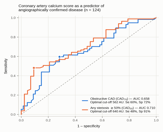
Two outcome definitions are shown: obstructive coronary artery disease (CAD_70 — defined as a stenosis of at least 50% in the left main coronary artery or of at least 70% in any other major epicardial vessel or dominant side branch) and any stenosis of at least 50% (CAD_50). Filled circles indicate the optimal Youden threshold for each curve. The dotted diagonal represents the line of no discrimination. AU — Agatston units.
| CAC threshold (AU) | Se | Sp | PPV | NPV | Accuracy | LR+ | LR– |
|---|---|---|---|---|---|---|---|
| ≥ 10 | 0.98 | 0.03 | 0.45 | 0.67 | 0.45 | 1.01 | 0.63 |
| ≥ 100 | 0.93 | 0.22 | 0.49 | 0.79 | 0.53 | 1.18 | 0.33 |
| ≥ 400 | 0.69 | 0.46 | 0.51 | 0.65 | 0.56 | 1.29 | 0.67 |
| ≥ 562 (Youden) | 0.60 | 0.71 | 0.62 | 0.69 | 0.66 | 2.07 | 0.56 |
| ≥ 1000 | 0.29 | 0.87 | 0.64 | 0.61 | 0.61 | 2.23 | 0.82 |
In univariable logistic regression, log10(CAC+1) was significantly associated with the presence of angiographically confirmed CAD_70. For each one-unit increase in log10(CAC+1), the odds of having CAD_70 were 2.28 times higher (OR 2.28; 95% CI: 1.13–4.60; p = 0.021).
Age, expressed per 10-year increase, was not associated with CAD_70 (OR 1.05; 95% CI: 0.69–1.59; p = 0.83). Male sex was not significantly associated with CAD_70 either (OR 1.37; 95% CI: 0.66–2.86; p = 0.40).
In the multivariable model, log10(CAC+1) remained the only independent predictor associated with CAD_70. The adjusted OR was 2.35 (95% CI: 1.11–4.95; p = 0.025; Figure 2). Age (aOR 0.90 per 10 years; 95% CI: 0.57–1.43; p = 0.65) and male sex (aOR 1.14; 95% CI: 0.52–2.50; p = 0.75) were not significantly associated with CAD_70.
The multivariable model explained little of the variation in the outcome. The McFadden pseudo-R² was 0.039 and the area under the ROC curve was 0.655. The likelihood ratio test showed no significant difference between the adjusted model and the null model (p = 0.082).
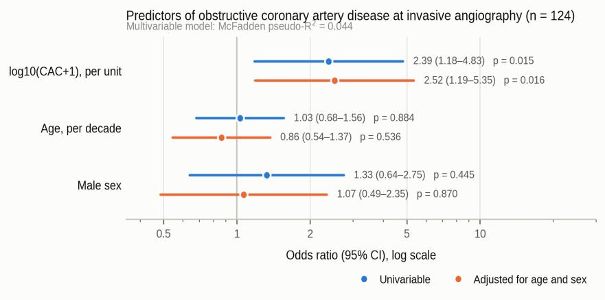
Circles indicate the point estimates and the horizontal lines represent the 95% confidence intervals. The dotted vertical line marks the reference odds ratio of 1. The x axis is logarithmic. The calcium score was the only independent predictor among the three available variables. The multivariable model showed limited explanatory power (McFadden pseudo-R² = 0.039). CAC — coronary artery calcium.
The pre-specified subgroup, made up of the patients referred for catheterization on the basis of a stenosis described on CCTA despite a low or disproportionately small calcium burden, included 33 patients (26.6% of the cohort). The median Agatston score in this subgroup was 135 AU (IQR 51–246). Agreement between CCTA and invasive angiography was markedly lower in this subgroup (Figure 3A). Of the 33 patients, two (6.1%) had confirmed obstructive disease; 12 (36.4%) had a stenosis of at least 50% somewhere in the coronary tree, and 21 (63.6%) had no vessel reaching 50%. The sensitivity analyses showed the same pattern at stricter calcium thresholds (Figure 3B): below 400 AU (n = 30), obstructive disease was confirmed in a single patient (3.3%), and in 21 (70.0%) no stenosis reached 50%; below 100 AU (n = 14), confirmation fell to zero, and in 11 patients (78.6%) no stenosis reached 50%. The three patients in the subgroup whose scores exceeded 400 AU had values of 474, 485, and 2,179 AU, which shows that the discordance is not simply an artifact driven by a few patients with extreme calcium values.
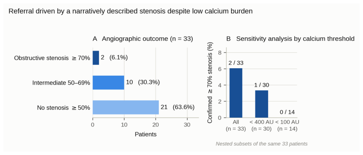
(A) Angiographic outcome: two patients (6.1%) had confirmed obstructive stenosis (≥70%), while in 21 (63.6%) no vessel reached 50%. (B) Sensitivity analysis narrowing the subgroup by calcium threshold, on nested subsets of the 33 patients. Confirmation of obstructive disease falls from 6.1% in the full subgroup to 3.3% below 400 AU and to zero below 100 AU, showing that the discordance is robust and independent of extreme calcium values. AU — Agatston units.
Post-catheterization management was medical in 77 patients (62.1%), percutaneous intervention was completed in 23 (18.5%), surgical consultation was recommended in 10 (8.1%), staged intervention took place in six patients (4.8%), and additional functional testing was indicated in eight patients (6.5%). Treatment allocation closely reflected the angiographic findings: all the patients who underwent percutaneous coronary intervention had confirmed obstructive disease, and 64 of the 77 treated medically (83.1%) did not (Table 3). In the discordant low-calcium subgroup, management reflected the low disease burden: 31 of the 33 patients (93.9%) received medical treatment, two were referred for functional ischemia testing, and none underwent PCI.
| Treatment decision | n (%) | CAD_70+, n | CAD_70+, % |
|---|---|---|---|
| PCI performed | 23 (18.5) | 23 | 100 |
| CABG consultation | 10 (8.1) | 10 | 100 |
| Staged PCI | 6 (4.8) | 5 | 83 |
| Functional testing recommended | 8 (6.5) | 4 | 50 |
| Optimal medical therapy (OMT) | 77 (62.1) | 13 | 17 |
| Total | 124 (100) | 55 | 44.4 |
This bicentric analysis of 124 Romanian patients who underwent both coronary CT angiography and invasive angiography yields three main findings.
First, the Agatston score was the only one of the three analyzed variables that was independently associated with angiographically confirmed obstructive coronary artery disease. The higher the calcium score, the higher the probability of CAD_70. This association had modest discriminatory power, however. The area under the curve was 0.653 for CAD_70 and 0.710 for CAD_50.
The multivariable regression results show that this association is not sufficient to identify patients with obstructive disease accurately. The model that included calcium score, age, and sex had low explanatory power (McFadden pseudo-R² = 0.039), and the difference from the null model was not statistically significant (p = 0.082). In a population already selected for catheterization, therefore, calcium score, age, and sex provide limited information for distinguishing patients with and without obstructive coronary artery disease.
Second, agreement between CCTA and invasive angiography was lower in the pre-specified subgroup of patients referred for catheterization for a stenosis described on CCTA despite a low calcium burden. Obstructive coronary artery disease was confirmed in only 2 patients (6.1%) of the 33, and in 21 patients (63.6%) no coronary vessel had a stenosis of at least 50%. In the patients with CAC <100 AU, no case of obstructive disease was confirmed.
These results must be interpreted in the context of patient selection. All the patients in this subgroup were referred for catheterization because CCTA had already indicated a possible stenosis. This subgroup therefore has a different profile from that of an unselected outpatient population. The results obtained here should not be extrapolated to all patients assessed by CCTA and do not contradict earlier data on the good ability of CCTA to rule out obstructive disease in unselected populations [4].
Third, the treatment decisions taken after catheterization were consistent with the angiographic findings. All the patients who were revascularized had confirmed obstructive coronary artery disease.
Comparisons with previously published results must be made with caution, because the performance of a test depends both on the characteristics of the test and on the population in which it is evaluated. The multicenter validation studies CORE-320 and NXT reported areas under the curve between 0.85 and 0.93 [3, 20]. These values, however, were obtained for CT angiography used together with myocardial perfusion or with CT-derived fractional flow reserve, and not for the calcium score used on its own. These studies also included patients with different levels of pre-test probability.
In the present study, all the patients had already been selected for catheterization. This selection reduced the variation in predictor values and may therefore reduce the area under the curve, even if the actual performance of the test is no different. For this reason, the AUC values in our study cannot be compared directly with those reported in CORE-320 and NXT.
A similar difference appears with regard to disease prevalence. In the outpatient cohort from Cluj-Napoca, which included unselected patients, CAD_50 was present in 41% of patients [11]. In our study, the proportion was 65.3%, which is to be expected in a population selected for catheterization.
The distribution of coronary disease was similar to that reported in the CONFIRM and PROMISE registries, with the lesions located most often in the left anterior descending artery and the right coronary artery [19, 6].
The low rate of disease confirmation in the low calcium score subgroup also raises the possibility that invasive angiography underestimated stenosis severity in this particular category of patients. This is possible because invasive angiography relies on twodimensional images, and the assessment of intermediate stenoses can vary between observers. The variability is greater when angiography is not complemented by measurement of fractional flow reserve or by intravascular ultrasound.
This explanation does not appear to be the main cause of the discordance observed, however. Differences in assessment between observers can mean that a stenosis is graded, for example, at 50% instead of 70% or the other way round. They do not explain the situation in which, in 21 patients (63.6%) of the subgroup, no coronary vessel had a stenosis of at least 50%, even though the angiographic assessment was made in several projections.
A more likely explanation is therefore that CCTA overestimated the severity of some stenoses. In patients with little calcium, the suspected lesions are often made up of non-calcified or mixed plaque. These plaques can cause outward remodeling of the arterial wall and can make the lumen border harder to define. CCTA images can also be affected by cardiac motion [8, 9, 10].
These results have important clinical implications. When CCTA overestimates the severity of a stenosis in patients with a low calcium score, the patient may be referred for catheterization unnecessarily. Every catheterization carries a certain procedural risk and additional exposure to contrast agent and radiation. It also involves additional costs.
The 2024 ESC guidelines recommend, with a Class I, Level B indication, the use of functional imaging to assess myocardial ischemia when CCTA shows coronary disease whose functional importance is unclear or when the CCTA examination is non-diagnostic [2]. The NXT study showed that using CT-derived fractional flow reserve can raise diagnostic specificity from 34% to 79% [20]. The ESC guidelines nevertheless recommend caution in using this method and classify it as a Class IIb, Level B option for intermediate stenoses located in the proximal or mid coronary segments [2].
In the Sibiu region, access to non-invasive functional methods is limited. Single photon emission tomography has only very recently become available under contract with the health insurance house (CASS), but with a considerable waiting time; stress cardiac magnetic resonance requires travel to another center, and CT-derived fractional flow reserve is not reimbursed by the state. Under these conditions, when a CCTA report indicates a suspected stenosis, direct referral to invasive angiography often remains the most accessible option.
Wider access to non-invasive functional investigations, and reimbursement of these methods, could therefore reduce the number of patients referred unnecessarily for catheterization. Developing a regional functional assessment capacity could also contribute to better selection of the patients who need invasive angiography.
One particular feature of this study is the availability, for the same patients, of both coronary CT angiography and invasive coronary angiography data. Patients were included on the basis of pre-specified criteria, and this pairing allowed direct comparison of the results of the two methods. Pre-specified sensitivity analyses were also carried out to check whether the results hold at different calcium score thresholds.
The study does have a number of limitations. The cohort of 124 patients was a population selected for invasive investigation and does not reflect the general population assessed by coronary CT angiography. Referral for catheterization was driven by symptoms plus the identification of an abnormality on imaging and was not random. All the accuracy estimates are conditional on this selection and cannot be transferred to an unselected referral population.
CCTA reporting at the institution the patients came from was narrative and did not systematically use the CAD-RADS 2.0 categories. For this reason, automatic re-adjudication of stenoses at the level of each coronary segment was not possible [21]. Introducing a structured reporting system in line with CAD-RADS 2.0, including the use of standardized modifiers, could be a quality improvement measure in regional centers and could reduce subjective overestimation of stenosis severity.
Conventional cardiovascular risk factors were not recorded systematically in the archive and, for this reason, could not be included in the multivariable model. This limitation may contribute to the low explanatory power of the model, reflected in the modest pseudoR² value.
Stenosis severity at invasive angiography was assessed visually, in several projections. Fractional flow reserve measurement and intravascular ultrasound were not used systematically. An independent functional or intravascular assessment of the stenoses was therefore not possible.
The interval between CCTA and catheterization was not recorded systematically in the available documentation. For this reason, we could not analyze whether the length of this interval influenced the agreement between the two methods. A longer interval between the examinations could reduce the agreement between the results.
Finally, the data were extracted by a single researcher and were not independently verified by a second researcher. Agreement between raters was therefore not formally assessed.
In a referral-selected cohort of 124 patients from Romania, assessed by both CCTA and invasive coronary angiography, the coronary calcium score was an independent but modest predictor of obstructive coronary artery disease. The discordance between the two methods was evident in the patients referred for catheterization for a suspected stenosis despite a low calcium burden. In this subgroup, only 6.1% of patients had confirmed obstructive disease, and in 63.6% no coronary vessel had a stenosis of at least 50%. This pattern suggests that the overestimation of stenoses on CCTA may be related to the features of non-calcified plaque and to motion artifacts, rather than to a general underestimation of stenosis severity at invasive angiography.
Access to non-invasive functional assessment methods within the public health system could reduce the number of cardiac catheterization assessments performed in patients in whom CCTA raises the suspicion of a stenosis but in whom obstructive disease is not confirmed. In patients with a low calcium score and an equivocal CCTA result, functional assessment performed before catheterization could help establish the ischemic relevance of the lesion and improve the selection of the patients who need invasive angiography.
The authors declare that they have no conflict of interest.
The authors thank the Sibiu Pediatric Clinical Hospital, as the source of the coronary CT angiography data, and Dr. Ciprian Radu Sofariu, Medical Director of that institution, for the scientific collaboration on this research. They also thank the Sibiu County Clinical Hospital as the source of the data on the patients assessed by invasive coronary angiography, Assoc. Prof. Dr. Minodora Teodoru, head of the Cardiology Clinic of SCJU Sibiu, and Dr. Cornel Ioan Bitea, head of the Catheterization Laboratory of SCJU Sibiu. In line with the ICMJE and WAME recommendations on the use of artificial intelligence in academic publishing, the authors declare that the assistance of a large language model was used for the language editing of a manuscript drafted by the authors, for checking the reference list, and for checking the internal consistency of the reported data against the study database. It was not used to generate data, to perform or interpret the statistical analyses, or to formulate the conclusions. The authors have read and verified every sentence of the final text and take full responsibility for the accuracy and integrity of the work.
Conceptualization, methodology, validation, A-M.B-B., D-F.C.M and I.M.; investigation, data curation, writing — original draft preparation, A-M.B-B.; writing — review and editing, visualization, A-M.B-B., D-F.C.M and I.M.; supervision, I.M. All authors have read and agreed to the published version of the manuscript. Ethics approval and consent to participate
The study was covered by four institutional authorizations. Ethics approval no. 31 / 24.04.2026 was issued by the Ethics Council of the Sibiu Military Emergency Clinical Hospital. Administrative and research approval no. 7969 / 07.11.2023 and institutional data use agreement no. 3697 / 07.05.2026 were granted by the Sibiu Pediatric Clinical Hospital for retrospective access to the anonymized CCTA records. Ethics approval for access to the catheterization archive was granted by the Ethics, Medical Deontology and Discipline Committee of the Sibiu County Emergency Clinical Hospital, through Decision no. 17160 / 29.05.2026. Because the analysis was retrospective and used fully anonymized records, already collected in the course of routine medical care, individual written informed consent was not sought; this complies with Order no. 1502/2016 of the Ministry of Health, with Law no. 46/2003 on patients’ rights and with Article 89 of Regulation (EU) 2016/679, which allows the secondary use of pseudonymized health data for scientific research purposes, subject to appropriate safeguards.
Individual written informed consent for publication was not required: the analysis was retrospective and used fully anonymized records, and no information allowing individual identification of patients appears in this article. The legal basis is set out above, in the Ethics approval and consent to participate section.