
Degenerative AS is one of the most important valvular diseases of aging. Population-based and modeling data show that AS becomes increasingly common after the seventh decade of life, and contemporary guideline frameworks emphasize that disease severity, symptoms, ventricular response, frailty, and procedural risk must all be integrated when selecting medical surveillance, SAVR, or TAVI/TAVR [1-3]. In parallel, ATTR-CM has evolved from a perceived rarity to a treatable cause of restrictive cardiomyopathy and heart failure in older adults. The recognition of non-biopsy diagnostic pathways and disease-modifying therapy has transformed clinical practice, but this benefit depends on identifying ATTR-CM before advanced irreversible myocardial dysfunction develops [4-8].
The intersection between ATTR-CM and AS is clinically compelling because both disorders are age-related, both may present with dyspnea, syncope, exertional intolerance, left-ventricular hypertrophy, atrial arrhythmia, conduction disease and HFpEF, and both may be misattributed to “normal aging” or hypertension. In an elderly patient with severe AS, the hypertrophied, stiff ventricle may be interpreted as a pressure-overload response, while amyloid infiltration remains unsuspected. Conversely, amyloid-related low stroke volume may modify the echocardiographic phenotype of AS, contributing to paradoxical low-flow low-gradient presentations and diagnostic uncertainty.
Nuclear cardiology is central to this field. The landmark non-biopsy diagnostic work by Gillmore et al. established that grade 2 or 3 cardiac uptake on bone-avid tracer scintigraphy, when combined with the absence of a monoclonal protein, can diagnose ATTR-CM with very high specificity and positive predictive value [6]. Subsequent expert consensus documents from cardiac imaging societies clarified acquisition, interpretation, reporting, and the need for SPECT or SPECT/CT confirmation of myocardial rather than blood-pool uptake [7,8]. The original visual scoring system introduced with DPD scintigraphy remains the foundation for clinical interpretation [9].
The aim of this review is to synthesize evidence on the prevalence of ATTR-CM in AS, emphasize the dual-pathology phenotype in elderly patients, and provide an academically grounded approach to nuclear diagnosis. The intended practical output is a diagnostic framework for clinicians evaluating severe AS—particularly TAVI candidates—in whom ATTR-CM may be clinically silent, prognostically relevant, and therapeutically actionable.
This manuscript was designed as an extended narrative review with structured search methodology rather than a formal systematic review or meta-analysis. The objective was to integrate evidence across epidemiology, cardiology, nuclear medicine, multimodality imaging, and therapeutics, while allowing contextual discussion of implementation and clinical decision-making.
A targeted literature search was conceptualized for PubMed/MEDLINE, Scopus, Google Scholar, and major society documents available up to 9 May 2026. Search concepts included: “transthyretin cardiac amyloidosis”, “ATTR-CM”, “ATTRwt”, “ATTRv”, “aortic stenosis”, “severe aortic stenosis”, “low-flow low-gradient aortic stenosis”, “TAVI”, “TAVR”, “SAVR”, “99mTc-PYP”, “99mTc-DPD”, “99mTc-HMDP”, “bone scintigraphy”, “Perugini score”, “heart-to-contralateral lung ratio”, “SPECT/CT”, and “RAISE score”. Reference lists of key reviews, consensus documents, and pivotal cohort studies were also considered.
Eligible material included peer-reviewed cohort studies, systematic reviews, meta-analyses, case-based reviews with substantial literature synthesis, nuclear cardiology consensus statements, cardiology guidelines, and major therapeutic trials relevant to ATTR-CM. Studies were prioritized when they included elderly AS populations, TAVI/TAVR candidates, explicit nuclear diagnostic protocols, or outcomes after valve replacement. Non-cardiac amyloidosis literature was included only when it supported systemic-recognition concepts relevant to cardiac diagnosis, as in digestive amyloidosis trends.
Calcific AS is an active fibro-calcific process involving endothelial injury, lipid infiltration, inflammation, valvular interstitial-cell activation, extracellular matrix remodeling, and progressive leaflet mineralization. Its clinical expression is not determined solely by valvular area or gradient. The LV response—concentric hypertrophy, fibrosis, diastolic dysfunction, impaired longitudinal mechanics, pulmonary hypertension, and reduced flow reserve—modulates symptoms and outcomes. Contemporary valve guidelines therefore distinguish high-gradient severe AS, classical low-flow low-gradient AS with reduced EF, and low-flow low-gradient AS with preserved EF [2,3].
Low-flow low-gradient AS is especially relevant to amyloidosis because both conditions can converge on a small, stiff, hypertrophied ventricle with low stroke volume despite preserved EF. In such patients, the calculated valve area may suggest severe AS while the mean gradient is lower than expected. ATTR-CM can contribute to this phenotype by reducing longitudinal shortening, impairing diastolic filling, and increasing myocardial stiffness.
ATTR amyloidosis results from misfolding of transthyretin, a tetrameric transport protein mainly synthesized by the liver. Because hepatic function is central to transthyretin production, liver diseases and systemic inflammatory conditions may influence hepatic protein homeostasis and should be considered in the broader context of transthyretin-related disorders [10]. Tetramer dissociation is the rate-limiting step in amyloidogenesis; misfolded monomers aggregate into oligomers and fibrils that deposit in the extracellular matrix. In ATTRwt, the transthyretin sequence is normal, and the disease is strongly age-associated, classically affecting older men but increasingly recognized in women. In ATTRv, a pathogenic TTR variant destabilizes the tetramer and produces hereditary disease with variable cardiac, neurologic, autonomic, and gastrointestinal phenotypes [4,5,11-14].
Although the present review focuses on cardiac disease, systemic clues matter. Bilateral carpal tunnel syndrome, lumbar spinal stenosis, biceps tendon rupture, peripheral or autonomic neuropathy, unexplained weight loss, and digestive symptoms may precede cardiac diagnosis. Bucurica et al. reviewed digestive amyloidosis and underscored the importance of recognizing multiorgan manifestations, biopsy confirmation with Congo red staining, and protein typing when gastrointestinal disease is suspected [13]. In elderly AS, such extracardiac clues can shift the clinical probability toward ATTR-CM and justify nuclear evaluation.
Most dual AS-ATTR cohorts identify predominantly ATTRwt, which is biologically plausible because both degenerative AS and ATTRwt are common in later life. Nevertheless, ATTRv cannot be excluded by age, sex, ethnicity, or cardiac phenotype alone. After a non-biopsy diagnosis of ATTR-CM, current consensus statements recommend TTR genetic testing to distinguish ATTRwt from ATTRv, because the result affects family counseling, cascade testing, and awareness of neurologic or mixed phenotypes [4,5].
For review framing, ATTRwt should be treated as the principal subtype expected in elderly severe AS and TAVI referral pathways, while ATTRv should be discussed as a mandatory differential subtype once ATTR-CM is confirmed. This distinction also avoids a common diagnostic error: a positive bone-avid tracer scan identifies ATTR-CM only after AL amyloidosis has been excluded and does not, by itself, determine whether the patient has wild-type or variant TTR disease [4-8].
The reported prevalence of ATTR-CM among patients with AS varies by population, age threshold, AS severity, referral setting, tracer, imaging protocol, and diagnostic definition. The highest-yield populations are older patients with severe AS referred for TAVI/TAVR, because advanced age enriches for ATTRwt and TAVI pathways often include systematic imaging, biomarker, and frailty assessment. Surgical cohorts and unselected AS cohorts are more heterogeneous.
Early pathologic and imaging work suggested that occult ATTR may be present in a substantial minority of older AS patients. Treibel et al. identified occult transthyretin amyloid in patients undergoing SAVR and reported adverse prognostic associations, supporting the concept that amyloid infiltration may be clinically relevant rather than merely incidental [15]. Castaño et al. then prospectively evaluated elderly patients with severe calcific AS undergoing TAVR using PYP scintigraphy and found ATTR-CM in 16%, with associations including low-flow low-gradient physiology and reduced longitudinal systolic velocity [16].
Scully et al. evaluated patients referred for TAVI using DPD scintigraphy and reported dual AS-amyloid pathology in approximately 13%, with higher biomarkers and structural/functional differences; importantly, TAVI was still associated with improved outcome compared with medical management in the dual-pathology group [17]. Nitsche et al. expanded this concept in severe AS cohorts by examining both AL and ATTR and by proposing structured screening possibilities. In the JACC cohort, DPD uptake compatible with ATTR-CM was present in a clinically meaningful minority of patients, and the RAISE score was introduced to identify patients with increased likelihood of concomitant amyloid [18,19].
Narrative and systematic reviews have since consolidated these observations. Ternacle et al. estimated that cardiac amyloidosis may affect up to approximately 15% of AS patients and a higher proportion of those with low-flow low-gradient AS, emphasizing that coexistence can contribute to treatment futility and adverse outcomes if unrecognized [20]. Ho et al. pooled 21 studies and reported an overall cardiac amyloidosis prevalence of 14.4% among AS patients, while noting higher mortality in the dual-pathology group [21]. Jaiswal et al. summarized the state of the art and described coexistence in the approximate 4% to 16% range among older AS/TAVI populations [22]. More recent meta-analyses specifically addressing TAVR/TAVI populations continue to support a nontrivial prevalence and the need for standardized diagnostic pathways [23-25].
Adam et al. provide a clinically useful bridge between case-based recognition and literature synthesis. Their report of severe AS with ATTRwt amyloidosis in an aging patient illustrates how AS may mask amyloid-related restrictive cardiomyopathy, and how low-flow low-gradient physiology, ventricular hypertrophy, biomarker elevation, and multimodality imaging should prompt ATTR evaluation [14].
Taken together, the literature supports the following practical statement: ATTR-CM is not rare among elderly patients with severe AS, particularly those referred for TAVI, and its prevalence is sufficiently high to justify systematic suspicion and selective or protocolized screening in enriched subgroups. However, prevalence should not be quoted without describing diagnostic criteria. Studies that include Perugini grade 1 uptake, do not perform SPECT confirmation, or do not fully exclude AL may overestimate or misclassify definite ATTR-CM.
| Study | Population/diagnostic approach | Key finding | Relevance to this review |
|---|---|---|---|
| Treibel et al., 2016 [15] | SAVR-oriented severe calcific AS cohort; histology and imaging-oriented assessment. | Occult transthyretin amyloid was identified in a subset and associated with adverse prognosis. | Established that amyloid may be clinically relevant in surgically treated AS, not merely an incidental autopsy finding. |
| Castaño et al., 2017 [16] | Elderly severe calcific AS patients undergoing TAVR; PYP scintigraphy with monoclonal-protein assessment. | ATTR-CM identified in 16%; associated with low-flow low-gradient AS and lower tissue Doppler systolic velocity. | Key prospective TAVR study supporting nuclear screening in enriched AS populations. |
| Scully et al., 2020 [17] | Patients aged ≥75 years referred for TAVI; DPD scintigraphy and clinical outcomes. | Dual AS-amyloid pathology in approximately 13%; biomarkers higher; TAVI remained beneficial compared with medical management. | Important for clinical decision-making: ATTR-CM should not automatically preclude TAVI. |
| Nitsche et al., 2020 [18] | Severe AS cohort examining AL and ATTR diagnostic pathways. | Emphasized screening possibilities, monoclonal workup, and outcome analysis. | Highlights that AL must be considered and excluded in AS patients with suspected amyloid. |
| Nitsche et al., 2021 [19] | 407 severe AS patients screened with DPD; introduced RAISE score. | Grade 2/3 uptake in 7.9%; one-year mortality higher in AS-CA; TAVR improved survival and should not be withheld. | Provides a pragmatic screening score and outcome-oriented evidence. |
| Ternacle et al., 2019 [20] | JACC review focused on AS and cardiac amyloidosis. | Estimated clinically meaningful coexistence, especially in low-flow low-gradient AS. | Frames dual pathology as a contributor to heart failure and treatment futility. |
| Ho et al., 2022 [21] | Systematic review/meta-analysis of 21 studies. | Pooled cardiac amyloidosis prevalence in AS: 14.4%; dual pathology associated with higher mortality. | Quantifies burden and supports systematic suspicion. |
| Jaiswal et al., 2023 [22] | State-of-the-art review. | Coexistence has been reported at 4–16% in older AS/TAVR populations. | Useful contemporary synthesis of imaging and management. |
| Fatima et al., 2024; Cannata et al., 2022; Jaiswal et al., 2023 [23-25] | Meta-analyses/systematic reviews focused on TAVR/TAVI outcomes. | Suggest altered procedural and post-procedural risk profiles, including conduction disease and heart-failure outcomes, with heterogeneous mortality findings. | Supports the need for standardized diagnosis and prospective outcome studies. |
| Adam et al., 2020 [14] | Case report and literature review of severe AS with ATTRwt. | Illustrates diagnostic vigilance in aging and the overlap between AS and ATTRwt phenotypes. | Clinically anchors the present review question. |
The diagnostic challenge is that AS already explains many findings that also occur in ATTR-CM. Therefore, suspicion should be triggered not by a single sign but by disproportion, clustering, or trajectory. Severe concentric LVH in a patient with only modest hypertension, small LV cavity, restrictive filling, right-ventricular thickening, biatrial enlargement, pericardial effusion, impaired longitudinal strain, or unexpectedly high NT-proBNP and troponin should not be automatically attributed to valvular pressure overload.
Low-flow low-gradient AS is a particularly important clinical phenotype. In ATTR-CM, stroke volume can be reduced by small ventricular cavity size, impaired diastolic filling, atrial dysfunction, and longitudinal systolic impairment despite preserved EF. This may produce discordant AS measurements and may lower gradients despite severe leaflet obstruction. Castaño et al. and later cohorts found enrichment of amyloid in low-flow low-gradient presentations [16,19,20].
Electrocardiography can provide additional clues. Classical low QRS voltage is not universally present in ATTR-CM, and AS-related LVH may further obscure voltage-wall thickness discordance. Nevertheless, pseudo-infarct pattern, conduction delay, atrioventricular block, bundle-branch block, atrial fibrillation, and pacemaker requirement are important red flags. The RAISE score incorporated remodeling, age, injury, systemic clues, and electrical disturbances to identify AS patients more likely to have concomitant amyloid [19].
Extracardiac features may be decisive. Bilateral carpal tunnel syndrome, lumbar spinal stenosis, biceps tendon rupture, neuropathy, autonomic symptoms, unexplained gastrointestinal dysmotility, proteinuria, or a history of monoclonal gammopathy should be actively sought. Bucurica et al. emphasize that gastrointestinal amyloidosis may present with nonspecific symptoms and requires pathologic and protein-typing confirmation [13]. In the AS clinic, such clues help identify patients who should undergo amyloid evaluation rather than routine valve-only assessment.
| Domain | Clinical finding | Diagnostic significance |
|---|---|---|
| Demographic | Age >75–80 years; male sex; frailty or disproportionate functional decline. | ATTRwt and degenerative AS are both enriched in advanced age. |
| AS phenotype | Paradoxical low-flow low-gradient AS; low stroke-volume index; discordant valve area/gradient. | Amyloid-related restrictive physiology can reduce flow and mask the severity of the gradient. |
| Echocardiography | Disproportionate LV wall thickness; small LV cavity; severe diastolic dysfunction; biatrial enlargement; RV thickening; small pericardial effusion. | Suggest infiltrative disease beyond pressure overload. |
| Strain imaging | Reduced global longitudinal strain with relative apical sparing; severe longitudinal impairment despite preserved EF. | Supports amyloid suspicion, although AS can reduce specificity. |
| ECG/conduction | Low voltage or voltage-mass discordance; pseudo-infarct pattern; AV block; bundle-branch block; atrial fibrillation; pacemaker. | Electrical disease is common in ATTR-CM and may worsen after TAVI. |
| Biomarkers | NT-proBNP or troponin elevation disproportionate to AS severity, renal function, or symptoms. | Reflects myocardial injury/stress and may aid risk stratification. |
| Extracardiac ATTR clues | Bilateral carpal tunnel syndrome, lumbar spinal stenosis, biceps tendon rupture, peripheral/autonomic neuropathy. | May precede cardiac diagnosis by years. |
| Systemic amyloidosis clues | Unexplained proteinuria, macroglossia, purpura, gastrointestinal dysmotility, weight loss, monoclonal gammopathy. | Raises concern for AL or systemic amyloid and mandates hematologic evaluation. |
Bone-avid radiotracer scintigraphy has become the cornerstone of non-invasive ATTR-CM diagnosis. The clinically used tracers are 99mTc-PYP, 99mTc-DPD, and 99mTc-HMDP. Although their exact molecular binding to amyloid is not fully reducible to one mechanism, they localize to ATTR cardiac deposits sufficiently reliably to support diagnosis when interpreted in the correct clinical and laboratory context [6-11]. Recent reviews also emphasize that nuclear medicine in cardiac amyloidosis includes validated bisphosphonate scintigraphy for ATTR-CM and emerging amyloid-specific PET methods for disease-burden assessment and follow-up [26].
This is particularly valuable in AS. Endomyocardial biopsy is definitive but invasive and not always feasible in frail elderly patients already undergoing valve assessment. Cardiovascular magnetic resonance can strongly suggest amyloidosis through diffuse late gadolinium enhancement, abnormal gadolinium kinetics, elevated native T1, and extracellular volume expansion, but it cannot reliably type amyloid and may be limited by renal dysfunction, arrhythmia, implanted devices, or access. Scintigraphy, in contrast, can noninvasively establish ATTR-CM when paired with a negative monoclonal-protein evaluation [4-8,27].
The Perugini visual score remains the most widely used semiquantitative approach. Grade 0 indicates absent myocardial uptake with normal bone uptake. Grade 1 indicates myocardial uptake less than bone uptake. Grade 2 indicates myocardial uptake equal to bone uptake. Grade 3 indicates myocardial uptake greater than bone uptake, often with reduced or absent skeletal visualization due to intense cardiac localization [9].
For ATTR-CM diagnosis, grade 2 or 3 myocardial uptake is considered strongly positive only after AL amyloidosis has been excluded. Gillmore et al. showed that radionuclide scintigraphy has excellent sensitivity for ATTR-CM, but specificity becomes effectively diagnostic when grade 2/3 uptake is combined with absence of a monoclonal protein [6]. Grade 1 uptake is not sufficient for a nonbiopsy ATTR diagnosis. It may reflect early ATTR, AL amyloid, blood-pool artifact, rib/valvular activity, or technical factors; such cases require careful SPECT review, repeat imaging, CMR, or tissue diagnosis depending on clinical probability [4-8,12].
The most important safety principle is that a positive bone-avid tracer scan does not exclude AL amyloidosis. AL can occasionally show cardiac uptake, and missing AL has major consequences because it is a hematologic emergency requiring urgent therapy. Therefore, every suspected cardiac amyloidosis patient should undergo serum free light-chain assay, serum immunofixation, and urine immunofixation. Serum protein electrophoresis alone is insufficient. If a monoclonal protein is present, ATTR-CM cannot be diagnosed non-invasively on scintigraphy alone; tissue biopsy with amyloid typing is generally required [4-8].
This point is especially relevant in elderly AS because monoclonal gammopathy of undetermined significance is common with age. A patient may have AS, ATTRwt, and an unrelated monoclonal gammopathy, or may have AL amyloidosis with AS. The diagnostic algorithm must therefore be designed to avoid both underdiagnosis and misclassification.
Planar images are useful for global visual assessment and heart-to-contralateral quantification, but they are insufficient as the sole basis for diagnosis. SPECT or SPECT/CT confirms whether activity is truly myocardial rather than intravascular blood pool, overlying rib, sternal, valvular, annular, or pulmonary activity. In severe AS, annular and valvular calcification are common, and blood-pool retention may be increased in heart failure or renal impairment. These issues increase the importance of tomographic confirmation [7,8,12].
Stan et al. demonstrated that implementation gaps remain clinically relevant. In their survey of Romanian nuclear medicine physicians, incidental diffuse cardiac uptake was frequently encountered, but awareness, interpretation, tracer selection, and multidisciplinary communication were variable [12]. Although the survey is regional, its message is general: diagnostic accuracy depends not only on tracer availability but also on standardized acquisition, correct interpretation, reporting language, and referral pathways.
The H/CL ratio has been widely used, especially in PYP imaging, as a semi-quantitative adjunct. Thresholds differ by acquisition timing and local protocols, and the ratio can be affected by blood-pool activity, body habitus, contralateral-lung region placement, pleural or pulmonary disease, and extracardiac overlap. Expert statements therefore recommend that H/CL should support but not replace visual SPECT-confirmed diagnosis [7,8]. A high H/CL ratio without definite myocardial uptake on SPECT should not be reported as diagnostic ATTR-CM.
For AS patients, reports should explicitly state the tracer used, injected activity, acquisition timing, planar and SPECT/SPECT-CT findings, visual grade, whether uptake is myocardial or blood pool, whether H/CL ratio was measured, and whether findings are compatible with ATTR-CM only in the context of negative monoclonal-protein testing. Suggested reporting categories are: negative for ATTR-CM, equivocal, positive for ATTR-CM if AL excluded, or positive cardiac uptake with monoclonal-protein/biopsy correlation required.
Although grade 2 or 3 myocardial uptake on bone-avid tracer scintigraphy, in the absence of a monoclonal protein, has very high diagnostic specificity for ATTR-CM, false-positive results can occur when acquisition, reconstruction, localization, clinical context, or amyloid typing requirements are not rigorously respected [7,8,28,29]. This point is particularly important in elderly AS patients, in whom renal dysfunction, heart failure with delayed blood-pool clearance, valvular or annular calcification, previous coronary disease, rib lesions, and complex thoracic anatomy may coexist and may complicate planar interpretation.
The most frequent interpretative pitfall is mistaking persistent intracavitary or vascular blood-pool activity for true myocardial tracer uptake. This problem is most relevant for early imaging and for planar-only interpretation, and it is magnified when myocardial uptake is assessed without the characteristic tomographic distribution of tracer within the ventricular walls. In a large clinical experience with 99mTc-PYP, Poterucha et al. showed that reliance on planar grade 2 findings can generate false-positive interpretations when SPECT is used as the reference standard, reinforcing the need for tomographic confirmation of myocardial retention [29]. Accordingly, SPECT or preferably SPECT/CT should be considered mandatory when cardiac uptake is suspected, because it distinguishes myocardial uptake from blood pool, ribs, sternum, valvular/annular calcification, and extracardiac activity [7,8,28].
| Step | Recommended approach | Clinical rationale |
|---|---|---|
| Tracer | 99mTc-PYP, 99mTc-DPD, or 99mTc-HMDP. | Use validated bone-avid tracers; do not extrapolate diagnostic criteria to non-validated tracers. |
| Acquisition | Planar plus SPECT; SPECT/CT where available. | Tomography is necessary to confirm myocardial uptake and avoid blood-pool/overlap artifacts. |
| Visual score | Perugini grade 0–3. | Grade 2/3 is strongly positive; grade 1 is equivocal and not diagnostic alone. |
| Laboratory exclusion of AL | Serum free light chains, serum immunofixation, urine immunofixation. | Mandatory before making a non-biopsy ATTR-CM diagnosis. |
| H/CL ratio | Optional adjunct, especially for PYP. | Helpful for reproducibility but must not override SPECT-confirmed visual interpretation. |
| ATTRwt vs ATTRv | TTR genetic testing after ATTR-CM diagnosis. | Determines hereditary status, family counseling, and systemic phenotype surveillance. |
| AS-specific pitfalls | Valvular/annular calcification, blood-pool retention, renal dysfunction, heart failure, rib uptake. | May mimic or obscure myocardial uptake; SPECT/CT improves localization. |
| Communication | The report should recommend a hematology/genetics/cardiology correlation as appropriate. | Prevents false reassurance, missed AL, and incomplete ATTR workup. |
Technical and general nuclear-medicine artifacts should also be considered before assigning a positive ATTR-CM diagnosis. Excess free pertechnetate, suboptimal radiochemical preparation, premature acquisition, patient motion, attenuation, extracardiac overlap, reconstruction artifacts, and SPECT/CT misregistration may alter visual interpretation or create apparent focal or diffuse activity in the cardiac region. Mititelu et al. provide a practical nuclear cardiology framework for recognizing artifacts arising from patient-related, equipment-related, and technique-related factors, including motion, attenuation, extracardiac activity, and acquisition and reconstruction problems. Their emphasis on raw-data review, correct positioning, artifact recognition, attenuation or overlap assessment, and systematic quality control is directly relevant to ATTR scintigraphy, because the same interpretative discipline helps prevent false-positive myocardial localization and false-negative examinations in bone-avid tracer imaging [30].
Several biological or disease-related causes of false-positive cardiac uptake have been described. AL amyloidosis remains the most clinically dangerous mimic, because it may occasionally show moderate or intense bone-avid tracer uptake and cannot be excluded by scintigraphy alone; this is why serum free light chains, serum immunofixation and urine immunofixation are mandatory before any non-biopsy ATTR-CM diagnosis [4,6-8,27]. Other reported causes include hypertrophic cardiomyopathy, hydroxychloroquine- or chloroquine-induced cardiotoxicity, myocarditis, post-radiation myocardial injury, doxorubicin-induced cardiotoxicity, myocardial calcification, and focal uptake related to rib or skeletal lesions projecting over the heart [28,31,32]. In AS patients, hypertrophic remodeling, coronary artery disease, and valvular/annular calcification are frequent background abnormalities, so a positive scan should be interpreted only after confirming that uptake is truly myocardial and that the clinical pattern is not better explained by another process.
Recent myocardial infarction deserves specific emphasis. 99mTc-PYP was originally introduced in the 1970s for radionuclide imaging of acute myocardial infarction, because myocardial necrosis is associated with calcium influx and formation of intramyocardial calcium complexes with affinity for phosphate-based bone tracers [28,33]. Therefore, a patient scanned shortly after an acute infarct may show intense myocardial tracer uptake that can mimic ATTR-CM. In contrast to the diffuse or circumferential ventricular-wall uptake expected in typical ATTR-CM, post-infarction uptake is often focal or regional and follows a coronary-territory distribution. Khor et al. illustrate this pitfall with a patient scanned 10 days after left anterior descending artery infarction, in whom intense septal uptake represented recent infarction rather than amyloid; they also note that PYP uptake after infarction may become less prominent after approximately two weeks but can occasionally persist for months [28]. Makivic et al. similarly described acute myocardial infarction with reperfusion-related myocardial edema as a differential diagnosis for cardiac ATTR, emphasizing that scintigraphic findings should be interpreted alongside clinical history, ECG, biomarkers, coronary anatomy, and CMR when appropriate [34].
In practical terms, a recent acute coronary syndrome, revascularization, a marked troponin rise, a new regional wall-motion abnormality, or a CMR pattern consistent with infarction should preclude an automatic ATTR-CM label. In such cases, the report should describe the distribution of uptake, state that recent myocardial injury is a potential cause of false positivity, and recommend delayed repeat scintigraphy, CMR correlation, or biopsy with amyloid typing if diagnostic uncertainty persists. This approach is especially relevant before TAVI/TAVR, because elderly AS patients often have coexisting coronary artery disease and may undergo amyloid evaluation during the same period as coronary assessment or acute decompensation.
Nuclear imaging should not be interpreted in isolation. Echocardiography remains the first-line imaging modality in AS and is indispensable for assessing valve severity, gradients, valve area, stroke volume index, LV wall thickness, diastolic function, pulmonary pressures, and coexisting valve disease. The challenge is that AS produces LVH and diastolic dysfunction even without amyloid. Thus, echo should be used to identify disproportionate features and to stratify amyloid probability rather than to “rule out” ATTR-CM.
Speckle-tracking strain can be useful. Relative apical sparing is a classic amyloid clue, described as a base-to-apex gradient of longitudinal dysfunction [35]. However, severe AS itself alters strain, and apical sparing is not universally present in dual pathology. In Castaño et al., conventional amyloid strain patterns were less discriminative than other functional parameters in the TAVR cohort [16]. Therefore, strain should be interpreted alongside age, AS phenotype, biomarkers, ECG, and extracardiac clues.
CMR adds tissue characterization. Diffuse subendocardial or transmural late gadolinium enhancement, high native T1, abnormal myocardial nulling, and elevated extracellular volume support cardiac amyloidosis. CMR is also useful when scintigraphy is equivocal, monoclonal testing is abnormal, or multiple cardiomyopathic processes coexist. Nevertheless, CMR cannot reliably distinguish ATTR from AL, and nuclear plus hematologic testing remains decisive for non-biopsy ATTR typing [4-8].
Biomarkers are particularly relevant in dual disease. NT-proBNP and troponin may be elevated in severe AS, ATTR-CM, renal dysfunction, and atrial fibrillation, so specificity is limited. However, disproportionate elevation should raise suspicion for infiltrative myocardial disease and may refine risk stratification. Nitsche et al. incorporated biomarkers into screening logic, and systematic reviews have associated dual pathology with higher biomarker burden and worse outcomes [19,21].
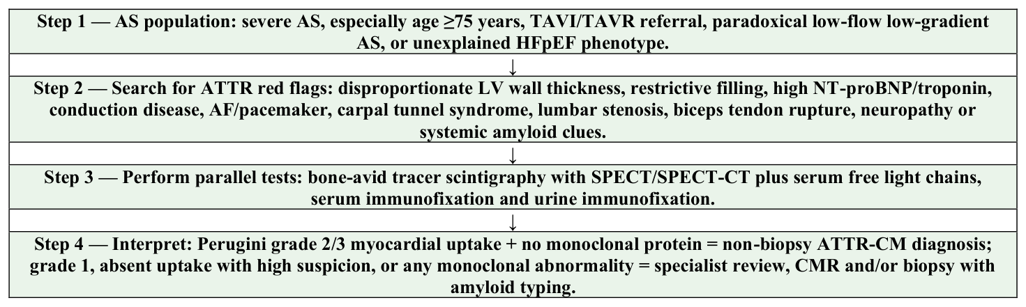
There is no universal mandate to perform bone scintigraphy in every AS patient. A balanced approach is to screen patients in whom the pre-test probability is meaningfully enriched. Reasonable candidates include: patients aged 75 years or older with severe AS referred for TAVI; patients with paradoxical low-flow low-gradient AS; patients with disproportionate LVH or restrictive physiology; patients with conduction disease or atrial fibrillation out of proportion to AS; patients with high biomarkers; and patients with extracardiac ATTR clues.
The pathway should be operationally simple. First, identify red flags during the valve-clinic evaluation. Second, order both nuclear scintigraphy and monoclonal-protein testing rather than staging them weeks apart. Third, interpret scintigraphy with SPECT/SPECT/CT confirmation and report visual grade. Fourth, if grade 2/3 myocardial uptake is present and AL workup is negative, diagnose ATTR-CM and perform TTR genetic testing. Fifth, if uptake is grade 1 or monoclonal testing is abnormal, proceed to specialist evaluation, CMR and/or biopsy with amyloid typing. Sixth, discuss valve intervention and ATTR-specific therapy in a multidisciplinary team.
The presence of ATTR-CM should not be interpreted as an automatic contraindication to TAVI. Observational data suggest that patients with dual AS-ATTR may have higher heart-failure burden, more conduction disease, and worse long-term prognosis, but valve intervention can still improve survival and symptoms compared with conservative management in appropriately selected patients [17,19,21-25]. A useful clinical interpretation is that TAVI treats afterload obstruction but not infiltrative cardiomyopathy; therefore, residual dyspnea, biomarker elevation, diastolic dysfunction, and arrhythmic risk may persist after technically successful valve replacement.
This distinction matters for patient counseling. In isolated severe AS, symptomatic improvement after TAVI may be dramatic. In dual pathology, improvement may occur but be incomplete. Recognizing ATTR-CM pre-procedurally helps avoid unrealistic expectations, guides post-TAVI monitoring, and may explain persistent HFpEF symptoms that would otherwise be misattributed to prosthesis dysfunction or deconditioning.
Both AS and ATTR-CM are associated with conduction abnormalities. TAVI itself carries risk of new conduction disturbance and permanent pacemaker implantation. Dual pathology may therefore identify a subgroup requiring closer rhythm surveillance. Existing meta-analyses suggest higher rates of conduction-related outcomes in cardiac amyloidosis patients undergoing valve intervention, although estimates vary by study design and diagnostic criteria [21,23-25]. Pre-existing PR prolongation, bundle-branch block, atrioventricular block, or pacemaker history should be integrated into procedural planning.
The therapeutic landscape of ATTR-CM has changed rapidly. Tafamidis, a transthyretin stabilizer, reduced all-cause mortality and cardiovascular-related hospitalization in ATTR-CM in ATTR-ACT, establishing disease-modifying therapy as a practical reason to diagnose ATTR-CM [36]. Acoramidis, another TTR stabilizer, improved clinical outcomes in ATTR-CM in the ATTRibute-CM trial [37]. Vutrisiran, an RNA interference therapeutic that reduces hepatic TTR production, improved outcomes in ATTR-CM in the HELIOS-B trial, further expanding the therapeutic rationale for early detection [38]. Stanciu et al. summarize this shift from molecular pathogenesis to radionuclide diagnosis and pharmacologic intervention [11].
In AS patients, evidence from trials specific to dual AS-ATTR is limited. However, once ATTR-CM is diagnosed, treatment decisions should be guided by ATTR-CM stage, functional status, renal function, comorbidities, drug availability, and goals of care. Valve intervention and ATTR therapy should be viewed as complementary rather than mutually exclusive in suitable candidates.
Optimal care requires coordination among valve cardiology, heart failure, nuclear medicine, hematology, genetics, geriatric medicine, imaging specialists, and, when needed, neurology or gastroenterology. Stan et al. demonstrate that nuclear medicine physicians may encounter incidental cardiac uptake without always triggering appropriate referral pathways [12]. This underscores the need for standardized report language and institutional protocols that connect nuclear findings to hematologic exclusion of AL, genetic testing, and cardiology follow-up.
First, prevalence estimates require harmonization. Future studies should distinguish definite ATTR-CM from equivocal grade 1 uptake, document SPECT/SPECT-CT confirmation, and report monoclonal protein testing. Without these elements, pooled prevalence estimates risk mixing true ATTR-CM with technical or biologic uncertainty.
Second, prospective outcome studies are needed to determine which AS patients benefit most from screening. Universal scintigraphy in all AS patients may not be cost-effective, whereas risk-based pathways using age, LFLG phenotype, biomarkers, conduction disease, strain, and extracardiac clues may be feasible. The RAISE score is a useful step in this direction, but external validation in diverse populations remains important [19].
Third, the impact of ATTR-specific therapy after TAVI is not established. Trials of tafamidis, acoramidis, and vutrisiran were not designed specifically for dual AS-ATTR populations. Whether early nuclear diagnosis before valve intervention improves post-TAVI quality of life, hospitalization, rhythm outcomes, or survival remains an important research question [36-38].
Fourth, implementation science deserves attention. Even when evidence supports nuclear diagnosis, real-world practice depends on tracer availability, acquisition protocols, SPECT/CT access, reporting templates, clinician awareness, and multidisciplinary referral. The survey by Stan et al. provides an instructive example of how awareness gaps can limit the diagnostic value of available imaging technology [12].
Finally, sex-specific and phenotype-specific issues require further study. ATTRwt has historically been considered predominantly male, but women may be underdiagnosed because wall-thickness thresholds, ECG voltage expectations, and frailty phenotypes differ. Similarly, ATTRv may be missed in elderly patients if genetic testing is omitted after a positive scan.
ATTR-CM, especially ATTRwt, is a clinically meaningful and likely underdiagnosed comorbidity among elderly patients with severe AS, particularly those referred for TAVI/TAVR. Its presence can mimic, amplify, or modify the AS phenotype by contributing to low-flow low-gradient physiology, HFpEF, disproportionate LV thickening, conduction disease, biomarker elevation, and persistent symptoms after valve replacement.
Nuclear diagnosis is central to modern recognition. Bone-avid tracer scintigraphy with 99mTc-PYP, 99mTc-DPD or 99mTc-HMDP should be interpreted using SPECT/SPECT/CT-confirmed myocardial uptake and Perugini visual grading. A non-biopsy diagnosis of ATTR-CM requires grade 2/3 myocardial uptake and mandatory exclusion of AL amyloidosis by serum free light chains, serum immunofixation and urine immunofixation. TTR genetic testing should follow to distinguish ATTRwt from ATTRv.
The practical message for the AS clinic is not to screen indiscriminately but to think systematically. In elderly severe AS, especially with low-flow low-gradient physiology, disproportionate myocardial disease, conduction abnormalities, high biomarkers, or extracardiac ATTR clues, nuclear evaluation can transform an apparently valve-only disease into a dual diagnosis with implications for prognosis, valve strategy, disease-modifying therapy, genetics, and follow-up.
The authors declare no conflict of interest. This research received no external funding.
Conceptualization I.G.I., V.C.M. and S.M.S.; methodology, I.G.I., L.E.M., M.G.; validation, T.S.M. and D.I.; data curation, D.I.; writing—original draft preparation, I.G.I., L.E.M., T.S.M.; writing—review and editing M.G., D.I.; supervision, S.M.S., V.C.M.; project administration, S.M.S. All authors have read and agreed to the published version of the manuscript. No generative AI was used in the production of this manuscript.
Not applicable.
Not applicable.